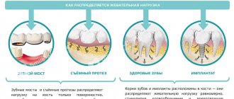
How to use prosthetic teeth for periodontitis and periodontal disease
Periodontitis and periodontal disease are gum diseases with different origins. The first pathology occurs due to pathogenic microorganisms, and the cause of the second is a metabolic disorder in tissues. The processes in both cases lead to loose teeth, so it is much more difficult to prosthetize teeth. However, modern dentistry can cope with such situations and return the aesthetics of a healthy smile to the patient.
Differences and characteristics of diseases
Periodontal disease occurs when there is a metabolic disorder, which leads to a failure in the process of renewal of gum tissue. This, in turn, causes loose teeth. In the early stages, the pathology is almost impossible to detect, since there is no pain or bleeding. Other signs include:
- bad breath is noticeable;
- gums become pale;
- the roots of the teeth are exposed.
Important: without treatment, the patient may completely lose teeth.
Periodontitis is different in that it is caused by gum inflammation and in most cases it is a consequence of bacterial gingivitis. The disease has characteristic symptoms:
- swelling and severe pain;
- bad breath;
- redness of the gums.
The acute form is manifested by bleeding and constant pain. Without treatment, gum tissue erodes and cannot hold teeth in place. These pathologies have several treatment options, which are prescribed by the dentist. Advanced stages definitely require one of the prosthetic methods.
How to choose a denture for periodontal disease
The disease cannot be completely cured at the moment, but medicine can achieve stable remission and a stable condition. If the disease is detected at an early stage, then therapeutic methods are used. If the condition is neglected, the teeth will need to be completely removed and replaced with dentures.
In any case, dentists carry out treatment to achieve remission of periodontal disease and stop the loss of bone tissue. Only after this, a prosthetic option is selected, using removable or non-removable orthopedic structures:
- zirconium crowns, which are placed on your own ground teeth. Usually they are placed in the smile zone;
- bridges fixed to crowns or abutment teeth. This solution is suitable only for the initial stages of pathology;
- splinting clasp dentures. This is a conditionally removable option that has minimal impact on the oral mucosa;
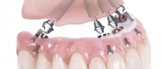
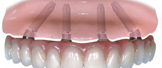
- implant-supported dentures (to avoid the risk of rejection, it is important to regularly treat periodontal disease).
The choice of orthopedic design depends on the clinical case and general health. If the patient still has his own teeth, the doctor will recommend a removable denture. In difficult clinical situations, two methods are suitable: installation of splinting clasps and implantation with immediate loading.
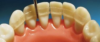
Removable tires
The splinting properties of removable splints are provided by various combinations of continuous support-retaining and reversible clasps, as well as different shapes of occlusal pads. Their spread was facilitated by the development of parallelometry techniques, precision casting on refractory models, and the use of cobalt alloys and precious metal alloys.
Removable splints can be used to splint one group of teeth or the entire dentition. When immobilizing the front teeth, it is advisable to bring the splint to the premolars, and when splinting the lateral teeth, to the canines.
Removable splints can be included in the design of the arch prosthesis as its integral part. These are prosthetic splints:
1) continuous clasp type tire;
2) splint guard;
3) a single splint for the entire dentition.
Orthopedic systems for periodontal disease
Before selecting the optimal option, it is necessary to evaluate the supporting teeth to see whether they can withstand the chewing load. They must not be allowed to fall out or become loose after prosthetics.
Important: before taking impressions, the gums should not be inflamed due to periodontal disease, otherwise the finished prosthesis, crowns or inlays will be uncomfortable to wear. In addition, the risk of root infection will increase.
The most commonly used constructions are:
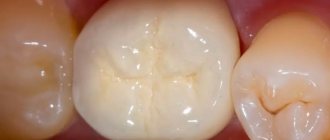
- Zirconium crowns.
For this pathology, metal ceramics are contraindicated and porcelain is not used. Therefore, the patient is offered products made from zirconium dioxide. They are more expensive than other crowns made from other materials, but are biocompatible with gum tissue and provide excellent aesthetics.
- A bridge that is used only in certain cases.
Its peculiarity is that installation requires supporting teeth, inlays or implants. The gums and other tissues experience pressure from such a prosthesis, which gradually leads to bone atrophy, so the system does not last long, about 5 years.
- Implantation, which is possible even in one stage.
Previously, it took time for artificial tooth roots to heal, and only six months later permanent crowns were fixed on them. The one-stage method involves making a small puncture into which a titanium base is inserted and the prosthesis is almost immediately put on. The method is also called momentary loading implantation.
- Clasp prosthesis.
Option suitable for periodontal disease and periodontitis. It does not have high aesthetics, but returns functionality when eating. With the help of a prosthesis, the load on the bone is distributed evenly. Abutment teeth will be required for installation.
Main types of splinting.
The direction of pathological mobility of any tooth is always cut out and depends on its location in the dental arch. For molars and premolars, the lines of their mobility lie in almost parallel planes, for incisors and canines - in planes located at an angle to each other. The best result of splinting is achieved if the splint unites the teeth and the fracture lines lie in intersecting planes.
For the anterior group of teeth, a splint is used to unite the incisors and canines. This is anterior immobilization. It is convenient because, firstly, the periodontium of the canines is less affected and takes on part of the pressure, unloading the weakened periodontium of the incisors, and secondly, the unity of a group of teeth that have the same is restored; thirdly, the teeth are located in an arc, due to this Shinan has greater stability. Immobilization of teeth, in which the splint is located in the anteroposterior direction, is called lateral (sagittal). Lateral immobilization allows you to create a block of teeth that are resistant to forces developing in the vertical, transverse and anteroposterior directions. With the slightest degree of atrophy of the sockets, this is enough to reduce the functional load and obtain a therapeutic effect.
Prosthetics for periodontitis
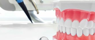
Before choosing one of the methods, the specialist conducts training. It will be approximately the same for periodontitis and periodontal disease. The set of activities includes:
- elimination of caries;
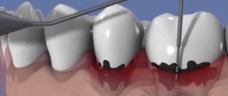
- cleaning gum pockets and teeth from stone and plaque;
- antibacterial and physiotherapy;
- removal of loose teeth.
Until recently, many types of prosthetics had contraindications for periodontal tissue diseases. The problem is that pathologies lead to thinning of the bone and loosening of the teeth.
But now prosthetics have become possible subject to certain conditions, including:
- installation of certain types of systems;
- stabilization of the pathological process;
- use of special materials.
As with periodontal disease, bridges are used at the initial stage. To restore chewing and aesthetic functions, clasps and prosthetics on implants are more often used.
Help: there is also the option of a complete denture (densary jaw) made of acrylic or nylon, but it, like the “bridge,” is rarely used because it does not prevent bone loss.
News
services » orthopedic dentistry » orthopedic treatment of periodontitis
The role of orthopedic methods in the complex treatment of periodontal diseases
Currently, there are two main groups of periodontal diseases:
1. inflammatory periodontal diseases – gingivitis, periodontitis;
2. dystrophic diseases – periodontal disease.
Inflammatory processes in the periodontium are widespread; periodontal disease is a rare disease and accounts for 2-3% of all periodontal diseases.
Systematization of periodontal diseases, data on the symptoms and genesis of each nosological form of periodontal disease made it possible to substantiate an integrated approach to the examination and treatment of patients.
Gingivitis is inflammation of the gum mucosa. Like any inflammation, gingivitis can be
considered as a protective-adaptive reaction of the entire organism to the action of a pathogenic stimulus, manifested at the site of tissue damage by changes in blood circulation, increased vascular permeability, edema, degeneration or proliferation of cells. In accordance with the classification of periodontal diseases, the following forms of marginal periodontal diseases are included in the gingivitis group: serous (catarrhal), hypertrophic (proliferative), necrotic. Of these forms, serous gingivitis is the most common. In the clinic of orthopedic dentistry, a type of gingivitis is encountered - papillitis - inflammation of the gingival interdental papilla.
One of the etiological factors of serous and hypertrophic gingivitis is anomalies in the development of the dental system and medical interventions.
Periodontitis is a collective concept of individual nosological forms of damage
dental system. They are characterized by a chronic or acute inflammatory process of periodontal tissue and atrophy of the bone tissue of the alveolar process of the jaw. The clinical manifestations of periodontitis are not diverse, despite the variety of causes leading to these diseases. One of the main methods of treating the disease at all stages is orthopedic.
Diagnosis and differential diagnosis.
The diagnosis is made based on the clinical picture, the degree of nature and extent of the process. The sudden appearance of symptoms, identification from the anamnesis of previous infectious diseases in the period immediately before going to the doctor, indicate acute serous gingivitis. A history of complaints of periodic bleeding, cyanosis and congestion, especially in the gingival papillae and marginal gums, are signs of exacerbation of chronic gingivitis. An acute onset on the 2-3rd day after fixation of a crown, bridge, or filling with localization of the process in the area of the supporting teeth reveals the cause of the disease. If the process is also widespread in the area of teeth that have not undergone orthopedic interventions, it is difficult to differentiate traumatic gingivitis from acute gingivitis of another etiology, which is an independent nosological form. It cannot be ruled out that the fixation of the prosthesis coincides with the development of gingivitis of various etiologies. It should also be remembered that fixation of both fixed and removable dentures in the oral cavity in patients with chronic gingivitis, as a rule, leads to an exacerbation of the disease.
Swelling of the gums in severe gingivitis can mimic a periodontal pocket. Therefore, in order to differentiate gingivitis from periodontitis, it is necessary to conduct an X-ray examination. With gingivitis, no changes in bone tissue are detected. In severe cases, if the presence of general somatic diseases is suspected, a request to the district clinic is necessary.
Establishing the diagnosis and etiological moment of the focal form of gingivitis, which developed as a result of the lack of approximal contacts, abnormal tooth position, and crowding of teeth, is not difficult. The presence of tartar indicates a chronic process.
In case of focal gingivitis and the presence of artificial crowns, it is necessary to find out and differentiate all possible causes that led to the development of the disease.
First of all, the correctness of the reconstructed anatomical shape and especially the presence and severity of the equator are established. Then, using a probe, the accuracy of the fit of the edge of the crown to the neck of the tooth, the depth of its immersion, and the presence of cervical carious cavities are determined.
With an elongated crown edge, in the elongation zone there are roller-like compactions of the gingival edge and a false gingival pocket. An attempt to reach the edge of the crown with a probe is unsuccessful and causes severe pain. A survey of the patient allows us to establish that when fitting the crown, pain was felt, which was repeated when the prosthesis was fixed with cement. With a wide crown, the gingival margin is loosened, and the edge of the crown is determined with a probe. When moving the probe from a vertical to a horizontal position and moving it towards the tooth, a distance greater than the thickness of the crown is determined. If the crown is wide but not long, there is no pain during fitting. The phenomena of inflammation after fixation of crowns occur after several days or even weeks.
In case of periodontitis, anamnesis data, clinical and radiological picture, indicating a generalized process in the periodontium of all teeth, occurring against the background of general somatic diseases, reflect the endogenous initial factor, determine the diagnosis and specific treatment.
When additional local factors, in addition to generalized damage, are added to this disease, the uneven degree of periodontal tissue resorption in individual teeth or a group of teeth is determined. Knowledge of the specifics of destructive processes under the influence of locally acting factors and a targeted examination of the general condition of the body make it possible to clarify not only the diagnosis, but also the severity of the process in the dentition and its individual links. The diagnosis can be clarified by the following additions: periodontal abscess, hypertrophic gingivitis, traumatic node in the area of the anterior teeth. In fact, these additions to the diagnosis should be regarded as complications of periodontitis.
To confirm the endogenous genesis of the disease, additional research methods should be used: a blood test for glucose content (for diabetes), determination of C-reactive protein, sialic acid content in the blood (for rheumatism, hepatitis, cholecystitis), i.e. it is necessary to conduct all studies to determine the severity of the general somatic disease. In these cases, it is necessary to maintain close contact with a general practitioner, endocrinologist, neurologist, gastroenterologist, which will allow you to correctly determine the general medical treatment tactics and eliminate repeated and often unnecessary research methods.
To diagnose the severity of the pathological process in the periodontium, indicators of hydroxyproline excretion in the urine, the level of citric acid in the blood serum, the content of glucoproteins in it and the redistribution of their fractions can be used.
Particular difficulties arise when deciding whether chronic periodontitis in a given patient is of endogenous or exogenous origin. These difficulties arise because in a number of individuals, during examination, etiological factors of local action are established against the background of general somatic diseases.
Determination of localized chronic periodontitis by clinical parameters predetermines the need for differential diagnosis with chronic osteomyelitis and eosyl granuloma.
When carrying out differential diagnosis with chronic osteomyelitis, an X-ray examination is crucial, in which the absence of sequesters, zones of sequestral separation in combination with the absence of fistulous tracts and scars from incisions on the mucous membrane, a careful analysis of anamnestic data indicate periodontitis.
The differential diagnosis of localized periodontitis and eosinophilic granuloma should be based on the primary complaint of constant, often unreasonably increasing pain in a certain group of teeth. This symptom should alert the doctor if a symptom complex characteristic of periodontitis has been identified, and obliges him to conduct an X-ray examination of the bones of the skull, phalanges of the hands and feet, and a blood test. The establishment of local changes in these bones in combination with eosinophilia speaks in favor of the presence of an eosinophilic granuloma in the subject. Manifestations in the oral cavity of this disease are often the first symptom of the disease, and the x-ray picture cannot serve as the basis for differential diagnosis; since the structural changes on the x-ray are similar to periodontitis. It is natural that patients diagnosed with chronic osteomyelitis, eosinophilic granuloma are not subject to orthopedic treatment methods during the period of detection of the disease,
When carrying out differential diagnosis of periodontal diseases, in addition to the currently accepted classification, it is recommended to use the following systematization of diagnoses of periodontal diseases, clinically justified, taking into account possible concomitant diseases in the dental system itself;
1) chronic generalized periodontitis (in the acute stage, remission) can occur with preserved dentition, combined with partial absence of teeth, abnormalities in the development of the jaws, and pathological abrasion;
2) chronic localized focal periodontitis - traumatic node (in the acute stage, remission).
When diagnosing traumatic nodes, it is necessary to determine not only the degree of periodontal damage, but also to establish the etiological factor in each specific situation using the method of questioning and examination and to trace the pathogenesis of the disease.
The role of orthopedic methods in the complex treatment of periodontal diseases
A comprehensive treatment method involves identifying etiological factors and clearly defining the pathogenetic mechanism and leading links of the disease. This is necessary to determine the means of etiotropic and pathogenetic therapy and to develop a specific plan for managing the patient.
Orthopedic methods used to treat periodontal diseases make it possible to relieve inflammation, improve blood circulation, and, consequently, tissue trophism by eliminating pathological mobility, normalizing occlusal relationships, removing the traumatic effect of chewing pressure, i.e. they can be classified as functional therapy methods. The theoretical basis for the use of these methods, fully confirmed by clinical observations, is as follows:
1. with periodontitis, there is a violation of the histofunctional correlation of the tooth with the surrounding tissues. Destruction of periodontal tissue leads to a decrease in the area of the ligamentous apparatus and the walls of the alveoli, a change in the topography of compression and tension zones under load, an increase in the specific pressure on the tissue, a change in the nature of the deformation of fibers and bone tissue due to a change in the direction of the spatial displacement of the tooth root.
2. The dynamic function of chewing is changed, but is an additional factor in the influence of the external environment on periodontal tissue.
3. There is a close connection between the chewing function and blood circulation in periodontal tissues.
4. A change in the chewing function causes a violation of histofunctional correlations in the tooth-periodontal system, manifested primarily by circulatory disorders due to changes in vascular tone, the development of reactive, and subsequently congestive hyperemia.
5. The term “injury”, “overload” of the periodontium, as well as “traumatic occlusion”, should be understood as such a change in the chewing function when a tooth or group of teeth is subjected to frequent, extended over time, similar effects of chewing pressure, causing distortion of vascular reactions.
6. The destructive effect of the unchanged function of chewing can manifest itself against the background of the inflammatory-dystrophic process of periodontal tissues developing under the influence of various etiological factors.
7. Pathological mobility of teeth in the initial stage of the disease by tissue edema and is subsequently aggravated by the ensuing destruction of the fibrous apparatus and periodontal bone tissue.
8. Pathological mobility, both unchanged and with impaired chewing function, is a leading factor in the progression of periodontal tissue destruction.
9. Destruction of periodontal tissues significantly reduces their endurance to vertical and especially loads directed at an angle to the long axis of the tooth, reduces the level of adaptation and compensation.
-Restoration of histofunctional correlations in periodontal tissues, -elimination of pathological mobility, -elimination of the destructive effect of the chewing function and normalization of the function itself, and, finally, - connection to the compensatory process of intact or partially damaged periodontal tissue of other teeth in order to normalize blood circulation and tissue trophism is only possible using orthopedic treatment methods.
Stages of prosthetics using implants
After preparing the oral cavity, classic one-stage or basal implantation is performed. The first option involves immediate installation of an implant after tooth extraction. Then the abutment is screwed onto it and a crown or prosthesis is put on. The procedure is different in that it does not require an incision in the gum and detachment of the periosteum; a small puncture is sufficient. After six months, instead of a temporary crown, a permanent crown is fixed; during this period, the implant should take root.
If the tooth has been removed a long time ago, tissue volume is increased or the basal implantation method is used. It lies in the fact that the artificial root is not implanted in the place of the previous one, but where there is enough tissue volume, that is, in a deeper layer (basal). Likewise, it requires no incision, just one puncture. This type of prosthetics is used most often when several adjacent teeth are missing. Immediate load on the bone will improve blood supply to the tissues, which will have a beneficial effect on periodontal diseases.