In this article
- Who is susceptible to developing interdental caries?
- How to detect caries in interdental spaces?
- Features of the treatment of interdental caries
- Interdental caries of the anterior teeth: what should be done?
- Specifics of treatment of interdental caries of chewing teeth
- How is caries between teeth treated when molars and premolars are affected?
- Prevention of interdental caries
Interdental caries is one of the most “inconvenient” and difficult to treat. In this article we will tell you why it appears, how caries between two teeth is treated, and whether its occurrence can be prevented using preventive measures.
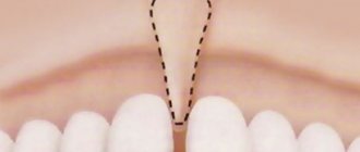
What is interdental caries called and why does it occur? Caries between the lower or upper teeth located next to each other, joint to joint, is called interdental. The denser the arrangement of adjacent teeth in relation to each other, the greater the likelihood of developing carious lesions of the tooth enamel in the area of the joints. When the teeth are tightly packed together, food particles become stuck in the spaces between the teeth, which are difficult to remove with a brush, even if a person regularly brushes their teeth twice a day. To clean the interdental spaces, you need to constantly use dental floss, floss or a special device - an irrigator.
Unfortunately, most people limit themselves to using a toothbrush and toothpaste, as a result of which high-quality cleansing of the interdental spaces does not occur. Accumulated food debris is a favorable environment for the development of microbes and the formation of plaque, which leads to the development of caries between teeth. Due to the location and anatomical features, interdental caries of the anterior teeth most often occurs. Its early detection and timely treatment play an important role from the point of view of not only health, but also aesthetics, because caries between the front teeth, which is located in the most visible place, spoils a person’s smile and appearance. Its specificity is also that the enamel of the front incisors is thinner than the enamel of other teeth, so carious lesions spread deeper and deeper, and treatment presents certain difficulties.
Who is susceptible to developing interdental caries?
Carious lesions on the lateral surfaces of adjacent teeth can occur in anyone, including children. But the following categories of people are especially susceptible to the formation of interdental caries:
- those who eat poorly: consume a lot of sweet foods and drinks, few solid vegetables, fruits and foods rich in fluoride, calcium, phosphorus;
- those who have problems with oral hygiene (do not brush or brush their teeth poorly, do not use dental floss or irrigator to clean the interdental spaces);
- those whose normal salivation process is disrupted;
- when there is a gap between the teeth that is too narrow or closed at the bottom and widened at the top, into which food easily gets clogged.
Manifestations of the disease
The onset of the disease may be asymptomatic. Then the caries spreads deeper, symptoms appear:
- Sensitivity to irritants (sweet, sour, cold foods). The discomfort goes away immediately after eliminating these irritants. Since the painful sensations are short-lived, the patient is in no hurry to see a dentist. But it is important to pay attention to this and see a doctor.
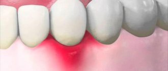
- In bright light, a change in the color of the enamel on the side surfaces is noticeable: grayness of the tissues is noted. Darkened or black enamel is visible between the teeth.
- Regular food getting stuck between teeth. If previously food did not get stuck between the gaps, then with the appearance of caries the patient notices this symptom.
- Bad breath, taste in the mouth.
- Advanced stage: chips of the side walls are observed. Fragile infected walls cannot withstand the chewing load.
How to detect caries in interdental spaces?
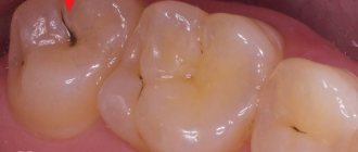
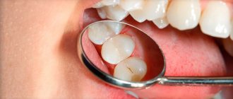
One of the specific features of interdental caries is that it is difficult to diagnose. Since enamel damage is located at the junctions of teeth, it can be quite difficult to detect it yourself. Therefore, caries between teeth is rarely diagnosed at an early stage, when it is possible to manage with conservative methods and not prepare the tooth. Much more often it is detected already in the presence of symptoms in the form of pain or a reaction to cold/hot.
The dentist diagnoses interdental caries using several basic methods:
- External visual inspection using a mirror and probe. Only an experienced dentist can detect areas affected by caries and dark spots on the enamel in hard-to-reach areas.
- If the teeth are very closely spaced, a diagnostic examination may be difficult, and then the dentist will order an x-ray, which will show the presence of a carious cavity in the tooth.
- Sometimes, as an alternative to x-rays, a modern sound diagnostic method is used.
After making a diagnosis, the question arises of how caries between teeth is treated.
Rules for using dental floss
Dental experts recommend the following procedure for cleaning interdental spaces [8]:
- Cut a piece of dental floss about 35-40 cm long. Its ends are wound around the first phalanx of the middle fingers of both hands.
- Pass the floss between the teeth, cleaning the side surfaces of each tooth in turn. You should try not to injure the periodontal papilla.
- Repeat the procedure for cleaning soft deposits for each tooth using a clean section of floss. When finished, the thread must be discarded.
If plaque is not removed regularly, it can gradually harden and turn into tartar. This mineralized deposit clogs the spaces between teeth and can lead to the development of gingivitis, an inflammatory gum disease.
Regular dental examinations (twice a year) and professional hygiene are also necessary conditions for preventing the development of interdental caries. It is these procedures that make it possible to detect the disease at the initial stage of the stain and avoid treatment of caries between teeth.
Features of the treatment of interdental caries
Treatment tactics depend on several factors:
- stages of the carious process;
- localization of caries.
In terms of the depth of damage, interdental caries can vary from the spot stage, when there is no carious cavity yet, but only a section of demineralized enamel is found, to medium and deep caries, when a large carious cavity forms in the dentin and there may be a risk of pulpitis and other complications.
At the stain stage, it is possible to treat teeth using conservative methods, without preparation. As a rule, this is remineralization by simple or deep fluoridation, infiltration method or ozone therapy. These methods strengthen tooth enamel, increase its protective functions and resistance to external influences.
At the stages of superficial, medium and deep caries, the disease will have to be treated surgically - through tooth preparation, removal of affected tissue and further filling of the affected areas.
At the same time, the main difference between interdental caries and other types is that it is necessary to treat not one, but two teeth at once in the area of contact surfaces.
Diagnostics
Whether it will be painful to treat caries on a front tooth depends on the degree of damage. To diagnose it use:
- Fluorescent radiation. Due to the uneven reflection (the carious spot does not glow), the presence of the disease can be determined. The method is suitable for preventive examination.
- Vital staining with a mixture of methylene blue (1%) and red (0.5%) solutions. A carious stain is colored differently than healthy dental tissue. The method is cheap, it also allows you to identify areas of caries, but it is not used for prescribing treatment, because does not show the depth of the carious cavity.
- Electroodontometry. Check the condition of the pulp using an electric current.
- X-ray. The image shows the depth of destruction and the protective reaction of the tooth. The most accurate method by which treatment is prescribed in the most severe cases.
Interdental caries of the anterior teeth: what should be done?
The most unpleasant and “ugly” caries occurs between the front teeth. If the disease is not detected at the spot stage, then treatment will be carried out approximately according to the following scheme:
- a drug with an anesthetic effect is applied to the affected area;
- clean areas affected by caries from dental plaque and other deposits to prevent the spread of pathology to neighboring areas;
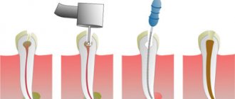
- isolate the affected teeth using a latex napkin (rubber dam) to prevent saliva from getting into them or being injured during dental treatment;
- carious cavities are prepared, the affected tissue is removed on the contact surfaces between the two front teeth using a drill;
- treat the interdental areas with antiseptic solutions (for example, chlorhexidine), and then with adhesive compounds, which improves the fixation of the filling to the tooth walls;
- teeth are filled, and then polished and ground in such a way as to restore their natural anatomical shape and the contours of the interdental spaces as much as possible;
- in case of severe damage, when it is not possible to restore the aesthetics of the front teeth with a filling, they are restored using crowns or veneers.
Both upper and lower teeth are treated in this way.
What kind of caries is this and why does it occur?
The front teeth include the incisors (denies incisivi) and canines (dentes canini), the purpose of which is to bite off individual pieces of food. The main difference from the lateral teeth (premolars (dentes praemolares) and molars (dentes molares)) is the absence of a tuberous chewing surface for grinding. Therefore, caries forms on the crown in the form of a chisel (incisors) or slopes (fangs), in the interdental space, at the junction of the visible part of the tooth and the gum (cervical caries of the front teeth). The tooth is gradually destroyed, “pockets” form at the “neck” - caries spreads deeper, destroying the root.
Modern dentistry is based on the theory of W. D. Miller (1883), who, while conducting experiments, noticed that if an extracted tooth was placed in a fermenting mixture of bread and saliva, something similar to caries appeared on it. At the same time, the density and structure of dental tissue provide protection of teeth from destruction.
Caries is, first of all, problems in the neurohumoral regulation of the body. Normally, the hypothalamus, with the help of the pituitary gland, controls the parotid salivary glands, which secrete parotin. With the help of this substance (in medicine “hormone”), calcium metabolism in the blood-bone tissue-organs system is controlled and stimulates the movement of dental lymph through microscopic tubules inside the teeth. This cleanses and strengthens the dental tissue. Therefore, the main cause of caries is a delay in the production of dental lymph and subsequent demineralization of the tooth. Then the weak tooth in the aggressive environment of the oral cavity is destroyed from the outside.
Caries on front teeth
The dental lymph contains monoblasts, with the help of which the enamel and dentin are nourished. In order for the tooth to be cleaned from the inside, the microscopic structures of monoblasts work like a pump. The nutritious fluid released from the blood is pumped through the dentinal tubules and, under capillary pressure, is pushed towards the enamel, strengthening it. In case of disorders, when capillary pressure decreases, the amount of mineral substances decreases, plaque is literally attracted to the tooth walls (the enamel is like a fine-mesh sieve and allows oral fluid to pass into the tooth).
According to classical dental textbooks, caries is classified as an infectious bacterial disease. Recent research confirms that caries:
- odontoporosis (decreased density of dental tissue);
- odontoclasia (destruction of all layers of the tooth).
Factors influencing the intensity of caries development on the front teeth:
- Excessive consumption of confectionery products without cleaning the mouth for 4 hours (maximum time for the formation of soft plaque). In addition, excess sugar inhibits the normal functioning of the pituitary gland.
- Inflammatory process or removal of the salivary glands (saliva contains a complex of antimicrobial factors, neutralizes the acids of dental plaque and is involved in enamel calcification).
- Environmentally unpleasant factors and work in the metallurgical, mining, coal, chemical industries.
- Reduced general resistance (resistance) of the body.
- Improper development of the jaw, crowded teeth, making it difficult to clean the mouth.
How is caries between teeth treated when molars and premolars are affected?
The algorithm is like this:
- First, anesthesia is performed. If the lesion is shallow, sometimes topical anesthesia is sufficient; for deeper stages, an anesthetic injection is given with a drug to which the patient is not allergic.
- The affected areas are cleaned of plaque and tartar, and a rubber dam is applied.
- Carious cavities are prepared; in most cases, not only the affected tissue is removed from the chewing teeth, but also some of the healthy tissue to provide the necessary access;
- The cavities are treated with disinfectants and treated with adhesives.
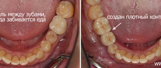
- The tooth is filled with modern photocomposite materials, which are illuminated with special lamps. To restore the side walls of the chewing teeth and ensure tight contact between them, the dentist uses special wedges and matrices.
- At the final stage, the restored surfaces are polished.
Case from practice
I remember a patient who came to see me with advanced caries between her teeth. The destruction was significant, the girl was nervous, afraid not only of the dissection, but also of the injection. It is difficult to work with frightened patients; fears must be dispelled. Before administering the anesthetic, I treated my gums with Lidocaine, the girl realized that it wouldn’t hurt, and she calmed down a little. Fortunately, the pulp was not exposed, and the treatment was successful. But due to deep lesions on the lateral surfaces, both dental units had to be restored with ceramic crowns.
Prevention of interdental caries
Interdental caries is difficult to diagnose and treat; it affects the contact surfaces of several teeth at once. Therefore, it is much easier to prevent its occurrence than to treat it.
The following preventive measures will help reduce the risk of developing caries between teeth:
- brushing your teeth twice a day with fluoride toothpastes (unless your dentist recommends a different toothpaste);
- After each brushing of your teeth, clean the interdental spaces with a special floss;
- use an irrigator: with a powerful stream of water it cleans not only the interdental spaces, but also gum pockets, areas under crowns and braces and other hard-to-reach areas where food debris can accumulate;
- It is recommended to reduce the consumption of foods rich in simple carbohydrates and sugars, because this is what promotes the growth of bacteria in the oral cavity;
- do not snack between meals, especially sweet and sticky foods;
- visit the dentist once every six months for a preventive examination and for professional teeth cleaning using special equipment;
- on the recommendation of a doctor, promptly remineralize the enamel with fluoride and calcium preparations in order to stop the process of carious lesions at an early stage.
Following these simple rules will help reduce the risk of developing caries between teeth several times and maintain a healthy and beautiful smile for a long time.
Treatment of dental discoloration
Before starting treatment, our specialists carry out sanitation of the oral cavity, if dental diseases have been identified, and professional hygienic teeth cleaning. In this way, they eliminate the risk of developing infectious diseases and achieve the best results. The main methods used for treatment are presented in our table:
| Treatment method | Her description |
| Whitening | CELT uses in-office whitening, or more precisely, the latest version of the popular “Zoom 4” system from Philips. During the procedure, the doctor applies a gel containing hydrogen peroxide and potassium phosphate to the tooth surface. The gel itself begins to act under the influence of UV rays: oxygen molecules are released and penetrate into the dental tissues, destroying coloring pigments, which provides the desired whitening effect. |
| Installation of veneers | They are special onlays made of composite or ceramic that are fixed on the outer surface of the teeth. Thus, they are able to disguise the dark color of teeth even if it cannot be removed by whitening. A veneer can be installed on one darkened tooth, or on all units of the frontal group to restore their whiteness. |
| In-canal whitening | The procedure is indicated for deep pigmentation at the root canal level. Hydrogen peroxide is used in the process. Just like in the case of “Zoom4”, under the influence of UV rays, oxygen molecules are released and penetrate into tissues, destroying coloring pigments. In this case, several visits to the dentist will be required to achieve the desired result. |
Painfulness of the procedure
Most people are afraid of the procedure precisely because of toothache, but modern technology and the use of anesthesia eliminate this ailment.
The session is completely painless. To reduce the likelihood of discomfort, all manipulations are carried out after the administration of an anesthetic. If there is no nerve, the procedure is performed without anesthesia. The patient may feel slight pressure on the gum.
An exception is intolerance to certain medications used for anesthesia. The choice is made in favor of a similar drug with a different active substance.
After the injection, no painful sensations are observed.
It hurts if there is a strong inflammatory process at the root apex and in the canals. Unpleasant sensations occur only when the drug gets into the periodontal tissues. They go away instantly after increasing the dosage of the painkiller.
Some pain is possible if there is a deep stage of carious lesions, depulpation or canal treatment is performed, or sensitivity is increased.