Content
hide
1 Pulp - structure, functions, features
2 Why is a tooth without pulp considered dead?
3 How long does a tooth live without pulp?
4 What can lead to the need to remove a “dead” tooth: 4.1 There are several rules by adhering to which patients can prolong the use of the tooth:
Inflammation of the “nerve” of the tooth (pulpitis) is not always accompanied by severe toothache. A chronic infectious process can occur in a tooth unnoticed, leading to significant tooth destruction, the formation of a cyst on the root of the tooth, and even tooth loss. This pathology is an indication for tooth depulpation—removal of its “nerve.” Sometimes such news can come as a surprise to a patient if he did not suspect the presence of chronic pulpitis. At the same time, we are often asked - what will happen to the tooth later, after pulp removal? How long will the tooth live after this procedure, how will its condition change? We'll talk about this in the article.
Tooth removal
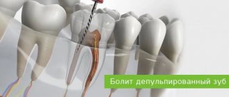
Depulpation (depulpation of a tooth) is the removal of pulp, connective tissue consisting of blood vessels and nerve endings. Removing the tooth nerve and cleaning the canals is designed to relieve the patient from pain, inflammation and further complications. However, the tooth, having lost its blood supply, becomes dead and, therefore, more fragile.
Dental treatment and nerve removal is one of the most common dental procedures. This is partly because patients turn to the dentist only in case of unbearable pain, when it is no longer possible to do without depulpation. Removing the nerve of the tooth and filling the canals allows you to preserve the natural tooth, which is a priority for endodontic treatment.
Indications for depulpation:
- extensive caries
- classic pulpitis and infectious pulpitis (bacteria penetrate through the root apex)
- trauma and damage to the tooth affecting the pulp
- if it is necessary to install crowns or classic bridges
Many patients who come to the dentist's office with toothache are interested in the question: is it possible to save the pulp? Today, there are methods of biological treatment (preservation of the entire pulp) and vital amputation (preservation of the root pulp), but many favorable factors must converge for them to be carried out. For example, biological treatment is not carried out after 25 years.
What ways to get rid of toothache are there?
- Let's start with the fact that if you are experiencing severe throbbing pain, you should not endure the pain. The dentist will conduct a detailed diagnosis and be able to identify the cause; it could be damage to soft tissues or incompletely cleaned canals. Next, you will be prescribed effective treatment.
- Under no circumstances should you search the Internet for methods to relieve pain at home and self-medicate. You need to identify the exact cause and undergo treatment. Otherwise, the disease will progress and inflammatory processes will spread to neighboring teeth.
- If, in addition to pain, you experience any other unpleasant symptoms, for example, severe swelling, discharge, fever, etc., you should also consult a doctor immediately.
Removal of tooth nerve with arsenic
Removing pulp using arsenic is considered an outdated treatment method, which is almost never used today. The procedure was quite simple: the tooth was prepared using a drill, then the root canals were expanded, medicine was applied to remove the nerve in the tooth, after which a temporary filling was installed for 2 - 3 days. The negative side of such treatment is that arsenic is a strong poison that has a detrimental effect on both the tooth and the tissues surrounding it. With the advent of new safe techniques, the need for arsenic has disappeared.
Is it possible to cure such a tooth?
You can cure, but you cannot resurrect. The dentist will remove all affected tissues, nerves, root canals, expand them, clean them and treat them with an antiseptic, and then seal them hermetically so that infection does not enter from outside. The crown, if more than half of it is preserved, will be restored with a filling. For more severe lesions, an artificial crown will be installed. However, in case of extensive inflammation, the tooth can be removed and replaced with a prosthesis - an implant.
A “dead” tooth must be shown to a doctor, otherwise there is a risk of complications from the infection, which will develop into gumboil, osteomyelitis. A cured tooth can perform its function fully, but it can withstand the load worse, so its service life is significantly lower than that of “living” teeth.
Modern method of depulpation
Today, depulpation is carried out using a pulp extractor - a thin metal rod with teeth that can penetrate even remote areas of the root canals. The apex locator device helps determine the depth of the canal to reduce the risk of injury. After the procedure, a temporary filling is usually installed, but a permanent one can also be installed if the doctor does not see the risks of complications and has high-precision instruments on hand, for example, a dental microscope.
Tooth after depulpation - complications
A fragment of the instrument remained in the dental canal
A common complication associated with negligence or inexperience of the doctor. Repeated treatment involves removing instrument fragments and refilling the canals. Due to a number of factors, removal of a foreign body can be very difficult. Pain after removal of a nerve in a tooth does not occur very often, so in some cases the doctor recommends leaving everything as it is if the complication does not manifest itself.
Incompletely removed nerve or insufficient filling of dental canals
The most common reason for pain after depulpation. In such a situation, an inflammatory process develops that spreads to the root apex, provoking the formation of periodontitis, gumboil and cysts. If your tooth aches after nerve removal (several days or more), then you need to consult a doctor who can check the quality of the tooth canal filling.
Resealing
Extraction of filling material beyond the root apex. In mild cases (with a small amount), the tooth hurts when pressed for some time after the nerve is removed, after which the pain goes away. If the excess material is large, then surgical intervention is necessary to avoid the development of complications.
Perforation of the wall or root
Mechanical damage that leads to the formation of a perforation (hole) in the wall, floor of the tooth cavity or root. After the nerves are removed, the tooth continues to hurt. The pain is usually aching and paroxysmal.
Darkened tooth after nerve removal
If the tooth darkens after removal of the nerve, this may be caused by poor filling material or poorly performed depulpation. Also, over time, a dead tooth can become cracked and more vulnerable to dyes. This is partly why the front tooth, after removing the nerve, is subsequently covered with a crown or veneer.
First visit:
Anesthesia or is it painful to remove a nerve from a tooth?
How painful is it to treat pulpitis: It is definitely very painful if you decide to do it without anesthesia. Fortunately, modern anesthetics can completely solve this problem. If you still feel pain after anesthesia, this may be due to the anesthetic not being strong enough or the anesthesia technique being used incorrectly. The latter usually happens when the doctor tries to anesthetize large molars in the lower jaw (mandibular anesthesia, which is complex in technique, is performed there).
An example of anesthesia (video) –
Drilling out all carious tissues with a drill -
Firstly, at this stage all carious tissue is removed. Secondly, healthy tooth tissues are also partially removed, namely all tooth tissues above the pulp chamber and the mouths of the root canals. This is necessary to ensure visualization of the root canal orifices and ease of their processing with instruments. In Fig. 6-7 you can see the boundaries of excision of hard tooth tissues in the treatment of pulpitis. Figure 8 shows a view of the root canal mouths after they have drilled into the required amount of tooth tissue.
Tooth isolation from saliva –
This is done using a rubber dam. Isolation is necessary to prevent infection from the oral cavity from getting into the root canals along with saliva. This is standard international practice, but in Russia a rubber dam can often be seen only when a doctor fills a tooth. Normally, any work with root canals should be carried out using a rubber dam.
Removal of pulp from the tooth crown and root canals –
It is carried out with special tools designed to work in canals.
In Fig. 9 you can see tooth pulp wound around such a tool. By the way, video 1, which we posted above, shows the process of pulp removal. The video below clearly shows the moment when the tooth pulp is removed from the root canal (time – 1 minute 5 seconds). Treatment of pulpitis: video of nerve removal from a tooth
Measuring the length of root canals in a tooth –
This is one of the most important stages, because... if the length of each channel is determined incorrectly, it will cause -
- or underfilling of the canals, which will lead to complications after the end of treatment,
- or refilling the canals, which can lead to long-term pain and injury to the mandibular nerve.
Measuring the length of the canals is ideally carried out using a combination of the x-ray method and the use of an “apex locator”. In this case, first, special K-file instruments are introduced into each root canal in turn (Fig. 10), which are connected to the apex locator using a thin electrode (Fig. 12). The K-files are gradually advanced deeper into the root canal until there is a signal on the apex locator screen that the tip of the instrument has reached the apex of the tooth root.
It is necessary to measure each channel in turn, because The length of each channel is unique and there are no exact standards. After the measurements are completed and the data are recorded, K-files are simultaneously inserted into all channels (each to its own depth), and a control x-ray is taken (Fig. 11). The apex locator sometimes makes mistakes, so the x-ray will show how accurately the length of the canal was measured and whether adjustments are needed.
Mechanical processing of channels –
A budget option for mechanical treatment of root canals involves the use of manual files (K-files or reamers) - in Fig. 13 you can see a K-file in the root canal. The dentist rotates this instrument by the handle with his fingertips, and the cutting edges of the instrument excise chips from the walls of the canal, expanding it. The purpose of mechanical treatment is to widen the canal so that later it can be properly filled.
A better and more expensive processing option involves the use of an endodontic micromotor and special nickel-titanium files with shape memory. Mechanical processing of each channel is carried out to the depth determined at the previous stage. This is necessary to ensure that each root canal is filled exactly to the root apex. During the expansion process, it is very important to constantly rinse the canals with antiseptics, which is necessary for disinfection, but first of all, to wash out the shavings from the canal (24stoma.ru).
Mechanical treatment of root canals:
In video 1, you can see in detail how the expansion of root canals is carried out with ordinary hand instruments (for this, hand-held K-files of different diameters are used - from smaller to larger). In video 2, the dentist processes root canals using an endodontic micromotor and ProTaper Gold nickel-titanium profiles.
Placing a temporary filling –
After the canals are washed and dried to remove excess moisture, turundas soaked in antiseptic are left in them, and a temporary filling is applied to the tooth. The cost of treatment is calculated based on the number of root canals in the tooth.


