Lipoma and skin atheroma are two common types of benign neoplasms. They require exceptionally attentive treatment, since in some cases (though, fortunately, not often) they can degenerate into malignant tumors. The appearance of atheroma may not cause suspicion - at first it usually does not cause much inconvenience. However, even if the tumor is not painful, you should still see a doctor. Often a lump (lipoma) on the neck or scalp gradually increases in size, in this case you need to visit a doctor urgently - the new growth will need to be examined to determine whether there is a risk of developing cancer.
Lipoma and atheroma are often similar in appearance, and patients often do not distinguish them from each other, defining them under the general name “wen.” Let's try to figure out what the difference is between a lipoma and an atheroma, and also what to do if you have one of these formations.
Lipoma
This is a benign formation consisting of adipose tissue. In essence, it is a local accumulation of adipose tissue under the skin. Lipoma is a benign tumor, although in rare cases, liposarcoma, a malignant formation, can develop under its mask.
Lipomas manifest themselves in the form of soft-elastic subcutaneous formations, mobile, painless, and can slowly increase in size. The skin over lipomas is not changed and easily moves over them. Small lipomas are not visible at all; they can only be detected by palpation. Larger lipomas stand out as “bumps” of round or oval shape. The size of lipomas is very variable - from 1-2 cm to 20 cm or more. Lipomas never become inflamed or suppurate.
Causes of lipoma
The general pattern of the appearance of wen is the accumulation of fat cells due to a violation of fat metabolism in the body. If measures to treat the problem are not taken in a timely manner, the formation quickly grows, pinching nearby muscles and blood vessels. Among the causes of lipoma that influence the unfavorable course of the pathology, it is worth noting:
- Hormonal disorders, a period of hormonal changes in the body.
- Metabolic failures.
- Disturbances in the diet with a predominance of food of animal origin.
- Diseases of the kidneys and liver, pancreas and thyroid gland.
- Bad habits.
- Diabetes.
- Genetic predisposition.
Also, the formation of excess adipose tissue is affected by physical inactivity, a sedentary lifestyle and refusal of full-fledged physical activity.
Atheroma
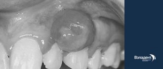
The origin of atheroma is fundamentally different from lipomas. Atheroma develops from the sebaceous glands of the skin. For various reasons, the gland duct becomes clogged, secretion accumulates in the gland, which gradually begins to increase in size. Atheroma is defined as a small (from 0.5 to 2 - 3 cm) formation, which always rises somewhat above the skin and is always fused to it (i.e. the skin above the atheroma does not move), and can grow slowly. Atheroma always has a capsule and contains atheromatous masses resembling crushed lard.
Because The atheroma is connected to the external environment by a duct; there is always a threat that it will become infected through the duct and suppuration will occur. In this situation, moderate pain appears in the area of the previously “quiet” atheroma, the formation quickly (over several days) increases in size, redness appears around it, and body temperature may rise. Suppuration of atheroma requires urgent surgery.
Symptoms of lipomas
The clinical picture of the pathology is quite sparse and is characterized by:
- the presence of a palpable formation, which is distinguished by its soft consistency, mobility, and elasticity;
- the appearance of pain due to compression of nerve trunks or growth in internal organs;
- stability of the compact size or its increase with weight loss;
- swelling of the limbs and disruption of their function (if the formation compresses nerves and blood vessels).
If a malignant process develops, general malaise and headaches appear, blood pressure rises and other characteristic symptoms of intoxication of the body appear.
In this case, depending on the exact location of the compaction, other, more pronounced symptoms and signs may occur:
- neoplasms in the esophagus cause nausea and cough;
- seals on the trachea and bronchi cause a painful dry cough that does not subside after taking antitussive drugs;
- a fatty tumor on cartilage and tendons causes pain in the joints and impedes movement;
- formation in the mammary gland provokes pain in this area;
- compaction in the kidney area causes increased blood pressure, colic and lower back pain;
- a formation in the head causes neurological symptoms – headaches, dizziness;
- lipoma of the neck is accompanied by hoarseness, hoarseness, difficulty swallowing;
- a neoplasm in the heart area causes the development of cardiac pathologies: arrhythmia, heart failure, etc.
Symptoms
Identifying the problem in both cases is usually not difficult.
Signs of lipoma:
- mobile and painless, sizes can range from a few millimeters to 10-15 cm;
- to the touch - doughy or dense;
- not fused to the skin - the skin easily moves over the formation;
- never becomes inflamed – i.e. There is no redness or swelling of the skin above the lipoma.
Most often, lipomas are located on the limbs, head and torso; they are almost never on the face.
Preparation for the procedure
In case of subcutaneous lipoma removal, no special preparation is required. The mini-surgery is performed on an outpatient basis, meaning it does not require hospitalization. The surgeon performs all necessary manipulations under local anesthesia. Thus, the procedure is painless for the patient.
Giant subcutaneous lipomas, as well as neoplasms of the intestines, internal organs, and peritoneum require more serious and thorough preparation. Operations of this type are carried out with hospitalization of the patient. Before the intervention, samples are taken and, if necessary, additional studies are done. Operations performed under general anesthesia require restriction of water and food on the eve of the operation.
Symptoms of atheroma
The formation is in the form of a tubercle, painless, mobile, fused to the skin, you can often see the opening of the excretory duct of the gland. Most often, the “bump” is located on areas of the body where there is hair: on the face, scalp, in the genital area, on the legs, on the back, and is found on the face. If a suppurating atheroma appears, the formation increases in size over several days, the skin on the affected area may turn red and become painful, and the temperature may also rise.
Types of lipomas
There are several main classifications of lipomas that are actively used in medical practice. Depending on the type of tissue that is involved in the pathological process, the following types of neoplasms are distinguished:
- perineural - localized around nerve trunks;
- intermuscular – located between the muscles of the body;
- lumbosacral - grow near the vertebrae or in the spinal canal;
- soft tissues - located on the surface of the skin, less often subcutaneous;
- joints - are located in the synovial membrane or vagina of the joints.
The formation can appear in almost any part of the body and internal organs. Depending on the location of the compaction , the following types of lipomas are most often diagnosed:
- mammary gland - forms in the glandular tissue and deforms the shape of the breast as it grows;
- breasts - a soft and mobile formation that appears in the subcutaneous fatty tissues;
- head - a frequently occurring pathology, which is mainly formed as a result of insufficient hygiene;
- back - one of the most common neoplasms, characterized by extremely slow development;
- neck is a hereditary disease that, during development, can impair the airways, cause weakness and angina.
There are also other, less common places where pathology forms, which include the brain, limbs, skin, peritoneum, eyes, lips and face.
Also, these compactions are divided into two large groups: single and multiple. The first represent a single formation in any part of the body. The latter, accordingly, are characterized by multiple manifestations in different areas of the body and are much less common.
Treatment
Now that we have figured out the difference between a lipoma and an atheroma, let’s move on to the next question: is it necessary to remove the lipoma or remove the atheroma? Let's start with the fact that conservative treatment of lipoma, as well as treatment of atheroma, is absolutely futile. Moreover, aggressive influence on these formations using various “folk” remedies can cause suppuration of atheroma, as well as malignancy (malignancy) of lipoma.
Tactical approaches to the treatment of lipoma are as follows: if the lipoma is small (2-3 cm), does not grow and does not cause any inconvenience (does not rub against clothes, is not a cosmetic defect, etc.), then it does not need to be removed. In case of growth (especially rapid growth), it is better to go for surgery. If the lipoma grows, then sooner or later you will still have to remove it, but it is better to do this while it is small in order to avoid large incisions and traumatic intervention. Any removed lipoma should be sent for histological (under a microscope) examination.
As for atheroma, it is recommended to remove it in any case, because practice shows that sooner or later they fester, and during surgery against the background of inflammation it is not always possible to completely remove the atheroma capsule, which is fraught with relapse (reappearance of the formation). In addition, when suppuration occurs, the wound is almost never sutured; it heals by secondary intention, which often leads to the formation of a rough scar. If, after removing the atheroma, it turns out that the formation does not have a capsule and does not contain atheromatous masses, it should be sent for histological examination to exclude dermatosarcoma, which is sometimes similar in appearance to atheroma.
Reasons for appearance
There are many factors that influence the formation of lipomas. Among the reasons are hereditary predisposition, impaired metabolism of fatty acids in the body, liver disease, pancreatic disease, non-compliance or violation of hygiene rules.
For a long time it was believed that soft tissue injury predisposes to the development of lipomas, but this fact was subsequently refuted in research. Thus, doctors agree that one reason that would explain all the processes has not yet been found. However, predisposition to gastrointestinal lipomas has a proven connection with a gene mutation on chromosome 12. In other cases, the reasons remain unknown.
To sum up all of the above, we can say
- Lipoma and atheroma are benign formations of different natures - lipoma simply consists of altered adipose tissue, and atheroma is made of a sebaceous gland with a capsule filled with secretion - sebaceous atheromatous masses.
- Conservative, incl. Treatment of lipoma with folk remedies, as well as treatment of atheroma, is absolutely ineffective and often harmful.
- A small (2-3 cm) lipoma can not be operated on, but observed. In case of growth, as well as any discomfort, surgery to remove the lipoma is indicated.
- Removal of atheroma is always desirable, because they tend to increase in size and fester.
- If you find a subcutaneous formation in yourself, you need to consult a doctor, because... under the guise of a lipoma or atheroma, other formations can develop - dermatosarcomas, liposarcomas, hygromas, lymphadenitis, etc.
Dr. Elshansky I.V. has been involved in the diagnosis and surgical treatment of benign formations of the skin and subcutaneous tissue for many years.
Methods for diagnosing lipomas
The long period of latent progression greatly complicates the diagnosis of soft tissue lipoma in the early stages of development. More often, pathology is detected when the wen reaches a size of 1.5-2 cm and is easily palpable under the skin. To carry out differentiated diagnostics and confirm the benign nature of the formation, you can:
- Blood tests to exclude viral or bacterial infections, as well as the absence of an inflammatory process.
- X-ray of the chest, limbs or abdomen. Allows you to detect a formation, clarify its location, calculate its size and assess the condition of surrounding tissues.
- Ultrasonography. It is used as an additional diagnostic method when the presence of a lipoma is confirmed by other examination methods.
- Computed and magnetic resonance imaging. Effective methods to determine the location and size of the lipoma, as well as confirm its benign nature.
- A biopsy of wen tissue to analyze it for the risk of cells degenerating into a malignant structure.
If the patient has other diseases of the internal organs, the diagnosis of lipoma is carried out taking into account their characteristics, and deciphering the results suggests the mutual influence of pathologies, making it possible to more accurately determine the cause of the appearance of the wen.
How are atheromas and lipomas treated?
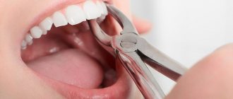
Atheromas are treated surgically; the type of operation will depend on the size of the formation. Small sebaceous cysts can be removed with a laser. Most often, atheromas are large in size and are removed with a scalpel. The “bump” on the skin is surrounded by two incisions, then the cyst is peeled out and removed along with a small piece of skin. Stitches are placed on the wound.
Atheroma cannot be cured by “sucking out” the contents with a needle and syringe. It is imperative to remove the walls of the stretched sebaceous gland - if they remain, they will begin to produce sebum again, and the cyst will grow again.
If atheroma suppurates, surgical treatment is performed, and the doctor may prescribe a course of antibiotics. Lipomas do not need to be treated. The operation is performed if:
- education is large and growing rapidly;
- the lipoma is in an inconvenient place and is constantly in the way;
- bothered by soreness;
- The patient himself insists on removing the lipoma.
Wen is removed in the classic way, using a scalpel. Relapses are possible, but extremely rare. Typically, atheromas and lipomas are removed on an outpatient basis; hospitalization is not necessary. Anesthesia is also not needed - local anesthesia is sufficient. The operation lasts on average 15–20 minutes.
Make an appointment by phone +7 (495) 120-08-07.
Prevention of lipoma
After lipoma removal, the following preventive measures must be observed:
- reduce the consumption of fatty meats and high-fat dairy products;
- introduce vegetable oil and fish into the daily menu;
- refuse late dinners;
- move as much as possible.
You need to follow these recommendations not only if a lipoma has already appeared, but also if there is a hereditary predisposition. Doctors say that the risk group for the formation of wen includes middle-aged and elderly people.
How a doctor can help you remove a wen
It is highly advisable that lipoma removal be performed by an experienced specialist. After a thorough examination, a dermatologist may prescribe the following procedures to remove wen:
- Mechanical cleaning. This method is considered the simplest. The procedure is carried out by piercing the wen with a needle, after which all its contents are removed. Sometimes the procedure is performed under local anesthesia.
- Removal of lipoma with laser . Laser therapy is considered the most progressive method for removing fatty tissue. The procedure is very quick and does not leave scars. In addition, the possibility of the wen reappearing in the same place is excluded.
How to remove wen using traditional methods
If it is not possible to visit a doctor’s office, you can try to remove the lipoma yourself. But this applies only to those formations that have appeared recently. If the lipoma is old, it needs to be treated only surgically.
Baked onion
Onions are considered one of the most effective means for removing wen. Do the procedure at least 2 times a day.
- Take one medium onion and bake in the oven.
- Grind the cooled onion using a meat grinder.
- Grate baby soap, add to onion and mix thoroughly.
- Apply the resulting mass as a compress to the wen.
Sour cream with honey
Before the procedure, you need to take a hot bath to steam the wen. Then the components of the prepared remedy for wen will better penetrate the adipose tissue of the tumor. Carry out the procedure every other day.
- Mix equal amounts of sour cream, honey and sea salt.
- Apply the resulting mixture to the problem area.
- After 20 minutes, rinse with warm water.