Home → Articles → Recommendations after gum surgery
The dental team at the Stomatolog 11 clinic strongly advises that you follow all recommendations after gum surgery. Safe dental treatment using modern dental devices in the clinic creates conditions for a comfortable postoperative period. Maintain your oral health after surgery by following the tips below.
Sign up for a consultation at the dental clinic “Stomatolog11” in the Northern Administrative District:
Make an appointment
What is drainage
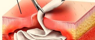
Gum drainage has been performed by surgeons for many decades. It is a tube made of dense materials and its role in the treatment of infectious diseases can hardly be overestimated.
The invention was invented by a surgeon from France, Chassagnac. To create the structure, the doctor used glass and rubber. With the help of such a device, doctors could remove excess fluid and remaining necrotic masses from inflamed tissues. To achieve the desired effect, doctors left the tubes in the patient's body for several days.
And nowadays, surgeons use drainage during operations. However, the appearance of the devices has undergone significant changes: the tubes have become smaller and thinner in size, and modern materials are used to create them. The external drainage has changed, but all its functions have been preserved. It does not allow the incision in the gum to close prematurely; promotes the rapid removal of pus, ichor and necrotic masses from the affected areas; Helps drugs reach deep layers of soft tissue.
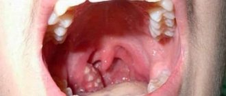
What drainage in soft tissues looks like in the photo
The drainage is left in the mouth until the swelling subsides. This sign indicates complete cleansing of the affected area. For speedy wound regeneration in the postoperative period, it will be necessary to use special ointments, gels and solutions for rinsing the mouth.
To create modern drainages, latex and rubber materials are used. Due to this, the patient practically does not feel discomfort when eating. Waterproof materials are firmly held in the gums and do not allow the tissues to tighten. A small section of the tube remains on the outside of the gum. Through it, pus is removed unnoticed by the patient.
Suction drainage from the pleural cavity
Suction tube
For suction drainage of the pleural cavity, various rubber and synthetic tubes are used.
For the most commonly used drainage, a rubber tube about 40 cm long with several side holes at the end is used. This tube is placed along the lung (from the base to the apex) and passed over the diaphragm from the pleural cavity to the outside. The drainage is attached to the skin with a knotted U-shaped suture. When the suction drain is removed, the threads are tied again, thereby sealing the hole in the chest. A three-barrel suction catheter (Viereck) is advantageous, allowing free passage of the tube inserted inside.
Insertion of suction drainage
In the chest between the two pleural layers, the intrapleural pressure is lower than atmospheric pressure. If air or liquid gets between the pleural layers, then the normal physiological state can only be restored by long-term suction drainage. A closed drainage system is used to suction pleural fluid for recurrent pneumothorax and to treat empyema. This drainage is now usually inserted into the intercostal space through a trocar. The thickness of the drainage tube is determined in accordance with the consistency of the substance being sucked out (air, as well as watery fluid or serous, fibrinous, bloody, purulent fluid).
On the drainage, mark with paint or thread the place to which it will be inserted. The size of the trocar must correspond to the size of the drainage. It is advisable to have at least three trocars of different sizes with suitable tubes of 5, 8 and 12 mm in diameter. Before inserting the trocar, you must make sure that the selected drainage tube passes through it easily.
The site of the skin incision is filtered with novocaine to the pleura. A test puncture in the designated area makes sure that the desired air or liquid is really there. The assistant gives the patient the necessary position: the patient must sit and lean on the highly raised operating table so that the puncture area protrudes as much as possible, and the selected intercostal space is, if possible, expanded. A scalpel is used to cut the skin over an area slightly larger than the size of the trocar. Then the trocar is inserted with a strong movement along the upper edge of the rib into the pleural cavity. After removal of the trocar, unimpeded release of fluid or free entry and exit of air indicates its correct insertion. Drainage is performed and the trocar tube is removed. If you are not convinced that the drainage is in the right place, you should, in order to prevent the trocar from puncturing the lung, heart or large vessel, perform the puncture again, taking all measures to localize it under X-ray control.
Before closing each thoracotomy hole, a drainage is introduced into the pleural cavity, which is brought out above the diaphragm through a separate hole in the intercostal space. Through a hole measuring about 1-2 cm, a forceps is inserted into the pleural cavity under the control of the eyes and under the protection of the left hand to ensure the correct position of the drainage from the inside. The drainage is pulled through the chest wall with a forceps from the inside to the outside. Pay attention to the fact that the drainage section, free from holes, is at least 5 cm in the chest cavity. If the fixation of the drainage to the skin is broken, then it slips out, and the first side hole appears outside the pleural cavity above the skin. In this case, the closed system turns into an open one, suction becomes ineffective, and pneumothorax often occurs.
Suction systems
There are so-called individual (“bed side”) and centralized suction systems. The suction action due to the hydrostatic effect can be obtained by a tube lowered under water, a water or gas pumping device (in this case the action is based on the valve effect) or an electric pump. Both individual and central systems must ensure individual regulation. If the release of air from the lung is insignificant, then due to its simplicity, the Biilau drainage system is still successfully used today, which can be sufficient to straighten the lung. A glass tube immersed under water (disinfectant solution) is equipped with a valve made from a finger cut off from a rubber glove, which protects against reverse suction. The Biilau system uses the physical law of communicating vessels to move bottles under the bed to create a suction effect.
The Fricar air pump best meets modern requirements. This device can work for many days without interruption and without heating up. The strength of the suction effect can be precisely adjusted.
Central suction devices are triggered by an oxygen canister system or a powerful suction pump. The system of outgoing tubes, if necessary, supplies hospital departments located on different floors. Depending on the need, the required number of hospital beds can be connected. An oxygen-powered system has the advantage that the suction and supply of oxygen to individual hospital beds is provided by the same tubing system. The suction effect is provided by a valve tube mounted along the oxygen flow. In this case, however, the effect produced by the central suction pump is not achieved.
Individual adjustment can be carried out using a dosimeter tap connected to a well-functioning pressure gauge, or through the so-called. three bottle system. The latter can be easily prepared by yourself. This system also has the advantage that it can easily and reliably create a very low suction effect (from 10 to 20 cm of water column). It is rarely possible to achieve such low pressure values using factory pressure gauges.
Indications for suction drainage
Spontaneous and traumatic pneumothorax, hemothorax
Spontaneous pneumothorax occurs at a young age, more often as a result of rupture of single pulmonary alveoli in the apex of the lung, in older people - as a consequence of rupture of alveolar vesicles with diffuse emphysema. Due to the fact that the number of patients with emphysema is constantly increasing, the number of cases of spontaneous pneumothorax is becoming more frequent. The same applies to traffic accidents that result in closed injuries in the chest cavity, which often occur with pneumothorax or hemothorax.
Correctly performed pleural puncture for spontaneous pneumothorax is practically safe, and its benefits can hardly be disputed. If the flow of air from the damaged lung is completely stopped and the perforation site is closed, then it may be possible to completely remove the air that created the pneumothorax with a simple closed puncture. If pneumothorax recurs after puncture (even repeated), then drainage with long-term suction should be used. Recurrence of pneumothorax, even after prolonged drainage with suction, can only be reliably eliminated by surgery.
Traumatic pneumothorax most often results from rib fractures. When a rib fragment injures the lung, most often a significant amount of air comes out of it, and a tension pneumothorax occurs. At the same time, subcutaneous or even mediastinal emphysema may occur. Spontaneous pneumothorax can also occur when the pulmonary alveoli rupture or due to blunt force on an emphysematous lung. Therefore, in patients with pulmonary emphysema, chest injuries are often associated with the occurrence of pneumothorax, often severe tension pneumothorax. The principles of treatment for spontaneous and traumatic pneumothorax are the same.
If clinical symptoms indicate tension pneumothorax (severe respiratory failure, subcutaneous emphysema, mediastinal shift), then drainage of the pleural cavity should be performed immediately. If these symptoms are not present, then a closed puncture is performed and the air is sucked out. After this, the needle is left inserted into the pleural cavity, and its nozzle is connected to a pressure gauge and the pressure in the pleural cavity is determined (whether it is higher or lower than atmospheric). If the pressure in the pleural cavity is indicated by the pressure gauge needle in the positive direction, it means that air continues to be released into the pleural cavity, and, therefore, drainage is necessary. This issue can, of course, be resolved by X-ray examination. If there is a total pneumothorax, then drains are inserted in two different places. One of them runs along the posterior axillary line above the diaphragm in the VII-VIII intercostal space, the other is inserted along the midclavicular line between the 1st and 2nd ribs. In our experience, drainage inserted under the collarbone performs the task of straightening the apex of the lung better.
In case of encapsulated limited pneumothorax, drainage should be inserted locally, under X-ray control after a test puncture.
Empyema of the pleura
Pleural empyema is a disease for which treatment with suction from the pleural cavity is absolutely indicated.
The principle of treatment of empyema does not depend on the causative agent of the disease. It consists of gluing the pleural layers and eliminating the empyema cavity through early drainage and suction of fluid. Treatment with suction from the pleural cavity is combined with targeted local chemotherapy, based on the identification of the pathogen and its resistance to the drugs used. Most empyemas result from infection of the exudate. In this case, incorrect and insufficient suction from the pleural cavity plays a certain role. In cases where pockets with delimited fluid form in the pleural cavity, their complete emptying becomes increasingly difficult, more difficult, and infection is more likely. In such cases, complete recovery can only be ensured by surgery.
Suction treatment may fail for two reasons: one is the presence of pleural cords, the other is a bronchopleural fistula.
Pleural moorings are often the result of insufficient emptying of the pleural cavity. When moorings have already formed in the pleural cavity and the walls of the empyema cavity are thickened, there is little chance of eliminating the empyema by suctioning out the fluid. The ability to expand the lung in this case is also very controversial. In this case, drainage with suction is a preparatory measure before the inevitable operation. Radical surgery (decortication) is performed only after the patient’s general condition has improved by washing the pleural cavity and targeted antibiotic therapy.
Bronchopleural fistula reduces the effectiveness of suction and thereby the prospect of lung expansion. In cases where there is a large bronchial fistula and its closure is contraindicated (for example, a rupture of the cavity, tumor disintegration, rupture of a cystic, emphysematous lung that has lost its elasticity), success cannot be expected from the use of suction. On the other hand, suction can also be used in cases where surgery is indicated. In elderly patients, with low general resistance and the possibility of severe complications, surgery becomes impossible. Then all that remains is to leave the patient with permanent drainage.
In case of chronic pleural empyema, drainage should be inserted into the pleural cavity at its lowest point. Large-diameter drains are used so that the thick liquid does not close the lumen and it is easy to wash the pleural cavity. Often, in the area where the drainage will be introduced, a rib resection (2-3 cm) is performed.
Postoperative suction from the pleural cavity
In order to remove fluid that accumulates after thoracotomy from the pleural cavity and maintain normal intrapleural pressure, a suction drain should be available.
If during pleural operations and mediastinal, transthoracic interventions on the esophagus, stomach, heart and large vessels there was no damage to the lung, then the chest can be closed with the introduction of one perforated drainage into the pleural cavity. Drainage is carried out above the diaphragm along the midaxillary line with its pleural end installed at the level of the apex of the lung.
Two drains are inserted into the pleural cavity if the lung was damaged during separation of adhesions, as well as after resection or excision of lung tissue. In such cases, one of the drains is inserted along the anterior and the second along the posterior axillary line. The use of a third drainage may be considered relatively appropriate when it is brought to the site of anastomosis of the esophagus or bronchus or when thoracoplasty is performed in combination with lung resection (for suction from the subscapular space).
After removing the lung, one drain with a diameter of 12-15 mm is inserted into the pleural cavity and placed in the lower part of the cavity so that a piece of drainage 10-12 cm long is equipped with 2-3 side holes. Active suction through this drain is prohibited.
After median sternotomy, a drainage is inserted retrosternally and its second end is brought out in the epigastrium.
Intensity and duration of suction
The intensity of suction through drainage from the pleural cavity depends on the cause of the disease, the condition of the lung and the nature of the operation. The flow of air from the lung into the pleural cavity is of decisive importance. If this occurs, then more air should be sucked out of the pleural cavity per unit time than is supplied there. Only in this way can gluing of the pleural layers be achieved. In practice, however, this is often not feasible. If the connection of the bronchus with the pleural cavity is significant (for example, in the case of a bronchial fistula), then intensive suction cannot achieve the goal. If you increase the suction force, then at the same time the patient will experience increased respiratory failure due to “air theft” from the tidal volume. Despite this, the lung will not be able to expand. In such cases, surgery is inevitable.
When the lung is damaged or after lung surgery, air most often escapes from a hole the size of a pinprick. In this case, specialized suction is indicated. In children and adolescents, due to the fact that their lung parenchyma is healthy and not affected by fibrosis and emphysema, it does not matter with what force the suction is performed. It doesn’t matter whether they suction with an intensity of 25 cm of water. Art. or simple underwater drainage, the lung will expand in 24-48 hours. The drainage can be removed after 48-72 hours. This is the advantage of elastic tissue capable of lung retraction in young patients. With emphysematous lung in an elderly person, the situation is different. The pinprick holes become gaping holes in the lung because the surrounding tissue is unable to contract. If you try to reduce the flow of air coming from the damaged lung by increasing the intensity of suction, you can easily get a paradoxical effect. The flow of air from the lung will increase. Small holes, due to prolonged suction, stabilize and turn into fistulas.
What to do in such cases? Begin gentle suction from the pleural cavity (5-6 cm of water column) and pay attention to ensure that tension pneumothorax does not occur. Thanks to this, the resulting fibrin seals small holes in the lung. Within 24 hours, a decrease in the release of air from the damaged lung begins to be detected. The intensity of suction can be increased slightly. On the fourth day you can already suction with an intensity of 10 cm of water. Art., if no unforeseen complications arise, then the drainage can be removed on day 4-5.
The same principles are followed when treating spontaneous and traumatic pneumothorax with suction.
If there is a significant intake of air from the emphysematous lung, they begin to carefully perform suction with a gradual increase in its intensity. If, after many days of treatment with suction, the release of air from the lung does not stop, then it is recommended to immediately undertake surgery, without waiting for the development of infection in the pleural cavity. If suction from the pleural cavity continues for more than a week, the development of infection becomes real.
In cases where the patient does not undergo surgery due to low general resistance, suction from the pleural cavity remains to be continued. Prolonged and specialized suction under the guise of drug treatment may be more or less effective. The pleural layers are glued together completely or partially. Only small limited cavities remain that do not lead to complications. The drain can be removed.
In the treatment of pleural empyema, long-term use of suction drainage is a common method. The empyema cavity gradually becomes smaller and smaller, the amount of fluid decreases, and in the end it can become bacteriologically sterile. If the daily amount of fluid extracted from the pleural cavity does not exceed 10-15 ml, then the suction is stopped, the drainage is shortened, but left until the residual cavity is completely closed.
When is the procedure scheduled?
Drainage is necessary to remove purulent contents after a gum incision or tooth extraction. The passage of pus through a rubber tube is called the drainage process.
The procedure has 2 main goals:
- complete elimination of purulent exudate;
- preventing gum healing.
Saliva contains special substances that stimulate the regeneration of damaged soft tissues. Because of this, pathogenic microorganisms can remain inside the gums without having time to come out along with the necrotic masses. The condition provokes repeated exacerbation of inflammatory processes. In addition, epithelial cells are characterized by rapid division, which also contributes to the rapid healing of the wound after gum excision.
Drainage is required in cases where purulent formations in the oral cavity have been removed - abscess, gumboil, phlegmon. In this case, the doctor cuts the gum and places a drainage
For complete release of exudate from the wound, it is necessary to slow down the healing of the epithelium. A tape or tube type device does not allow the edges of the wound to converge prematurely.
How long does the drainage last? The answer to this question can be given by the surgeon after assessing the condition of the gums in the postoperative period. The device is removed after signs of swelling and inflammation of the soft tissue disappear.
If the tube is removed prematurely or not installed at all, the exudate will spread to nearby tissues and subcutaneous fat. The condition can cause dangerous infectious complications that lead to health problems, even death.
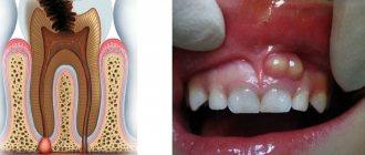
How long does drainage last after a gum incision? On average, this period is 1-2 days. The period can be extended after removal of abscesses and phlegmons to 1 week. If an infectious-inflammatory disease is accompanied by fever, then usually after installation of drainage this symptom disappears within a few days.
The installation is used not only after a gum incision, but also after tooth removal. Extraction in the first few days is also dangerous for the development of infectious complications. Installing a drainage tube reduces the risk of negative consequences and prevents gum infection.
The scheme is also used to accelerate the delivery of drugs to the pathological focus. This method simplifies the treatment of soft tissues located in hard-to-reach areas.
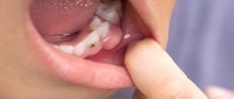
Flux is one of the common complications after tooth extraction performed with gum cutting
If the first signs of an abscess or flux occur, you should immediately consult a doctor. The methods of modern dentistry allow you to quickly and painlessly cope with the problem, while traditional medicine and self-medication can only aggravate the course of the disease.
How long can drainage last in the gum?
The drainage structure can be installed for a period of 3 to 6 days. This time is usually sufficient for the complete release of the pathogenic fluid. If after a week the swelling does not disappear, additional examination and a change in medical procedures will be required. This situation may arise after the removal of a wisdom tooth in the upper jaw. The drainage system is removed when the swelling completely subsides, which indicates complete cleansing of the wound area. Now the dentist’s main task is to speed up the healing process.
The doctor determines when to remove the tube or strip. It depends on many circumstances.
- In addition to a visual examination, the dentist may refer the patient for repeat images.
- It is important that yellow and whitish pus, as well as ichor and translucent liquid come out of the wound.
- The patient must close his jaws freely and chew food.
- The redness of the mucous membrane should gradually change to a normal pink color.
The speed of release of pathological fluid is influenced by the patient’s compliance with the doctor’s recommendations. He should eat liquid pureed food, drink enough clean water, and avoid sweets and solid foods.
Contraindications
Dissection of soft tissues with further installation of drainage is not possible in all cases. Among the prohibitions to the procedure are:
- pathologies associated with blood clotting disorders;
- allergy to painkillers used before the procedure.
In the latter case, the doctor must select a drug that does not cause adverse reactions in the patient. There are no other contraindications to drainage. If the cheek is swollen, and necrotic masses have accumulated in the periodontal tissues, then the easiest way to deal with the problem is by installing drainage.
Stages of drainage
The procedure can only be performed by a specialist. Trimming the gums and installing a rubber tube occurs in several stages:
- Examination of the oral cavity by a dentist.
- Taking an x-ray to determine the depth of the tumor and the extent of its spread to adjacent tissues.
- Injection of an anesthetic into the problem area. Does it hurt to cut your gum? The procedure is accompanied by significant discomfort, so anesthesia is used to minimize pain.
- Resection of the seal using a scalpel.
- Cleaning the purulent cavity using antiseptic agents.
- Fixation of a rubber tube to drain pus.
Using an X-ray of the oral cavity, the doctor can examine the condition of the roots of the teeth and, if necessary, prescribe extraction of problematic units
In severe cases, the drainage is left in the oral cavity for 3-5 days, until the swelling subsides and the ichor stops oozing from the wound. The drain may fall out on its own or be removed by a doctor. If swelling and inflammation persist 5 days after the intervention, you should immediately contact your dentist.
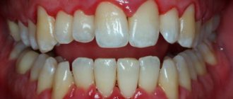
The gum has moved away from the tooth: treatment
If the gum has moved away from the tooth, treatment will largely depend on the severity of the inflammation. For example, if a patient seeks help at a fairly early stage of inflammation (when the depth of periodontal pockets does not yet reach 3 mm and there is no tooth mobility), significant success can be achieved in the treatment of periodontitis and the process can be completely stopped. Advanced cases of gum inflammation will require much more complex treatment and serious financial costs. Next we list the main stages of treatment.
1) Consultation and x-ray diagnostics –
It is necessary to begin treatment with a consultation with a periodontist and a panoramic photograph of the teeth, which will allow you to create an optimal treatment plan taking into account the condition of your teeth and gums. The image will allow you to determine the amount of destruction of bone tissue around each tooth, the location and depth of periodontal pockets, and will help guide the patient in the need for splinting of mobile teeth, the need for prosthetic replacement of missing teeth, and make the correct diagnosis.
An example of a panoramic image of a patient with periodontitis –
Looking closely at the image, you may notice that the level of bone tissue (the bone looks in the image as fine-mesh looped tissue and normally should reach the necks of the teeth) is reduced in different teeth by 1/4 to 4/5 of the length of their roots. The patient has decayed teeth that need to be removed, as well as carious teeth that require treatment (24stoma.ru). The level of bone tissue is most reduced in the front teeth of the upper and lower jaw, which in this case was the result of not only inflammation of the gums, but also the absence of the chewing group of teeth.
To treat gums, it is best to contact not ordinary dental therapists or hygienists, but periodontists. These are the dentists who specialize in gum treatment. The first and most important stage of treatment will be ultrasonic teeth cleaning. Next, a course of anti-inflammatory therapy is carried out, which in most cases can be successfully carried out at home.
2) Removal of dental plaque –
First of all, it is necessary to remove the cause of inflammation - microbial plaque and dental deposits. They are removed from the teeth using an ultrasonic scaler (Fig. 15), usually over several visits. It is simply impossible to remove all dental deposits in just 1 visit in a patient with periodontitis, because... It takes a lot of time to remove subgingival dental plaque, which is localized in periodontal pockets below the gum level.
It is subgingival dental plaque that poses the main danger for further progression of inflammation, so sometimes it is necessary to prescribe the patient even 3-5 times. Learn that without professional cleaning, all other stages (for example, anti-inflammatory therapy, splinting) will be completely meaningless.
Most dentists and hygienists won't bother with finding and removing subgingival calculus...as experience has shown. Therefore, it is very important to find a competent specialist. Anti-inflammatory therapy is prescribed immediately after 1 session of removing tartar, and within the first 24 hours you will be able to notice changes in the appearance of the gums. In parallel with the reduction in gum swelling, their volume will also decrease, which will allow you to see new portions of dental plaque and remove them during subsequent visits.
Removing dental plaque with ultrasound: video
You can see in the following photos what happens when subgingival tartar is removed poorly. The first photograph shows that the gums are visually in good condition, although a periodontal probe revealed the presence of a periodontal pocket about 5 mm deep. The second photograph was taken after the gums were detached from the teeth, and it shows a very large amount of destruction of bone tissue, which arose due to the presence of small subgingival tartar on the surface of the tooth root.
3) Anti-inflammatory therapy –
The course of anti-inflammatory therapy usually lasts 10 days. In most cases, it is carried out by the patient himself at home, after doctor’s prescriptions and patient education. However, if the patient has deep periodontal pockets with purulent discharge, the doctor may additionally prescribe washing the periodontal pockets, which is done using a syringe and antiseptic solutions. In some cases, antibiotic therapy may also be prescribed for periodontitis.
Anti-inflammatory treatment regimen - usually a complex is prescribed, consisting of an antiseptic mouth rinse and application of gel to the gums. The procedures are carried out 2 times a day (morning and evening, after meals and subsequent oral hygiene), for 10 days. Most often, a complex of the following drugs is prescribed:
- rinsing with Chlorhexidine (instructions)
- “Cholisal-gel” applications (instructions)
In patients with periodontitis, the correct choice of treatment agents is very important. For example, a standard 0.05% solution of chlorhexidine, sold in pharmacies for 40 rubles, is advisable to use only for superficial inflammation of the gums (gingivitis), but for periodontitis it is better to use a 0.2-0.25% concentration of this antiseptic. It is a big plus when such a solution contains not only a good concentration of chlorhexidine, but also other active ingredients (for example, aluminum lactate for bleeding gums or extracts of medicinal plants).
Antiseptic mouth rinse is carried out after breakfast and oral hygiene, rinse your mouth for 1 minute. After this, the gums need to be dried with a dry gauze pad to remove excess saliva, and an anti-inflammatory gel should be applied to the gum edge. The gel is applied in front of a mirror using a finger, and usually only on the front side of the teeth. After such treatment, it is not recommended to eat for 2-3 hours, and also not to drink for 30 minutes. There are a lot of remedies for treating gums, and we hope that our next articles will help you understand their diversity -
→ The best antiseptics for mouth rinsing, → Rating of the best gels for treating gums.
4) Oral hygiene training –
If you have not yet forgotten the beginning of the article, then remember that the cause of periodontitis and destruction of the dental-gingival attachment is unsatisfactory oral hygiene, which leads to the accumulation of microbial plaque and tartar on the teeth. Therefore, in addition to the basic treatment that we described above, it is very important to constantly maintain high-quality oral hygiene.
Good hygiene doesn't mean just brushing your teeth twice a day, that's not enough. Good oral hygiene includes brushing your teeth after every meal, not snacking on cookies or candy between meals, and regularly using dental floss. You can read about absolutely all the recommendations in the article at the link above. Below, you can watch the video on how to properly use a toothbrush and dental floss.
How to use dental floss and brush correctly -
In the presence of periodontal pockets and mobile gingival margins, it is important to carry out hygiene below the gum level. This can be done using special devices - home irrigators. Special attachments allow you to rinse at home not only areas of the oral cavity that are difficult to reach for hygiene, but also periodontal pockets below the level of the gingival margin. Irrigators can use both ordinary boiled water and special medicinal solutions.
What to do if the drainage falls out?
What to do if the implant falls out of the gums prematurely? The answer to the question depends on the characteristics of the clinical case. If the swelling has subsided and the patient is not bothered by pain, then there is no need to contact the clinic to re-install the device. You cannot remove drainage from the oral cavity on your own even if the problem signs have completely disappeared.
Among the reasons for the installation to fall out on its own, the following should be noted:
- active rinsing of the mouth and thorough brushing of teeth;
- improper fixation of the structure over the tooth.
Re-installation is required if the gums still hurt after the rubber tube falls out. It is better if the procedure for installing the system is carried out by a doctor in a clinic. Only in rare cases do specialists allow patients to remove the drainage themselves when signs of swelling and inflammation disappear.
Blister on the gum - what is it?
The procedure is carried out independently, taking into account the rules:
- Hands are washed thoroughly with soap.
- The oral cavity is treated with antiseptic solutions.
- The tape or tube is removed in front of a mirror. Grasp the free edge of the material with your thumb and index finger.
The procedure is accompanied by minor pain and blood loss. All of these signs are normal. You cannot interrupt treatment immediately after installing the tube.
The wound left after drainage should be regularly treated with antiseptics
How to rinse your mouth? Miramistin, Chlorhexidine solution, and hydrogen peroxide solution are suitable for this purpose. The procedure is necessary to prevent recurrence of the problem.
An abscess or gumboil is treated comprehensively. Pathology cannot be treated with medications alone. Therapy should include the installation of drainage, local treatment of the affected area and physiotherapeutic procedures carried out after opening the purulent focus.
Incision healing time
How long will it take for the gums to heal after drainage is removed from it? On average, tissue regeneration takes 1-2 weeks. The process largely depends on the complexity of the previous operation and the individual characteristics of the patient. For example, in older people, wounds take longer to heal. This fact is explained by the slowdown of metabolic processes with age.
Complete gum regeneration may take more than 6 months. The wound healing process after opening the abscess takes place in several stages:
- the formation of a blood clot that protects the wound from the introduction of pathogenic microorganisms;
- formation of new granulation tissue (within 3-4 days from the moment of intervention);
- maturation of new epithelium (7-10 days from the moment of opening of the purulent focus);
- soft tissue regeneration;
- formation of young bone tissue.
After the operation, the dentist gives the patient a number of instructions that help maintain drainage in the gums and avoid the development of complications after the intervention. In the first 3-4 hours after the procedure, it is prohibited to consume food. During treatment, it is advisable to redistribute the chewing load to the healthy side of the jaw. Doctors also advise giving preference to dietary products.
In the postoperative period, it is necessary to avoid intense physical activity and avoid visiting saunas, swimming pools and baths. It is also important to give up bad habits - smoking and drinking alcohol.