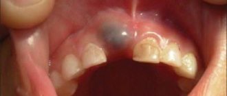
An even row of teeth is a sign of beauty and health. Although many people dream of a Hollywood smile only for aesthetic reasons, in fact, a correct bite is very important for the whole body. Therefore, dentists recommend resorting to orthodontic correction immediately after identifying a problem. Protruding teeth can be either a congenital defect or an acquired one. Modern medicine has something to offer in both cases.
What is a gap from a dental point of view?
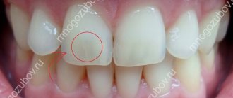
In dentists' language, the gap between teeth is called diastema (from Greek - “gap, distance”). According to statistics, varying degrees of this defect are present in every fifth adult on the planet. In childhood, increased interdental space is often temporary; it is observed in 50% of preschool children. Diastema refers to an abnormally large empty space between the incisors (can reach 10 mm).
Patients often confuse a diastema (a gap between “units”) with a trema (a gap between any teeth in a row), although formally these are completely different defects, although similar in essence. Accordingly, the approach to treatment/elimination is different in both cases.
Blind man regains vision after tooth implantation in eye
Englishman Martin Jones was blind for a decade. However, in 2009 he saw the light. This happened after a very unusual medical procedure: doctors implanted a fragment of a tooth into his eye. It was the fang that doctors removed from Jones' mouth. Doctors inserted an artificial eye lens into the base of the fang and placed this entire structure in the eye, thus allowing the lens to grow to the desired size. A small piece of skin was also taken from Martin’s mouth, from which a special flap with its own circulatory system was then made. In addition, doctors had to reduce the hole in the cornea of the eye so that it lets less light into the eye. This procedure has helped restore vision to six patients.
Reasons for the appearance and enlargement of gaps between teeth
The gap between the front teeth is a result of internal or external factors. Most often it occurs as a result of:
The cause may also be the presence of microdentia, which inherently affects the symmetry of the teeth.
An increase in an existing gap occurs for the following reasons:
- abuse of chewing seeds;
- progressive problems with the root system;
- the appearance/development of various types of orthodontic pathologies;
- Frequent chewing of hard food (particularly with the front teeth).
Correct and recommended prevention will help maintain the diastema at the existing level (prevent the distance between the incisors from becoming larger), and the orthodontist will select an individual program for eliminating the defect.
You might be interested in:
Types of teeth bites
Straightening teeth without braces
Treatment and straightening of teeth
Clear braces
Rare tumor in baby's brain turns out to be a tooth
A 4-month-old Maryland baby may be the first person to have a brain tumor that turned out to be a tooth. Doctors first suspected something was wrong when the child's head seemed to begin to grow faster than normal for children his age. A brain scan revealed a tumor that contained structures closely resembling human teeth, usually found in the lower jaw. The tumor was removed and the boy is now feeling well.
Types of diastemas
How the gap between the teeth can be removed depends on the reasons why the gap appeared. Therefore, it is very important to correctly identify the species. Diastemas are classified according to three main characteristics:
- By time of occurrence of the defect
:- False
. It is diagnosed in children when the bite is not yet fully formed. The problem disappears on its own without third-party intervention after replacing baby teeth with permanent ones. - True. Diagnosed in adolescents and adults in the period after the formation of the bite. It occurs due to periodontal pathologies, injuries or the absence of some frontal teeth in a row.
- By location
:- Symmetrical
. The location is determined relative to the conditional center of the dentition (usually oriented along the frenulum). The position of the incisors relative to each other is also taken into account. - Asymmetrical
. Most often, one of the incisors is in its place, and the second is deviated relative to the conditional center. - According to the position of roots and teeth
:- Literal deviation of crowns
. The defect is most often observed in people with a superset of teeth. In this case, the position of the roots is correct, but the incisors themselves deviate. Usually the gap in this case is no more than two millimeters. - Hull literal offset
. A common cause of the defect is excessively compacted bone tissue of the middle suture, which does not allow teeth to erupt in the place where they are needed. It is expressed in the lateral displacement of both the root and the incisor. - Medial tilt
. The most complex defect that can occur with the central incisors. As in the previous case, teeth and roots shift, but they can grow not only to the sides, but also with a displacement around their own axis.
To understand how to remove the gap between the front teeth and which method is best to choose, the dentist must correctly determine the type of diastema. Keep in mind that it is impossible to get rid of a gap on your own or at home - you cannot do this without the help of a specialized doctor.
Teenager has 232 teeth removed
Doctors from JJ Hospital found 232 teeth in the mouth of a 17-year-old boy. They were removed during an operation that lasted more than 6 hours. The hospital's dental department decided to report this case to the Guinness Book of Records as the largest number of teeth extracted from a human mouth. The boy, Ashik Gavai, lives in one of the villages located in the Amravati region. About a year ago, his family noticed a tumor on the boy's lower jaw, but local doctors were unable to determine what the problem was. The boy's family was even afraid that he had cancer. And then Ashik was diagnosed with odontoma, that is, a benign tumor consisting of teeth. It could cause difficulty eating, swallowing, and lead to unsightly, although not dangerous, swelling of the face. Doctors extracted 232 teeth from a tumor in Ashik’s jaw. Calling it a “medical miracle,” doctors said the boy now has a normal set of 28 teeth.
Ashik Hawaii
How can a gap be dangerous?
A small gap between the teeth can be a highlight and be regarded solely as an aesthetic defect. This problem does not bother some people, even those who are often under the gun of video cameras (for example, E. Temnikova, Madonna, K. Novikova, V. Paradis, K. Raikin, etc.). However, in some cases, diastema can cause serious functional disorders. Without therapeutic intervention, the size of the gap will increase, which has the following consequences:
- speech defects (their degree depends on the size of the diastema);
- violation/change in bite (due to uneven chewing load on the upper and lower jaw);
- caries (pathogenic bacteria and hard deposits accumulate in the interdental space; regular home cleaning is not always enough to combat them);
- inflammation and other gum diseases (due to direct contact of the gums with chewed food).
An experienced orthodontist can determine exactly how to remove the gap between the front teeth and whether it is necessary for medical reasons. If the defect is purely aesthetic, then only you can decide whether it needs to be eliminated.
Consequences of malocclusion
If the defect does not create physical inconvenience, then you can ignore it. This is a misconception held by many people who experience protruding front teeth. In fact, this is not only an aesthetic flaw.
Incorrect bite leads to very disastrous consequences, various kinds of complications affecting all organs and systems.
Here are some of them:
- 1. Accelerated tooth decay. Uneven chewing load leads to grinding of incisors, formation and development of caries.
- 2. Deformation of the facial part of the skull. An aesthetic defect that affects the beauty of a smile, the appearance of the profile.
- 3. Disruption of the gastrointestinal tract. With an incorrect bite, chewing food is not of sufficient quality, which leads to diseases of the digestive system.
- 4. Development of periodontal disease. Inflamed gums cause physical discomfort. In addition, they can cause tooth loss.
- 5. Impaired functioning of the temporomandibular joint. If the problem is ignored for a long time, neuralgia and arthritis develop.
Protruding front teeth are the tip of the iceberg, underneath which lies a huge number of problems. The consequences of malocclusion worsen over the years. They concern not only the oral cavity, but the whole body. With protruding front teeth, beautiful diction and correct articulation are impossible. In addition, they can make breathing difficult. If the bite is incorrect, prosthetics become more difficult.
How to remove a gap between teeth
The main task that a dentist faces when solving the problem of diastema is to achieve the most even alignment of the central incisors. Depending on the chosen method, this process may take 2-3 dental procedures or last several years (the duration of treatment depends on the complexity and causes of the problem, the age of the patient and other factors).
Modern dentistry offers as many as 10 progressive methods of combating the gap between the front teeth:
- Surgical plastic surgery.
- Bracket systems.
- Records.
- Veneers.
- Lumineers.
- Cosmetic correction.
- Artistic restoration.
- Crowns.
- Implants.
- Mouthguards.
It would not be superfluous to study in detail the features of each technique, comparing their effectiveness and other characteristics.
Surgical plastic surgery
This method is used in the case of a congenital defect associated with an incorrect position of the labial frenulum (it is because of this that the incisors cannot close correctly). Surgical plastic surgery of the frenulum will not remove the gap, but will create conditions under which the teeth will be able to take the correct position. And after this, the dentist selects an effective method of correction. This technique has several nuances related to the patient’s age:
- This procedure is most effective for children 5-8 years old - after the operation, the incisors will close on their own without additional dental intervention.
- After surgery, older patients will be forced to wear orthopedic structures (some of which are fixed not temporarily, but permanently).
The plastic surgery itself is safe for health and painless. Does not require long time for rehabilitation and recovery.
Bracket systems
They are non-removable orthodontic structures designed to straighten the dentition. Parts of the structure are fixed directly on the teeth (from the outside or inside). The use of braces to eliminate gaps between teeth has its own nuances:
- The method is most effective for patients under 16 years of age, when the jaw tissue is not yet fully formed. For older patients, wearing braces may not help (depending on the individual characteristics of the problem).
- The duration of treatment can be up to three years. Braces cannot be removed until the end of the period prescribed by the doctor. In addition, regular visits to the dentist are required (to tighten the braces and change the arches).
It is worth noting that to eliminate a diastema, it will not be possible to place braces only on the incisors - the structure is installed on the entire dentition at once.
Records
One of the most affordable and effective methods for correcting interdental space. These removable structures are made of high-quality plastic and are secured to the teeth using screws, hooks and springs. Due to the created load, the incisors are gradually attracted to each other. The features of this technique include the following facts:
- Efficacy is guaranteed in patients under 12 years of age. It is up to this age that the bone tissue of the jaws is perfectly amenable to correction.
- The plates are not able to cope with significant defects. For example, when the gap is caused by serious dental diseases.
It is worth keeping in mind that, in fact, the plates do not move the teeth like braces, but simply hold them in a given position.
Veneers
Installing veneers can be considered the most efficient option for solving the problem with a chip. They are thin ceramic plates that are used to cover the front surface of teeth. That is, formally the gap is not removed, but closed. The procedure has its own characteristics:
- Veneers do not correct, but disguise the gap, therefore they are suitable only for those cases where the diastema is not a medical, but an aesthetic defect.
- Average service life is about 10 years. After this, the veneers will have to be changed.
- Before installing veneers, a small grinding of the incisors will be required.
Ceramic plates come in a variety of colors, so you can choose ones that will not differ from natural enamel.
Lumineers
Orthopedic onlays are as similar as possible to veneers, but thinner. They are also fixed on the teeth, closing the gap. Features include:
- longer than veneers, service life - up to 20 years;
- installation does not require extensive grinding of teeth.
As in the case of braces, lumineers do not solve, but mask the problem. In addition, they are not suitable for everyone, so you need to pay attention to the list of contraindications.
Cosmetic correction
A budget option that allows you to get quick results. The essence of the procedure is that the gap between the teeth is filled with a filling, which makes the diastema visually invisible. This correction is safe for health, painless and is carried out in one visit to the dentist. The features include the following facts:
- the problem is solved temporarily - the seal has a relatively short service life (5-7 years), so it will need to be replaced;
- The filling material changes color over time, which visually begins to differ from the native enamel;
- There is a risk of developing caries at the junction of the incisors.
Those who choose cosmetic correction to eliminate the gap will have to control the load on the “units” so that the filling remains unchanged and does not break.
Artistic restoration
The method is similar to cosmetic correction, but guarantees a longer lasting result. Using composite materials, the dentist builds up the tissue of the front teeth, closing the gap. As a result, the visual impression is created that the incisors are tightly closed. Features of artistic restoration:
- the most natural appearance after the procedure (photopolymers are matched to the color of the native enamel);
- Just one visit to the dentist is enough to get the desired result;
- the procedure is safe for health and has no contraindications.
The main similarity with cosmetic correction is that after artistic restoration you will also have to control the load on the incisors when eating.
Crowns
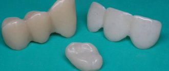
Plastic, ceramic or metal-ceramic crowns perfectly solve the problem of diastema. In this case, the cap-shaped design completely covers the teeth (in the case of a gap, it is necessary to apply crowns to both) and does not stand out in any way in the row. Features of installing crowns:
- average service life 10-15 years (depending on the material);
- safe for health;
- preliminary turning of the cutters is required.
The installation of crowns for diastema is not used as often as other methods.
Implants
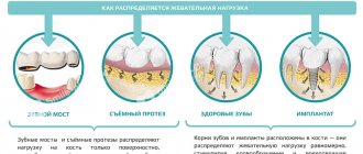
Implantation is used if the cause of the gap between the incisors is the absence of one or more teeth in a row. Filling the empty spaces will not allow the “ones” to continue to “spread out.” But, even if the dentition is replenished, the gap will remain and will have to be eliminated using some other of the listed methods.
Mouth guards
They are removable orthopedic structures that are manufactured using 3D technology. They look like original “cases” that are put on the teeth and are visually invisible. The mouthguard creates pressure, forcing the “ones” to move in the desired direction, which leads to their closure.
The duration of use of mouth guards (depending on the complexity of the problem) is from three months to three years. During this period, sequential replacement of the aligners is carried out, carried out as the “ones” move. From time to time, the mouthguards are allowed to be removed.
Preventing protruding teeth
It is easier to prevent any problem than to treat it. Therefore, at the first signs of dental deformation, it is recommended to consult a doctor.
To achieve a positive result you must:
- 1. Correctly use braces, veneers, Herbst apparatus and other modifications of orthodontic devices. Do not remove them ahead of time and if you suspect a violation or breakdown, immediately contact a specialist.
- 2. Listen to your doctor's advice. The orthodontist sees the big picture, so he or she will know better which treatments to use and for how long. It is important to attend all appointments to ensure timely adjustments to the devices and monitor changes.
- 3. Provide a psychologically healthy climate. Stressful situations, poor sleep, excessive physical and mental stress negatively affect all organs and systems, including bone tissue that needs to be corrected.
- 4. Eat right. A balanced diet and the intake of vitamins and minerals have a positive effect on regeneration processes in bone tissue and help eliminate malocclusions.
Beautiful teeth are not always a gift from nature. Most often, the painstaking work of an orthodontist is hidden behind a Hollywood smile. It is necessary to understand that the older the patient, the more advanced the problem, the longer it will take to correct the defect. After 25 years, processes in bone tissue slow down. Therefore, if there is such an opportunity, you need to start treatment in childhood or adolescence.
Also, we must not forget about the retention period. It lasts no less than the period of wearing braces. Consolidation of the result is necessary, because otherwise the ligaments will return the teeth to their original place. Thanks to the methods of modern medicine, the process of teeth straightening occurs relatively quickly and painlessly.
Preventive actions
As you know, preventing the onset of a disease is always easier and cheaper than treatment. To prevent the appearance of gaps between teeth, you must follow basic rules:
- control the habit of chewing hard objects (for example, seeds, nuts, crackers, threads, wire, etc.);
- carry out proper hygienic care of the oral cavity (to maintain the health of the gums and prevent the spread of bacteria, it is very important to brush your teeth correctly, carefully brushing not only the chewing units, but also the canines and incisors);
- Conduct timely examinations with a dentist (this allows you to diagnose any deviation in time and begin treatment at an early stage).
If the cause of the appearance of interdental gaps is a genetic predisposition, it is very important to begin correction as early as possible. Maximum effectiveness is possible during the period when the teeth are still mobile in the gums and can be rebuilt.
Now you know exactly how to remove the gap between your front teeth, what methods are used for this and how long it takes to eliminate the defect. All that remains is to consult with an orthodontist and, after examination and research, choose a method for eliminating the diastema that is right for you.
Doctors extracted a tooth from a man's nose
Nosebleeds are very common among both children and adults, but one young man's frequent nosebleeds had a very unusual cause: he had a tooth growing in his nose. A 22-year-old man from Saudi Arabia suffered from nosebleeds twice a month for three years. He finally decided to see a doctor, who discovered an ivory-white bony mass about 1 cm long in his nose. The doctor consulted with dentists, and they came to the conclusion that the bony mass was actually a tooth, which for some reason it grew in my nose. After this, the tooth was removed.
Read also
How to place veneers
The installation of veneers is performed for the purpose of aesthetic restoration of the dentition.
How long do you wear braces and what affects the timing?
When deciding on the use of orthodontic structures, patients are often interested in how long they wear braces on their teeth.
Correction methods
It also happens that large teeth interfere with others, disrupt the aesthetic appearance, or lead to some kind of disease. In this case, there are different types of correction:
- Grinding. The teeth are shortened and ground down.
- Restoration. Turning with composite compounds.
- Veneers. If grinding cannot be done, then veneers are used on adjacent teeth to make the appearance more aesthetic.
- Braces. They will help correct your bite and straighten your teeth. The result will be noticeable on your face.
- Tooth extraction. Sometimes this is the only option; instead of the extracted tooth, an implant of a more suitable size is placed.
In any case, you need to consult a dentist. Only he will tell you what will be the most optimal solution in your case. Also, hygiene is important before any correction method. Come to us for a consultation at Family Dentistry, our dentists will tell you what you need to do. And if you use the code word “FAMILY”, the consultation will be free.
Fish with human teeth
Pacu is a South American freshwater fish found in most rivers and tributaries of the Amazon and Orinoco River basins. It is also known that this fish is found in Papua New Guinea, but it is artificially bred there in order to help the local fishing industry. The pacu is a relative of the piranha, although the food preferences of these fish are different. Piranha is an exclusively carnivorous fish, while pacu is an omnivorous fish, preferring plant foods. The difference is also visible in the structure of the teeth of these fish. The piranha's teeth are razor-sharp, while the pacu's teeth are straight, square, and eerily human-like. The pacu is not nearly as carnivorous and aggressive as the piranha, but its jaws also have crushing power. One baby required surgery after a pacu bit his finger at Edinburgh Butterfly and Insect World in Scotland. Commenting on the incident, Matthew Kane, chief zoologist at Deep Sea World, said: "The pacu will eat anything, even a child's wiggling fingers."
A man had a tooth removed from his ear that had plagued him for 3 decades.
A Sheffield man who suffered from ear infections and pain for decades says his suffering is now behind him as doctors have finally removed a tooth from his ear canal. Stephen Hirst, 47, initially complained of piercing pain in his right ear, and as a teenager saw countless doctors over the years trying to discover the unknown cause of the mysterious illness. Stephen says the pain was sometimes so intense it made him scream. This continued until doctors at the Royal Hallamshire Hospital discovered a tooth growing in his ear. Then they deleted it. Doctors are still perplexed as to how the tooth could end up in the external auditory canal of a 47-year-old man.
Baby born with 28 teeth
In 2010, mother of many children Martha Matoni shocked residents of Nyandarua County in Kenya by giving birth to a healthy boy with 28 teeth. According to Peter Mumero, a local clinical officer, Martha gave birth to her son James Mwangi at home. But after a surprising abnormality was discovered, mother and child were taken to a local medical facility. Mwangi is now four years old, growing without any complications, and at the age of 8 months he was already eating Githeri (a traditional Kenyan dish).
Causes of dental defects
Lengthening or shortening of one of the incisors is usually a congenital anomaly. This feature of dentition development is associated with genetic factors. Experts also include the following as possible causes of the problem:
- periodontitis,
- malocclusion,
- presence of supernumerary teeth,
- abnormal shape of the alveolar process,
- disruption of the process of resorption of the roots of baby teeth.
Changes in the length of the central incisor can also be a consequence of jaw trauma. If a tooth is mechanically damaged, its lower part may chip. Because of this, the appearance of the smile deteriorates, discomfort occurs when chewing, and the risk of developing various diseases increases.
Surgeons discovered 2 teeth inside an eye tumor
Nagabhushenam Shiva, a 23-year-old woman from India, was one day as surprised as her doctors when they discovered that two fully formed teeth were located right inside a tumor that this woman had in her eye since birth. The tumor gradually increased in size over many years. When the woman’s vision began to deteriorate, she turned to doctors. Since the tumor was treated late, it had already affected the optic nerve. This resulted in the woman becoming permanently blind in one eye. However, surgeons removed the tumor and the procedure revealed the teeth.
