The central ratio (CR) can rightfully be considered the most controversial and controversial concept in dentistry. The concept of C.S. arose in the process of searching for a reproducible and tooth-independent position of the mandible (LM). Research in the field of CA has remained controversial for 80 years; one of the first definitions of CS was proposed by Hanau (1929); Currently, according to domestic and foreign literature, there are more than 30 definitions of CS [1]. Definition of Ts.S. jaws is the most important clinical skill required for predictable and effective orthopedic rehabilitation [2–4].
Numerous studies confirm that occlusal interference, which prevents the displacement of the temporomandibular joint (TMJ) condyles in the CS, significantly affects the coordination of the function of the masticatory muscles. Errors associated with the determination of the CS can most likely lead to occlusal-muscular dysfunction and the appearance of pain not only in the craniomandibular system, but also in the craniocervical region [5-8].
The relevance of studying this issue is associated with the ambiguity of approaches to assessing CA and the difference in determining the indications for it.
The purpose of the study was to study the precision of determining the CV of the jaws using different methods used in clinical practice.
Occlusion form
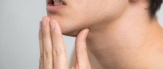
The shape of the bite can be determined by knowing the size and height of each individual tooth. During the measurement, both jaws should be in a state of complete rest. This is a condition when the lower jaw is as close to the upper jaw as naturally as possible. Only such an arrangement will make it possible to obtain accurate measurement results. A normal distance is considered to be 2-5 mm.
The height of the bite is the localization of parts of the dentition, and its size is the main factor in the normal position of the teeth. Thus, its incorrect height is one of the main signs of disorders in the structure of the jaws.
In addition to special methods, height is determined even by ordinary observation, but correct measurement can only be done by a highly qualified orthodontist with many years of experience.
Results and discussion
Analyzing the results of the study in terms of assessing the precision of determining the central relationship of the jaws using digital technologies, it should be noted that not a single method showed 100% accuracy of reproduction. We determined the most stable reproducibility of the position of the lower jaw during CS relative to the first registration in the method using a frontal deprogrammer (our own design) and amounted to 0.119±0.012 mm, which is statistically significantly less than when using methods of bilateral manipulation (0.225±0.028; p
≤0.05) and multileaf template (0.207±0.02;
p
≤0.05).
We observed a similar value of average values of reproduction accuracy when determining the CS by recording the Gothic angle (0.120±0.013; p
≤0.05), which is also statistically significantly less (
p
≤0.05) compared to other methods for determining the CS of the jaws.
Thus, the average discrepancy between the positions of the lower jaw during bilateral manipulation ( n
= 45) was 0.225 ± 0.028 mm, when using a sheet calibrator (
n
= 45) - 0.207 ± 0.02 mm, in the case of using a frontal deprogrammer (
n
= 45) - 0.119 ±0.012 mm and when recording the Gothic angle (
n
=45) - 0.120±0.013 mm (for all comparisons
p
≤0.05).
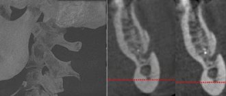
It should be noted that the maximum discrepancy when applying digital models of the lower jaw was determined during bilateral manipulation (0.717 mm) and in the case of using a multi-sheet calibrator (0.568 mm), and also that in the first case the difference between the maximum and minimum (0.004 mm) values obtained is the most pronounced (Fig. 13).
Rice. 13. Graphs of the reproducibility of the method of bilateral manipulation and the device for recording the Gothic angle (a), as well as the method using a sheet calibrator and a frontal deprogrammer (b). The abscissa axis shows the number of observations, and the ordinate axis shows the deviation in millimeters. Probably, this may be due to the peculiarity of the method, namely the formation of a certain manual skill, the practical experience of the doctor and the peculiarities of the state of the muscular component of the dentofacial system.
Causes and types
Reasons that have a negative impact on the intermaxillary distance:
- greater abrasion of teeth, due to which the strength of hard tooth tissue is lost;
- functional overload of individual teeth, which can be caused by improper bridge prosthetics;
- disorders of the nervous system that are accompanied by grinding of teeth;
- with a fight in the gastrointestinal tract;
- disturbance of phosphorus-potassium metabolic processes.
There are high and low pathological bites. The reason for the overestimation may be an incorrectly made denture, when the implant turns out to be slightly higher than the rest of the teeth.
Low bite, as a rule, is diagnosed when the hard tissues of the tooth are severely abraded, and in rare cases, when the prosthesis is installed incorrectly.
Causes
Reasons why the distance between the jaws becomes smaller:
- changes in the surface of the teeth;
- teeth grinding, which changes the condition of the enamel;
- habit of chewing on one side;
- removal of molars;
- lack of substances important for the normal functioning of the body;
- unqualified installation of prostheses.
To professionally make dentures, many measurements will be required, including the patient’s jaw at rest.
Height Determination Methods
The height of occlusion is determined using parameters such as the size and height of each individual tooth. During measurements, both jaws should be in a state of complete rest. This is a condition when the lower jaw is closest to the upper jaw. Only such an arrangement will make it possible to obtain accurate measurement results. A variant of the norm is a distance of 2-5 mm.
Anatomical method
The method is based on determining the normal position of the lower jaw. Experts in this field of dentistry, scientists Gysi and Keller, believe that this method must be guided by certain anatomical features, namely:
- The lips remain mobile and touch each other without tension during measurements.
- The corners of the mouth remain raised, the nasolabial folds remain clearly defined.
Orthodox practitioners believe that this method is not absolutely reliable, and therefore is not widely used in practice.
Hence, the desire of orthodontists to use objective data, which includes anthropometric measurements, is understandable.
Anatomical-functional method
The technique of this method is quite simple. The patient is asked to pronounce phrases containing labial sounds. After this, the patient closes his lips, and the muscles involved in speaking go into a state of relative rest—they should calmly touch each other. Nasolabial folds should have a normal appearance, and wrinkles in the area of the corner of the mouth should not be pronounced. If the position of the face takes the written form, then the patient’s height of relative rest has been established. It is measured with a ruler, and then templates with occlusal ridges are inserted into the patient’s oral cavity. Since the resting height is generally 2-3 mm greater than the height of the central occlusion, the ridges are cut or increased until the height at closure becomes 1-2 mm less than the previously determined resting height. This method is also subjective, but better than the previous one.
Preparation for prosthetics for malocclusion
| Click to sign up for a FREE consultation |
Depending on the type of pathology, a correctional scheme of preparation for future manipulation is determined. In the case of under-occlusion, an acceptable occlusion is first determined. For this purpose, supradental aligners or bite plates are used, that is, the reduced indicator is artificially brought to the normal level. In case of hyperabrasion of the tooth surface, the optimal correction is the installation of a Dahl device. This is a permanent plate made of chrome. It is recommended to wear the design for 3 months. An effective option for bringing the bite height to normal is artificial lengthening of the tooth crowns. In particularly advanced cases, they resort to surgical manipulations - the root of the tooth is exposed, the gum area is given the desired shape and relief.
Central ratio
A. I. Budovsky orthopedic dentist, private practice (St. Petersburg)
The term, which has become firmly entrenched in Russian dental practice, actually has many disadvantages. First of all, semantic ones. It is difficult to imagine around which center this ratio revolves. Secondly, purely practical ones - scientists and dentists often disagree in their understanding of the meaning of this term, record it differently and, accordingly, give different definitions.
To give the most complete and accurate formulation, we will not turn to the dictionary of dental terms, but will try to highlight the most important elements of this term for practice.
- This is a static position, registered for medical purposes.
- Repeatable and stable.
- Position independent of occlusion.
- It is preserved during the initial opening movement until the moment of displacement (translation) along the slope of the articular tubercle - pure rotation.
- Anatomically characterized by the anterosuperior position of the condyles in the joint with minimal activity of the masticatory muscles.
- Determined by the structures of a healthy joint.
In case of dysfunction or pathology of the joint, the CS is also recorded, but in the “sick” joint. Therefore, these provisions are given appropriate names (will be described below). With the invention of the first articulators, the need arose to accurately transfer the relationship of the jaws. I think that in those days they simply did not think about the problem of CS in patients with a complete dentition.
But the task of the “starting point” for prosthetics, of course, was the treatment of patients with an unfixed bite. Even Gysi (Alfred Gysi, 1907), using a recording device fundamentally similar to the mandibular arch with colored scribes of Campion (Georg Campion, 1905), drew the course of movement of the condyles in order to analyze the upper and lower jaws in the central ratio “in zentraler relation”.
He created his own articulator (Trubyte) with an incisal plate and pin and an articulator with average angles of inclination of the articular paths (Simplex).
Bite registration was carried out using an extraoral device and a “face bow” oriented along the Camper horizontal. He also described the "Gothic arc".
McCollum in 1920, using Gysi's instrumentation, determined the rotational axis and transferred the jaw relationship to the articulator. Using a Snow arch attached to the mandible, McCollum established that the hinge axis at a certain terminal position was constant and reproducible.
This is how he defined the CS: “...when both condyles are firmly seated in their most posterior positions in the glenoid fossae.”
Literally: “...if both condyles are firmly seated in their most posterior position in the glenoid fossa.”
Over the more than 100-year history of the development of gnathology in dentistry, a lot has been done and discovered. But the basic principles were laid down at the beginning.
In this regard, to put it mildly, the “attacks” on contemporary authors dealing with this issue sound strange, as well as statements about the lack of need for axiography or individualization of these patients, etc., etc.
The concept of “central” came to us from a direct translation of the English term centric relation.
Of course, the concept of “central” in this case does not correlate with the midline of the face or the concept of “medial” (from the Latin medius - middle) in anatomy: located closer to the median longitudinal plane of the body.
The implication is that the joint condyles are positioned centrally in the glenoid fossae during CS. But the closer, semantic meaning of the translation of the term “centric”, in my opinion, lies in the description of the process of “centering”, and not a static position.
Also, one of the reasons for describing the position of the condyles as “central” is that they are located relative to the central part of the disc.
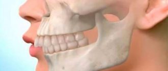
For a more visual representation of the CS, let us turn to the anatomy of a healthy temporomandibular joint.
The spatial position of the condyles is largely determined by the anatomical structures of the joint.
Pay attention to the histological preparations of the joints:
- Condyle.
- Disk.
- Glenoid fossa.
- Articular tubercle.
- Bilaminar zone.
- Lateral pterygoid muscle.
- Glaser's gap.
- Medial and lateral poles of the head of the condyle.
- Superior articular chamber (translation).
- Lower articular chamber (rotation).
Despite the limited space, there is quite a lot of controversy surrounding this “battlefield”.
Our task is to obtain a position without displacement along the articular surface of the tubercle, i.e. the condyles should be at the base of the tuberculum glenoidalis:
- The upward and forward displacement is limited by the glenoid fossa itself and the disc, which is composed of dense fibrous tissue.
- Medially - there is 0.5-1.2 mm to the temporal bone.
- Stays back. There we have a bilaminar zone - the upper and lower plates. Contains fiber, elastic fibers and venous plexus. And it is limited to the postglenoid tubercle of the squamous part of the temporal bone.
But this is a different reproducible position - the posterior position of the lower jaw, not the CS, but the ZP. Which is essentially what McCollum got. He needed a starting, reproducible point.
The jaw in the CS is characterized by the ability to rotate without moving forward along the slope of the tubercle, but up to a certain point. The CS is preserved during the initial opening of the mouth (pure rotation) by approximately 10-20 mm; with further opening, the condyle moves forward and down the slope of the articular tubercle through the lateral pterygoid muscle and Lig. Temporomandibulare.
Important in understanding the CS is its differentiation from other positions of the lower jaw.
The count in the joint is in millimeters and their fractions, but with displacements we get not only different definitions, but also different occlusal relationships. That’s why I don’t like manual methods for determining CV so much.
CS is a non-forced position; when determining, the chin is controlled by the doctor and not directed. With active manipulation, you can get a shift in one direction or another. Often dorsal in ZP.
The same applies to the Dawson technique. This is more of a stress test (positive if pain occurs). We seem to press the condyles into a superior anterior position, bringing the joints into compression. Since it is quite difficult to compress the disk too much, we will obtain a generally accurate CA.
When they talk about a more distal superior position of the condyles (until 1987 this outdated formulation was used), they still mean the ZP.
At the moment, most definitions of the central nervous system refer to the anterosuperior position. But this position is determined not by the location of the condyles, but rather by the anatomy and position of the glenoid fossa itself.
Note that the position of the condyles is influenced by the shape and severity of the articular tubercle.
Muscle activity is very important for determining CV - it should be minimal. In this sense, the central point should not be confused with another widely known concept - the resting position. The resting position is characterized by relaxation of the muscles, but at the same time their activity remains. The muscles seem to be balanced, but gravity (gravity force) also plays a role in the formation of this position.
Therefore, we often talk about the resting height, the mouth opens slightly (2.7–5.6 to 7 mm). At the same time, muscle activity must be maintained so that the jaw does not sag. There is probably another position - the “position of surprise” and “extreme surprise”, depending on the degree of jaw sagging.
This activity is much higher than the muscle activity during “speaking”, so using the resting height to determine the CO is a mistake. Also, the resting position is an adaptable value and focusing on it when determining the bite height can lead to an error.
Why do I often repeat: in a healthy joint?
With changes in the joint caused by a pathological process, the position will also differ from the CS.
Adapted centric posture (Dawson 1995) When a disc is displaced without reposition in the joint, functional adaptation may occur through proliferation of the bilaminar zone.
In a similar way, the joint adapts to permissible compression loads.
A CS that is “in disarray” - deranged referenz position (DRP) - when determining the CS in patients with dislocation (with complete or partial displacement of the disc from the condyles). When analyzing the CA, it is necessary to mention a few more concepts.
Long centric - “long centric”. Along with the centric position of the lower jaw in the form of a certain starting point (point centric), there is free space (Spielraum) for intercuspidation (free movement of the tubercles during articulation). This space allows you to avoid interference of the incisors when the lower jaw slides forward from the CS into the usual occlusion. There is also a lateral free space (wide centric). Both of these terms are combined under the concept of freedom in centric. And according to Schuyler (1969) they are 0.5-1 mm.
Power centric - “power centric”. Occurs with maximum “biting” (maximum muscle force) by the patient. Does not coincide with the CS, since with maximum closure the condyles are positioned more cranially.
To summarize the above, CA can be defined as follows.
CS is a repeatedly reproducible, stable relationship of the upper and lower jaws, regardless of occlusion, obtained for medical purposes through unforced, controlled rotational movements of the lower jaw without its displacement. It is characterized by a spatial anterosuperior position of the condyles in a healthy joint with minimal activity of the masticatory muscles.
Proportional ratio of teeth sizes
In most cases, specialists determine the proportions of a tooth by correlating its height and width. A result of about 0.75 is considered ideal. The most accurate diagnosis is carried out through the use of formulas.
- Gerlach's formula.
The method is based on the proportional ratio of the sizes of the front teeth and dental units of the chewing zone. The width of the crowns of the upper central incisors should correspond to the width of the four lower incisors. The canine, two premolars and one molar of both jaws are normally equal to each other. The width of the lateral part of the dentition is 10 mm greater than the width of the anterior segment. - Pon's formula.
The distance between the first premolars is equal to the sum of the widths of the four incisors multiplied by 100 and divided by 80, and the distance between the first molars is the sum of the widths of the four incisors multiplied by 100 and divided by 64. - Corkhouse formula.
The length of the segment from the midline to the first molar of the upper jaw should be 2 mm greater than the same distance on the lower jaw.
What is a state of physiological rest?
A position known as physiological rest means the position of the jaws when the muscles relax. In this case, the teeth do not come into contact with the antagonists; the retention of the lower jaw is based on the anti-gravity effect. Under normal conditions, the distance between rows is allowed at two to five millimeters.
There are two types of pathology:
- Overpriced. A common cause of the problem is an incorrectly selected prosthesis. In this situation, the teeth are constantly in contact, the distance between the dental rows is no more than one or two millimeters. There are difficulties in closing the lips tightly, and joint overstrain.
- Understated. The reason is abrasion of the enamel, violations in the installation of bridge crowns. The difference is three to four millimeters, the contours of the face change. The folds become pronounced, the corners of the mouth droop. Pathology of jaw closure, increased salivation gradually develops, and angular cheilitis appears.
Causes of anomalies
The causes of malocclusion are:
- pathological abrasion, destruction of incisors, molars, loss of tissue density;
- bruxism, which causes such consequences as a decrease in the central bite, subsidence of individual units;
- the chewing load is uneven, the Patient has the habit of chewing food only on one side;
- use of bridges with incorrect preliminary design calculations;
- loss of molars on one or both jaws;
- extraction of several units, displacement of elements from the central region to the side ones;
- deficiency of phosphorus and calcium, which reduces the strength of bone tissue and leads to the development of atrophy.