Addresses where you can take a panoramic photo of your teeth in dental clinics in Izhevsk
- Central
- Lenina street, building 5
- +7 3412 222 707 show all
- Mon-Fri 8:00-20:00, Sat 8:00-14:00, Sun
4.0 ratings: 10
- Your dentist Nemirova
3.8 ratings: 7
- Lada-esthete
4.4 ratings: 9
- VIP-dent
3.7 ratings: 7
- Sky
4.2 ratings: 5
- Maestro
4.0 ratings: 4
- Kazmaska
4.2 ratings: 6
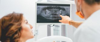
Dental X-ray is the basis for making an accurate diagnosis
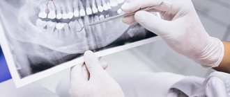
Dental X-ray is one of the mandatory stages of diagnosis and treatment. Often, only the results of dental photographs clarify the situation; it allows making a final diagnosis and, accordingly, prescribing appropriate therapy or performing the necessary surgical manipulation. Moreover, the doctor often prescribes x-rays not only before, but also directly during the treatment process in order to assess its correctness and adjust as necessary. This research helps make treatment high-quality, with long-term results. There are no special contraindications, only during pregnancy you should warn your doctor about this.
The work of a radiologist in regional dentistry in Astrakhan is well established, the queue moves quickly. The study is carried out using modern equipment - we strictly comply with the standards of radiological control and patient safety. Sensitive devices have low radiation levels.
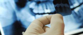
You can take a dental x-ray in Astrakhan in our clinic not only to see a local doctor, but also to confirm the opinion of another specialist. We take pictures of baby teeth, molars, filled canals, after resection and other types of surgical intervention. If wisdom teeth are not erupting correctly, an x-ray is indispensable. The image reveals cysts, cracks in teeth, and clarifies the degree of inflammation during periodontitis. It shows how deeply the teeth are affected by caries, and reveals jaw anomalies invisible to the eye.
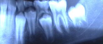
Panoramic photograph of teeth: importance and necessity
It is convenient for us to take an X-ray of a tooth in Astrakhan - both a targeted image and a panoramic one, when a three-dimensional image is obtained. A panoramic photograph of the teeth includes the upper and lower jaw, sinuses, maxillary joint, etc. Orthopantomography (as specialists call the picture) is needed for the work of an orthodontist, orthopedist, when installing dentures, crowns, implants and braces. It makes it possible to assess the condition of the periodontium, impacted teeth, roots and adjacent tissues.
Cost of dental photographs in dentistry
Making dental x-rays in Astrakhan as accessible as possible for dental patients is our task. The price of images is truly affordable for all categories, this also applies to panoramic x-rays. The exact price of the images is indicated on the website of the clinical dental center - each category has its own price.
View prices for paid radiology services (code 300) >>
Make an appointment with a dentist >>
Fresh questions about the service panoramic dental imaging in Izhevsk
- E
Ekaterina asks a question to the Avangard Baby Clinic, October 27, 2022, 20:59 Hello, is it possible for you to treat all of a child’s teeth under general anesthesia and how long will it cost? The child is 2.6 years old. - D
Dmitry asks a question to the Lada-esthete clinic, March 12, 2022, 18:40 Hello. I need to remove two teeth under general anesthesia, since the last time local anesthesia did not take me and they removed me alive. The teeth are level with the gums. Tooth 8 is the upper jaw and 5 is the lower jaw. What do I need for this...
- D
Dmitry asks a question to the Lada-esthete clinic, March 12, 2022, 18:14 Hello. I need to remove two teeth under general anesthesia, since the last time local anesthesia did not take me and they removed me alive. The teeth are level with the gums. What do I need to provide you with for this and how much will it cost?
- D
Denis asks a question to the Avangard Baby Clinic, March 08, 2021, 04:32 The gumboil needs to be removed and two teeth cured. I’m afraid to treat it in the usual way, but in a dream I want to try it. Health allows no chronic diseases. How much will it cost and when can I come, preferably this morning.
Panoramic shot
Panoramic images directly at the Medfodent Plus clinic
An orthopantomogram (OPTG) is a diagnostic panoramic image of the lower and upper jaws, which allows you to assess the condition of the teeth, temporomandibular joints, maxillary sinuses and other areas of the temporomandibular region. Important: Due to low radiation doses, orthopantomography is used in pediatric dentistry and in other cases where it is necessary to minimize exposure to X-rays.
Description of the procedure
You can undergo first-class diagnostics at a preliminary stage during treatment planning or already during treatment to obtain more accurate results. Using an orthopantomogram, a thorough examination is carried out so as not to miss the slightest pathology that interferes with effective treatment. X-ray examination of the dental system using this modern diagnostic equipment allows for gentle dental treatment.
Orthopantomogram is used:
- to identify all areas affected by caries;
- when identifying periodontitis;
- to identify features of teething, various anomalies in the development of jaws, teeth, etc.;
- in preparation for prosthetics or dental implantation;
- in periodontal and orthodontic treatment.
How is the procedure performed?
During filming, the patient's head is held with special clamps to prevent displacement. This is necessary to obtain more accurate images. As a rule, a high-quality photo is obtained the first time. The process takes just minutes.
A comprehensive digital examination is most complete and visual, promotes high-quality and effective treatment, and virtually eliminates erroneous diagnoses and procedures.
Patients see existing problems “from the outside”, it is easier for the doctor to explain possible treatment options, and at the end of the consultation they receive: examination results: two treatment plans - recommended and alternative.
Treatment plan
After passing all stages of a comprehensive examination, the patient receives 2 options for a treatment plan with a clear explanation: recommended and alternative.
- The plan clearly reflects all stages and their costs.
- The patient receives the results (plan) and has the opportunity to make the right decision.
- The patient can agree on the date and time of the appointment to undergo the recommended procedures (based on the accepted version of the treatment plan).
ATTENTION!!! The cost fixed in the plan does not change during the treatment process.
Interpretation of the orthopantomogram
But not only the methodology of the procedure is important, but also the analysis of the panoramic image (OPTG).
Interpretation of an orthopantomogram in dentistry is carried out in a certain order, namely:
1. The description of the image begins with an assessment of its quality - taking into account sharpness, contrast, complete coverage of the jaws, the presence of certain projection distortions (elongation, shortening, change in shape).
2. Next, the bone tissue is assessed. The doctor looks at what the temporomandibular joint looks like, evaluates the relationship between the articular heads and fossa, and examines in detail whether there are any pathological changes. A panoramic photograph also allows you to assess the condition of the interdental septa - their shape, height, degree of mineralization, condition of the spongy substance, compact endplate and cortical layer. When assessing bone tissue, the condition of the mandibular sinuses and mandibular canal is also taken into account, the presence of impacted teeth and changes in the intraosseous structure are determined. The analysis of pathological shadows is also important.
4. Definitions of the object of study.
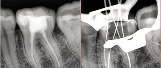
5. Tooth shadow analysis - a panoramic photograph of the teeth, or OPTG, makes it possible to determine the condition of the crown, roots, root canals, and periodontal fissure. Thanks to it, the doctor can see the presence of carious cavities, filling material, its defects, the number, shape, size of tooth roots.
What can the new tomograph do?
- excellent quality of 2D panorama images
- 3D scanning with the lowest possible radiation dose - Low Dose Technology - ensures high safety, which is very important when examining children, for postoperative monitoring, and when planning the installation of implants
- 5 scanning area formats in 3D mode will allow you to most reliably plan treatment for any task in the maxillofacial area: 5 x 5 cm - optimal when planning the installation of a single implant or for local diagnostics, the lowest possible level of radiation for the patient
- 6 x 8 cm – covers the entire dental arch when planning multiple implant placements and allows the use of surgical implant guides
- 8 x 8 cm – covers the entire dentition, including the lower and upper jaws, as well as part of the maxillary sinuses. Used for implantation on the upper and lower jaws; for the use of surgical templates in implantology; for differential diagnosis of sinusitis in ENT practice
- 8 x 15 cm – covers the upper and lower jaws, including the upper respiratory tract, cervical spine or sinus area. Both maxillary joints and sinuses can be examined. It is used both in dentistry for a 3D panorama, and in the diagnosis of the cervical spine and respiratory tract, in the ENT diagnosis of diseases of the paranasal sinuses (sinuses)
- 13 x 15 cm – covers the entire maxillofacial area: both jaws, both joints, cervical spine, respiratory tract and sinuses can be examined simultaneously. Application: maxillofacial surgery; gnathic surgery; diagnosis of TMJ (temporomandibular joint), diagnosis of injuries to the maxillofacial area; ENT diagnostics.
Sign up for a consultation
to clinic specialists Mon-Fri: from
09
to
20
, Sat-Sun: from
10
to
16
(4852)
230-631
Make an appointment
What does a 3D dental x-ray show?
3D dental tomography is a highly accurate diagnostic method that makes it possible to obtain a three-dimensional image of the dental system in different projections. Volumetric images obtained with CT allow the specialist to enlarge, rotate and examine the area of interest from all sides and at different depths:
- The entire maxillofacial apparatus.
- A dentition or an individual tooth.
- Paranasal sinuses.
- Bone and periodontal tissues.
Dental CT allows you to detect inflammation, assess the homogeneity of the filling material and check the quality of installation of a filling, crown or implant, see the number of dental roots and their fragments, identify neoplasms, assess the degree of curvature of teeth, determine the exact parameters of bone tissue (height, width, density, etc.). d.). The information obtained allows the doctor to optimize treatment measures and predict the result.
Preparation measures
In order to properly prepare for the examination, you need to clarify whether it will be carried out with contrast. This depends on the indications for the procedure; contrast is necessary to identify neoplasms in bone or soft tissues, or suspected tumor processes. If contrast is not needed, the procedure does not require preparation.
When you schedule a dental cone beam computed tomography scan with contrast, you must come to the clinic on an empty stomach. You will have to refrain from eating 6 hours before the scheduled procedure, so most often it is carried out in the morning.
Before the examination, you must remove jewelry, metal parts and objects.