When visiting a dentist, you have probably heard them more than once say such phrases as, for example, “lower left five” or “16th tooth” or “2nd premolar.” Experienced patients know that this is the numbering of the teeth, which is necessary for the doctor to describe the condition of the oral cavity when filling out the card. There are several systems used in dental practice for this purpose.
What is tooth numbering and why is it used?
If your doctor tells you that there is a problem with one of them, you will most likely want to understand which one. Of course, he can show it to you using a mirror or otherwise explain its location. But entering information related to diagnosis and treatment into the patient’s record requires greater accuracy and a professional approach. The same can be said if a doctor orders an x-ray - the x-ray room worker must know which area needs to be examined. This is what tooth numbers are used for.
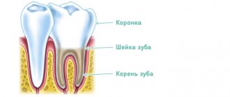
Normally, an adult has 32 of them (if there are wisdom teeth), and a child has 20. Each of them takes its place in the jaw, belonging to a specific type. Numbering implies that each has its own serial number, by which a specialist accurately determines its location.
There are various systems, most often used in dentistry, and each of them involves its own algorithm for generating a serial number.
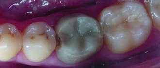
Three-channel pulpitis
Three root canals most often have large chewing teeth - molars, sixth through eighth. Each branch contains connective tissue - the pulp; it contains many nerve endings and vessels. When inflamed, the pulp swells and becomes compressed, which is accompanied by severe pain. Three-channel pulpitis has important features in treatment. Startsmile will tell you more about them.
The number of canals in each tooth varies, usually one or two, in molars – most often three. There are record-breaking patients in whom up to eight processes were discovered during treatment. Three-channel pulpitis, as the name implies, is diagnosed in a tooth that has three channels. Inflammation of the pulp or, as is often said, the dental nerve, manifests itself in severe pain.
- The doctor spends an hour to an hour and a half on a multi-channel tooth;
- under a microscope - about two hours.
This is due to the fact that 3-channel pulpitis requires treatment in each process.
The dental canals are very thin, tortuous, no more than 1 mm in diameter. The microscope magnifies them to 2.5-3 cm. The dentist does not treat blindly, under magnification he clearly sees all the branches and foci of inflammation. Treatment of pulpitis of the 3rd canal takes place in good lighting. The patient lies down, the doctor looks not into the mouth, but into the microscope camera. This has a calming effect on dentophobes. The microscope reveals the most difficult to reach areas for the doctor. This is especially true for patients who have hidden dental canals that are not visible to the human eye.
And during endodontic treatment, it is important to remove all foci of inflammation to avoid relapse
To treat three-channel pulpitis, the vital extirpation method is used. The pulp is removed, the canals are thoroughly cleaned, disinfected and sealed. Let's look at each stage of treatment of root pulpitis 3 in detail.
Pain relief using local anesthesia.
Expansion of the mouths of three canals and removal of pulp.
Flushing the canals with sodium hypochlorite as an antimicrobial treatment.
Drying and filling the canals and installing a temporary filling on the crown of the tooth.
The main treatment takes place during the first appointment. A break is needed so that the filling material in the canals hardens well. The second stage will take less time: the tooth will be processed again and a permanent filling will be placed.
The number of appointments may increase to three if it is impossible to immediately remove the pulp from the canals. In this case, the nerve will be “killed” by a special paste. It will be added to the cavity and left until the next visit. Soviet patients said this: “They put arsenic.” This substance is not used in modern toothpastes, but many, out of habit, believe that the dental nerve is removed with the help of arsenic.
Names of teeth
It is worth familiarizing yourself with them before starting to study numbering algorithms.
Each row of teeth is symmetrical, and it consists of left and right parts. There are several groups of teeth that differ in structure, function, and location.
For adults they are:
- Incisors. These are the front (central, medial) teeth, designed for biting food. They are flat and have sharp edges. There are 8 of them in total - 4 each in the bottom and top rows. In numbering they are often called ones and twos;
- Fangs. They follow the incisors. If you count from the first central incisor, then the canine will be the third from it - both to the right and to the left. For those who are not familiar with dentistry, the name of this type of teeth may seem funny. They wear it not in vain. These are strong teeth with a wide crown and sharp edges, having a conical shape. They are designed for biting off high-density food (for example, meat). Fangs have the longest roots. In total, a person has 4 fangs - 2 above and 2 below, and dentists call them triplets;
- Premolars. They follow immediately after the fangs and have a prismatic shape. On each side of the jaw - both upper and lower - there are 2 premolars, i.e. there are 8 in total. They are the 4th and 5th, and they are intended for chewing food;
- Molars. Their numbers are 6, 7, 8, if you count from the central incisor. Thus, the total number of them is 12 - 6 on each jaw (3 on each side). These are large chewing teeth with a wide surface. They are designed to literally grind food, being strong and powerful.
A child's dentition differs from an adult's in the number of its components. Babies have 5 of them on each side, i.e. 10 each on the upper and lower jaws. The last ones in each row are fives. But when their dentition begins to change, permanent molars are one of the first to appear.
The name of teeth used by dentists is one of the “beacons” by which they determine their location. But this is not enough for the most accurate description, and therefore an error-free diagnosis and treatment. Therefore, different numbering systems are used.
Haderup system
It is used in dental practice quite often, but doctors are divided on its convenience and the degree of risk of erroneous decoding. This algorithm involves designating teeth by a serial number with an index indicating their specific location.
Their numbers are formed as follows:
- Numbers 1-8 are used to indicate the number. The number 1 represents the central (front) incisor, and the number 8 represents the third molar. Accordingly, 2 – second front incisor, 3 – canine, 4 and 5 – premolars, 6-8 – molars;
- To indicate the location, the indices “+” and “-” are used, where the first is the upper jaw, and the second is the lower jaw;
- Considering that the report goes from the front to the molars, it is necessary to indicate the side - right or left. This is done through proper index placement. If the side is right, it is placed after the number, and if it is left, before it.
For example:
- 5+ is the upper second premolar on the right;
- -2 is the second lower front incisor on the left.
According to this system, children’s tooth numbers are indicated in a similar way, but a 0 is placed before the number and numbers from 1 to 5 are used.
For example:
- 02+ is the primary front incisor on the upper right;
- -01 is the primary front incisor on the lower left.
Square-digital system
Its other name is the Zsigmondy-Palmer system. It also provides for numbering using numbers 1-8, as in the Haderup system, but the location is indicated using the corner icon, in which the desired number is indicated. According to this algorithm, each jaw is divided into two segments, and the countdown, as in the previous case, begins with the central incisors. Those. 1 is the front incisor, and 8 is the third molar.
How to decipher the symbols?
- The angle directed downwards and to the left is the lower right jaw;
- The angle looking down and to the right is the lower left jaw;
- The angle depicted up and to the left is the upper right jaw;
- The angle directed upward and to the right is the upper left jaw.
For example:
- The number "5", located in the corner looking down and to the right, indicates the second premolar in the bottom row on the left;
- The number “2”, depicted in the corner directed to the left and upward, indicates the anterior second incisor on the right jaw from above.
In the case of designating baby teeth in children, the same algorithm is used, but they are designated by Roman numerals IV.
Two-digit system
Today it is increasingly used in dentistry, being one of the most convenient and modern. It involves dividing the jaws into four segments. In this regard, the algorithm is similar to the previous ones, but it differs in the designation of the segment - each of them has its own number:
- Upper right – 1;
- Upper left – 2;
- Bottom left – 3;
- Bottom right – 4.
The teeth themselves are numbered using numbers from 1 to 8. As in previous cases, 1 is the front incisor, 8 is the third molar.
The number consists of two digits:
1 – segment;
2 is its serial number.
For example:
- 36 – first molar on the left in the bottom row;
- 15 – second premolar on the right in the upper row;
- 43 – canine on the lower jaw on the right.
To designate baby teeth in children, the segments are designated by numbers 5-8:
- Upper right – 5;
- Upper left – 6;
- Bottom left – 7;
- Bottom right – 8.
The number of the teeth themselves is indicated by numbers 1-5.
For example:
- 62 – second front incisor on the left in the top row;
- 84 – first premolar on the right in the bottom row.
Alphanumeric system
It differs from others primarily in that it involves the use of letter designations. They correspond to the name of the teeth, and it is indicated in Latin letters. For clarification, a digital index is added to them in all designations except fangs.
In the case of adult dentition, capital letters are used:
- I1 – central first incisor (unit);
- I2 – central second incisor (two);
- C – fang (three);
- P1 – first premolar (quad);
- P2 – second premolar (five);
- M1 – first molar (six);
- M2 – second molar (seven);
- M3 – third molar (figure eight).
To designate children's milk teeth according to this system, Latin lowercase letters are used:
- i – incisors;
- c – fang;
- m – molars (4ths and 5ths).
Modern specialists today very rarely use the alphanumeric system, since it does not allow specifying the exact location - upper or lower jaw, side. Since dentists’ tooth numbers are designed to perform this function, such an algorithm is not informative for diagnosis and treatment.
Universal system
It is comfortable and light. This numbering method does not involve division into segments, the use of letters, or the use of several symbols. The principle is simple - each tooth has its own serial number, which is determined by its location:
- The count starts from the 3rd upper right molar. He, according to this system, has the number 1;
- Next, the markings proceed in order to the upper left 3rd molar, which is numbered 16;
- Number 17 is the lower left 3rd molar, and the numbering continues from there towards the 3rd lower right 3rd molar, which is numbered 32.
Capital Latin letters are used to designate baby teeth in children:
- AE – right upper dentition (from 5 to 1);
- FJ – left upper dentition (from 1 to 5);
- KO – bottom left row (from 5 to 1);
- PT – bottom right row (from 1 to 5).
Features of numbering of baby teeth
Unlike the molars that adults have, children have only 20 teeth, whereas in the first case there are 32. But at the age of 5-6 years they begin to be replaced by molars, which happens gradually, and this process is completed by about 13-14 years . Thus, during this period, a child, as a rule, has both one and the other at the same time. This may make them somewhat difficult to describe.
In this regard, not all numbering systems are convenient to use. Most often, dentists in this case use a two-digit algorithm, which implies the following designations:
- 51-55 – right segment of the upper jaw;
- 61-65 – left square of the top row;
- 71-75 – right segment of the lower jaw;
- 81-85 – left square of the bottom row.
Numbering of teeth is a necessary condition for an accurate description of diagnostic and therapeutic dental procedures, since without a description of their location, errors may be made.
Kostyuk Hanna Volodymyrivna
Orthopedic dentist of the first category
Did you like the article?
Rate: ( 4.50 out of 5)
Loading…
Temporary filling
This type of procedure involves filling the canal with special compounds for a certain period of time. Why do they do this?
- To achieve a disinfecting effect.
- To stop the inflammatory process during periodontitis.
- To protect the dental canal from various infections and external influences, when it needs to be treated over several visits to the doctor.
Sometimes temporary filling is done to protect the dental canal from various infections.
Temporary filling is done for the following problems:
- acute/chronic periodontitis;
- traumatic injuries;
- perforation of walls.
Therapeutic non-hardening pastes for temporary use include antibiotics and antibacterial agents.
Products that are injected into the root canal contain antibiotics and antibacterial agents
How is root canal treatment performed?
An important step in the process of treating root cavities is determining the working length of these canals. Not everyone knows the definition of tooth root length. So, the working length of the root canal is the distance from the edge of the frontal units to the apical constriction preceding the apical foramen. There are several methods for determining the working length of the root canal. The most commonly used are the calculation method, x-ray and electrometric methods.
Endodontics treats tooth root canals. When an endodontist treats a root canal, he performs the manipulations in the following sequence:
- diagnostics;
- X-ray;
- preparing the dental cavity for treatment;
- anesthesia;
- chemical treatment of instruments;
- opening of the tooth cavity;
- determination of the working length of root canals;
- medicinal treatment, cleaning and expansion of root canals along the entire working length;
- filling a tooth cavity.
READ ALSO: What should be the sequence and pattern of teething for children?
Diagnostic methods
The first stage of tooth root canal treatment is diagnosis, which will help the doctor make the correct diagnosis and decide on a treatment method. To do this, the patient needs to undergo an x-ray to examine the part of the crown that the doctor cannot see. This procedure allows you to understand how many roots and canals the tooth cavity has. If the X-ray examination is ignored, then the cavity of the diseased tooth will have to be opened again (we recommend reading: X-ray of a child’s jaw: is it possible to take an X-ray with baby teeth?).
INTERESTING: How many teeth should an adult healthy person have?
https://youtube.com/watch?v=aar2229bPhg
Preparatory procedures
After the X-ray of the dental cavity has been carefully studied, the diagnosis has been made, and the stages of the upcoming therapy have been planned, it is necessary to tell the patient about everything in detail. Next, you need to obtain documented consent for the opening and further treatment of the tooth cavity.
An important point in preparing for root cavity treatment is for the doctor to obtain information about the presence of allergic reactions in the patient to anesthetics. If such information is not available, an allergy test is performed. At this stage, chemical treatment of the instruments with which the manipulations will be performed is carried out.
Administration of anesthesia and application of anesthetic
Before treatment begins, the patient is anesthetized in the area of the jaw where the intervention will be performed. Anesthesia can be superficial or in the form of an injection. The first type of anesthesia blocks sensitivity not only in the dental cavity, but also on the mucous membrane. It is usually used to numb the area where the doctor is about to inject an anesthetic.
The following drugs are used for superficial anesthesia:
- 0.5% Promecaine ointment;
- Anestezin;
- Lidocaine;
- Dicaine.
Opening a molar tooth
What is the opening of a tooth cavity? In order to remove the pulp and clean the root canals, the dentist needs to provide good access to them. Opening the tooth cavity can begin immediately after grinding the caries and removing sawdust from the dentin. The process of opening the tooth cavity begins with the smallest bur, after which a large spherical one is used.
INTERESTING: the structure of the human oral cavity and the functions of the organs that are located in it
Medicinal treatment of canals
Canal treatment is divided into mechanical (scraping out the contents using special tools) and chemical (medicinal treatment of root canals with disinfectants injected with a thin needle). Today, the following scheme for medicinal treatment of the root canal is used: sodium hypochloride is applied after using each instrument and completing mechanical cleaning, then hydrogen peroxide, and after that distilled water. Drug treatment of root canals is carried out immediately after the opening of the dental cavity is completed.
Sealing
The final stage of tooth root canal treatment is sealing the cavity. The root cavities are filled with a special filling material (usually gutta-percha). The filling helps the tooth remain strong and prevents pathogenic bacteria from entering its cavity.
Filling a tooth cavity can be:
- temporary;
- permanent.
With temporary filling, the tooth cavity is filled with a non-hardening paste that has medicinal properties. This type is used in cases with advanced three-channel pulpitis or periodontitis.
Wrong worldview
Because a tooth consists of roots and a pre-coronal part, from time to time there is a misconception that there are as many canals in teeth as there are roots . This is far from true, because the canals quite often branch off and bifurcate near the pulp. Moreover, several channels can run parallel to each other in one root. There are also cases of their bifurcation at the apex, which is why it turns out that one root has two apexes and this, naturally, complicates the work of doctors when filling similar teeth.
Taking into account all the individual characteristics of the teeth, dentists need to be very careful when treating (the process of alleviating, removing or eliminating symptoms and diseases)
and filling, so as not to miss any branch. Indeed, from time to time, without an x-ray, it is very difficult, even during an autopsy, to identify how many canals are in the teeth.
Prevention after healing
After the procedure for healing the root system you should avoid putting stress on the treated tooth for some time; moreover, you should not eat food earlier than two hours after therapy (therapy is a process to relieve or eliminate the symptoms and manifestations of the disease)
, otherwise the filling is not completely hardened It might just fall out. But the same thing can happen if the doctor uses low-quality drugs or performs incorrect treatment (for example, the canals were over-dried or not dried before filling).
Also, after filling, for some time (up to several days) the tooth may feel pain (an unpleasant sensory and emotional experience associated with actual or potential tissue damage or described in terms of such damage) when pressed, or simply ache, cause discomfort, or have excessive sensitivity. This is usually a common condition if pain (physical or emotional suffering, painful or unpleasant sensation)
powerful, you can take painkillers.
If pain (physical or emotional suffering, painful or unpleasant sensation)
does not go away after a certain time, this may also be an indicator of disgusting healing (insufficient cleaning of infection
(The term means various types of interaction of foreign microorganisms with the human body)
or infected pulp, leaky filling, introduction of low-quality pharmaceuticals or materials).
From time to time, there are cases of allergic reactions , which are also accompanied by incessant pain, and from time to time itching and rashes appear on the body. It may be caused by a reaction to a pharmaceutical product or the material that was used for the filling. In this case, it must be changed to something else that will not cause allergies.
In all these situations, it is imperative to consult a doctor as soon as possible for a re-examination and dental prophylaxis in order to identify the cause of the defects.
Healing
The development of modern medicine and dentistry in particular now makes it possible to increasingly preserve those unhealthy teeth that only yesterday had to be removed due to the impossibility of healing. The procedure for healing root canals in teeth is in itself quite difficult, because they are filled with soft tissue (a collection of different and interacting tissues that form organs)
- pulp, which contains a huge number of nerve
endings (an animal organ that serves to transmit important information for the body to the brain) , blood vessels and other connective tissues. Now this is dealt with by a separate branch of dentistry - endodontics, the development of which makes it possible to improve the condition of a person’s teeth and cure even complex problems in more than 80% of cases, preserving the tooth itself.
The goals of this healing are:
- Removal of developing infection (The term means various types of interaction of foreign microorganisms with the human body)
from inside the root system; - Preventing pulp decay or removing it;
- Removal of infected dentin;
- Preparing the canal for filling (giving it a suitable shape);
- Increasing the effect of pharmaceutical drugs.
The difficulty of such healing of the root system is that it is quite difficult for the dentist to get to the unhealthy canals and supervise the progress of the procedure. After all, if you do not remove even a microscopic part of the infection (The term means various types of interaction of foreign microorganisms with the human body)
, after a while it will be able to develop again.
One of the main characteristics of such healing is the inflammatory process, which leads to damage to the soft tissue of the pulp inside the canals. Most often, this is caused by various diseases (disturbances in normal functioning, performance)
such as caries and pulpitis, but canal healing can also be useful for periodontitis.
The first symptoms (a symptom is one individual specific complaint of the patient)
the need for such healing is pain
(physical or emotional suffering, painful or unpleasant sensation)
in the tooth or swelling of the gums.
But it is worth considering that in the event of a disease (impairment of normal functioning, performance)
to an acquired stage, pain
(physical or emotional suffering, painful or unpleasant sensation)
may not be observed, but the disease develops and will ultimately lead to tooth loss . This is why it is so important to have regular dental checkups.
Channels in baby teeth
There are as many nerves in baby teeth as there are in molars—one. In addition, temporary units are similar to permanent ones in the structure of the root system. That is, a milk tooth such as the upper six or second molar has a canal system similar to its molar brother, the second premolar.
Nerve endings perform standard functions:
- signal about developing caries;
- responsible for the growth and development of teeth;
- control the flow of water and nutrients to dentin and enamel.
The root canals of baby teeth are also treated and filled, but the tactics of their treatment depend on how long ago they erupted. Under the temporary units, permanent ones are formed, so treatment should be aimed at preserving them. Milk teeth can only be removed if the permanent teeth are ready to emerge.
Read also: How to treat a broken jaw
The roots of permanent incisors, canines and molars do not form immediately, but over the course of about 3 years. Treatment of permanent teeth with unformed roots also differs from the standard one. The canals in the teeth of patients four, five, six years old (depending on the rate of formation of the dentoalveolar apparatus) are filled with a special paste with calcium and fluoride, which helps close the roots.
Dentists' tips for disease prevention
The development of dental pathologies can be prevented only by following the advice of trained doctors and observing the rules of oral hygiene.
So, for prevention purposes, dentists advise:
- do not abuse the rules of hygiene, brush your teeth only in the evening and in the morning. The most common effect on tooth enamel is its abrasion;
- hygiene procedures must be carried out half an hour after eating;
- use rinses to destroy bacteria remaining in the mouth after cleaning;
- Cleaning should be carried out for at least 3 minutes, performing radial movements.
The basic rule is in case of detection of the first signs of the disease (disruption of normal functioning, performance)
, you must contact your dentist immediately. This will help prevent the upcoming development of pathology and preserve teeth.
Video: tooth anatomy
Upper canine
It is the longest tooth in the mouth, averaging 26.5 mm (range 20-38 mm). It is extremely rare to have more than one root canal. The pulp chamber is relatively narrow and has only one horn; it is much wider in the vestibulo-oral section than in the mediodistal section. The root canal is type I and becomes round only in the apical third. The apical constriction is not as pronounced as in the incisors. This fact, and the fact that often the apical part of the root is significantly narrowed, causing the canal to become very narrow at the apex, makes it difficult to determine the length of the canal.
Upper canine
The canal is usually straight, but sometimes at the apex it bends towards the distal (in 32% of cases) and, less often, the lateral side. In 13% of cases, vestibular deviation of the canal was recorded. The frequency of occurrence of lateral (side) canals is about 30%, and additional apical ones - 3%. The apical foramen is located in 70% of cases in the range from 0 to 1 mm in relation to the root apex, and in 30% in the range of 1 - 2 mm.