Fangs protruding from the dentition - the third pair of teeth, counting from the center - is a fairly common dental pathology, occurring in approximately 30% of dental clinic clients.
The canines are located at the corners of the jaws, directly behind the incisors, and differ from them in having a longer crown and root. Canine dystopia can manifest itself in different ways:
- their protrusion from a generally even row;
- hiding behind other teeth;
- an excessively long or, conversely, short crown;
- turning around an axis.
The “vampire smile” not only affects appearance, but also seriously impairs chewing function, and therefore requires qualified orthodontic treatment.
Formation of permanent teeth
Molars are formed from the epithelial dental plate. The appearance of their rudiments occurs only closer to the 5th month of fetal development inside the mother’s womb.
There are two groups of molars:
- Substitutes. This includes canines, incisors and premolars, which have temporary analogues.
- Additional. This group includes molars that do not have milk predecessors.
The growth of the primordial teeth of the replacement type occurs in the same alveolus as the temporary teeth; they are located behind the lingual surface of the primary teeth. Only after some time does the volume of bone tissue increase, ensuring their insulation.
Additional teeth are formed only after a year, since for this the jaw must reach the appropriate size.
Crooked teeth
If permanent teeth begin to grow unevenly, you need to come for a consultation with an orthodontist at the A-Medic Network of Medical Clinics as soon as possible. The sooner such a decision is made, the easier it will be for the dentist to correct their growth. An incorrect bite is not only inconvenient, it can lead to the development of a number of diseases, for example, caries, stomach diseases, and cause childhood and teenage complexes and psychological problems.
The easiest way to straighten your jaw is to wear braces. It is very important for parents, together with the attending physician, to explain to the child that temporary inconveniences when using braces are justified and will bring invaluable benefits in the future. You need to take good care of it, because it will stay in your mouth for at least six months. This design will work most effectively during adolescence. Small children can more easily tolerate plates or trainers, which look something like boxing mouthguards, rather than braces. Children's enamel is quite delicate, so trainers that do not damage it will be the best way to correct a child's malocclusion.
At what age do they appear?
Statistics show that the beginning and completion of the change from temporary teeth to molars in most children occurs at approximately the same time. There are minimal differences only among children in different regions. The warmer the climate, the sooner the child will have permanent teeth.
The table shows age parameters that can be used to determine the approximate beginning of the eruption of molars according to several well-known authors.
| Set of teeth | Period of eruption of permanent teeth in children (in years) | ||
| according to Vinogradova | according to Lukomsky | according to Novak | |
| Central incisors | 5-6 | 6-9 | 6-9 |
| Lateral incisors | 7-9 | 7-10 | 7-10 |
| Fangs | 12-13 | 9-14 | 9-14 |
| First premolars | 9-11 | 9-13 | 9-13 |
| Second premolars | 9-11 | 9-15 | 10-14 |
| First molars | 4,5-7 | 7-8 | 5-8 |
| Second molars | 12-13 | 10-15 | 10-14 |
| Third molars | 18-25 | 15-24 | 18-20 |
The differences in the age at which permanent teeth appear depending on the author are due to the fact that they present the results of studies in different regions that were carried out with a serious difference in time (several decades).
Typical problems.
NORMALLY, the permanent incisors are located “evenly” in the dentition without “protrusion” or “sagging” to the side. This indicates a harmoniously occurring process of physiological replacement of milk teeth with permanent ones.
If there has been early removal of baby teeth in children and, as a result, permanent chewing teeth are displaced forward, improper closure of the dentition occurs and crowding of teeth appears in the area of the frontal and chewing teeth.
A common problem in the period from 9 to 12 years is poor oral hygiene, which is associated with the characteristics of the psychological development of children. As a result, caries develops in “young” permanent teeth. Most often, the 6th chewing teeth (1st molars) are affected in the area of fissures (natural depressions of the teeth located between the cusps of the tooth).
Fissure caries, remaining unnoticed, develops rapidly and is quickly complicated by inflammation of the nerve of the tooth (pulpitis).
PHOTO: The child's lower permanent incisors erupted in the second row. In this situation, it is necessary to free up space by removing the mobile baby teeth so that the permanent teeth can take the correct position in the dental arch.
Teething sequence
Almost all parents believe that the first molars should be incisors, which replace temporary elements of the dentition. But this opinion is wrong. Even before baby teeth fall out, at the age of 5-6 years, children receive their first molars, which are not on the list of primary teeth.
After this, the sequence of formation of a permanent bite is almost no different from the order of eruption of primary teeth:
- the lower and upper central incisors grow;
- lateral incisors appear on both jaws;
- lower and upper first premolars;
- fangs;
- upper and lower second premolars;
- second and third molars (you must understand that the so-called “wisdom teeth” sometimes do not penetrate the surface of the gums at all).
Teeth cutting in this order does not happen just like that, because it ideally corresponds to the speed of development and formation of the maxillofacial system. If the optimal sequence is followed, correct bite development occurs.
Why do fangs erupt incorrectly?
In children, under the milk teeth in the jaw bone in the second row there are the rudiments of permanent teeth, and even deeper, in the third row, permanent fangs. Therefore, during the period of change in occlusion, the “three” canines erupt last, and the first permanent teeth become “sixes”, at the site of the eruption of which there were no milk teeth at all.
- If for any reason a child prematurely loses a milk “four” or “five,” the sixth tooth will move into the vacant space, and there will simply be no room left for the canine to erupt. He will have to crawl out either outside the row or on the palatal surface.
- The cause of pathology can also be crowding of teeth, caused by the small size of the jaw, when there is simply no room in it for the physiological placement of large complete teeth.
Duration of growth
Typically, children say goodbye to their last temporary teeth at about 12-13 years of age, although the roots of baby teeth dissolve even earlier. By that time, the oral cavity already has molars that are actively growing, and their roots are at different stages of formation.
It is necessary to understand the approximate timing of growth and root formation in case of deviations. It is these indicators that are taken into account when choosing a treatment method.
Experts distinguish two main stages of development of the roots of permanent teeth:
- Stage of unformed apex.
- Unclosed apex stage.
At the first stage, the length of the root becomes maximum, but its walls are parallel to each other. The channel is of sufficient width; it ends in the area of the future apex with a bell. The periodontal gap can be seen only on the sides of the tooth root.
At the next stage, there is a gradual formation of the upper part of the root, the convergence of the root walls and the release of the periodontal fissure, the apical region of which is slightly enlarged.
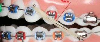
Features of canine correction with braces
The problem can be solved with or without tooth extraction. Both methods help to obtain the space that is missing, but necessary for the dystopic canine to be returned to its place. The choice of the optimal option depends on the clinical situation.
- If removal is not performed, free space is formed due to the gradual lengthening of the dentition with braces. The doctor must assess in advance how the process will affect the proportions of the face.
- If changes in appearance turn out to be negative (for example, the front teeth deviate noticeably forward), a pair of teeth will have to be removed - usually the first premolars.
There is no need to be afraid of removing completely healthy teeth. This method will be suggested by the doctor only if absolutely necessary - to improve chewing function, balance load distribution and maintain the health of the remaining teeth. Due to the free distribution of tooth roots, in this case a stable correction result that is not prone to relapse will be achieved.
Orthodontic patients often ask: is it possible to put braces on one jaw? Dystopic fangs are just that case. If there is a need for significant straightening of the canines with braces on the upper jaw and minor correction of the lower dentition, the lower bracket can be installed later, which is more financially beneficial for most patients.
The completion of tooth root formation occurs at approximately this age:
| Teeth | Upper jaw, age | Lower jaw, age |
| Central incisors | 9-13 | 7-11 |
| Lateral incisors | 9-12 | 8-11 |
| Fangs | 9-12 | 9-12 |
| First premolars | 11-13 | 11-13 |
| Second premolars | 11-13 | 11-13 |
| First molars | 9-12 | 9-12 |
| Second molars | 14-15 | 14-15 |
Since the eruption of third molars does not occur at a specific time, it is impossible to establish a clear age at which their roots are formed.
X-ray results confirm the completion of the process of tooth root formation. The key signs are the absence of an opening at the apex, as well as a pronounced periodontal contour.
Thus, completion of dental growth, including full maturation, usually occurs only between the ages of 15 and 18 years. It is at this time that the maxillofacial apparatus already has the same dimensions as in adults.
Deviations in timing and problems when changing teeth
One or another problem with the loss of baby teeth and the germination of molars occurs in at least every tenth child. Fortunately, dentists are ready to correct the bite during the growth stage, remove extra teeth, and even insert an implant if there is no point in waiting for a root one.
Let's look at the main deviations that may cause concern:
"Shark's Mouth"
Sometimes baby teeth do not have time to fall out, but the molars have already grown “second row”. The “shark’s mouth” is not worth admiring. You need to go to the dentist as soon as possible and remove unnecessary temporary teeth, otherwise your bite may be significantly damaged.
Untimely change of teeth
If the first baby teeth begin to leave the baby’s mouth before 5 years of age or after 8 years of age, then this is a deviation from the norm, which means you need to understand the reason. Trauma, caries, ecology, nutrition, congenital problems with bite or other heredity, various infectious diseases, hormonal problems (for example, thyroid disease), as well as diseases due to which the child is at risk for underdevelopment and delay may be to blame. formation of permanent dentition (diabetes, leukemia, immunodeficiency and others).
The molar does not grow in place of the lost milk tooth
There is no need to panic, but it is worth consulting with a specialist. For example, if a molar tooth does not erupt, although the baby tooth has fallen out a long time ago, the reason cannot be determined with ordinary eyes, but an x-ray will help. If there is no tooth germ in the picture, this is called adentia. This problem is extremely rare - the tooth simply has nothing to grow from. The decision will require the help of a prosthetist.
With retention, the germ of a new tooth is present, but it is directed incorrectly or is deep in the gum. All that remains is to wait for the tooth to grow. This problem occurs more often in the lower third molars, as well as in the upper incisors and canines.
With impaction, the tooth cannot come out due to the close fit of neighboring teeth. Here, too, you cannot do without the help of professionals.
We also advise you to consult a doctor if:
- molars appear darkened or grow crooked;
- the order of tooth loss and growth differs significantly from the above graphs;
- the child’s gums hurt and his cheek is swollen;
- baby tooth is corroded by caries;
- a tooth fell out, but blood from the socket does not stop oozing for more than an hour (not to be confused with ichor).
Supernumerary teeth in children
The appearance of additional tooth buds is rare, occurring in only 2% of cases. The anomaly is associated with a disorder of embryonic development. A tooth that appears above a child's tooth is usually removed. But this does not always happen: if the tooth does not disturb the shape and aesthetics of the dentition, the dentist can leave it.
Sometimes supernumerary teeth are located outside the dental arch. The appearance of a tooth on the palate of a child is rare, however, this is no exception. There is no need to be afraid of this: it does not affect your health in any way. The Natadent clinic has a pediatric orthodontist who will help solve this problem quickly and painlessly.
How to ease the baby's suffering?
It is impossible to ensure completely painless eruption of eye teeth. However, you can help your baby in several ways.
A light massage of the gums slightly dulls the pain. In any case, this stops the kids from crying. To do this, you need to gently stroke the gum just above the eye tooth for a couple of minutes. This massage can be done two to three times a day.
Now pharmacies sell special teethers. Before using, keep them in the refrigerator for a while. These simple products are filled with distilled water. If the baby bites through the shell, then nothing bad will happen.
You can dull the pain with the help of anesthetic gels Dentinox, Kalgel or Kamistad. They begin to act a few minutes after application to the gums.
If the baby’s nose is stuffy during the teething of the eye teeth, then in this case it is worth using drops of Otrivin, Nazivin or Quix. They tend to constrict blood vessels. It also happens that the body temperature of babies rises to 38 degrees or higher. In this case, you have to resort to antipyretic children's drugs paracetamol or ibuprofen. They are available in the form of syrups or candles.
If the baby is having too much trouble with the teething of the eye teeth, then in such a situation it is better to call a pediatrician at home.
Which baby teeth fall out first?
According to the standard pattern for replacing baby teeth, the lower incisors are the first to fall out. They are located in the center of the jaw. They can loosen one by one or almost simultaneously.
Following them, the opposite upper units begin to move. This is why you can often see children five or six years old with a funny toothless smile.
Aesthetic objectives: canines in the lateral incisor position
Very often, dentists do not think about the long-term consequences of their treatment. An incomplete treatment plan or poor treatment choices (often dictated by the patient) usually lead to more serious problems years later. If only we could know what will happen in the future, it would make the task much easier.
However, dentistry can be a predictable subject if approached with proper function and aesthetics in mind. Each dental student receives a phantom - a model that shows the ideal condition and position of the teeth. All dentists are taught the principles of balanced occlusion and the importance of reducing destructive forces.
This article will demonstrate what happens when the factors of time and aging are not taken into account when choosing a treatment method. Very often, what patients themselves do not notice or consider unimportant, about which they do not complain, can turn into a real problem only several years later. If we, dentists, approach the patient from the standpoint of individuality and at the same time as a component of one population, if we carry out careful planning and elaboration of parameters, striving for that same phantom model, then we come to a predictable and long-term quality result. The clinical case discussed here concerns a woman with congenital absence of lateral incisors. When she was a teenager, the maxillary canines were orthodontically moved into position as lateral incisors. In her younger years, her appearance was quite harmonious and attractive, but over time the picture began to change.
Absence of lateral incisors
It is reported that approximately 2% of the population are congenitally missing one or two lateral incisors. Paired absences are more common, and if only one is present, it is usually a microdent. Performing OPTG at an early age makes it possible to find out which of the permanent teeth have not formed.
It is very important to learn about the congenital absence of a tooth at an early age, so that the entire sequence of therapy can be correctly coordinated to restore aesthetics and function. Treatment of congenital absence of lateral incisors is an orthodontically and therapeutically complex task, which is based on the tooth-dental arch size relationship. Numerous studies have been published comparing the method of mesial repositioning of canines and distalization of canines followed by prosthetic restoration of missing incisors.
From a modern point of view, the most logical way is, of course, to open up space and replace missing teeth with prosthetics. The phantom from student life, at least, definitely had his own lateral incisors.
Moving the canine to the lateral incisor position may have several aesthetic and functional disadvantages:
- The canine, a fairly wide tooth, begins to replace the naturally narrow incisor. The color of the canines is usually somewhat darker than that of the lateral incisors, so they stand out from the general plan if they are not in the corners of the smile.
- The level of the gingiva of the canines is approximately similar to that of the central incisors, so moving the canine to the place of the lateral incisor causes visual disharmony.
- Insufficiency appears in the processes of occlusion and articulation, since after movement the canine guidance of the lower and upper canines is not carried out together.
- In the absence of canine protection, the risk of abrasion of the remaining teeth increases, usually consisting of small cracks and microfractures. Over time, periodontal problems and increased sensitivity may appear.
- Long-term retention with an orthodontic retainer will be required to retain the canine in the lateral incisor position.
- The patient may begin to experience TMJ discomfort, muscle tension, grinding, and headaches. This may occur due to effects on muscles that are not anatomically intended.
- The tissues of the vestibule of the oral cavity and the bony prominence in the area of the canine root do not look natural in the area of the lateral incisor. Without the presence of a tubercle in the corners of the mouth, the tissues do not have sufficient support, which leads to recession of the cheeks and narrowing of the buccal corridors. Over time, fewer and fewer teeth become noticeable when smiling.
Orthodontic vision
Traditionally, orthodontists have not encountered people in need of restorations, as they have primarily worked with younger patients. Young people rarely need major restorations. However, in the 21st century, orthodontists often began to take on patients with the need for restoration or due to periodontal disease in the latter. The absence of a lateral incisor is an aesthetic indication, so the orthodontist should treat such a case in this aspect.
The goals of orthodontic treatment often vary depending on the final goal and the need for restoration:
- Positioning of teeth can occur in order to improve the performance of any restoration, for example, to replace a lateral incisor or another tooth.
- There are some advantages to performing permanent or temporary restorations before, during or after orthodontic intervention. This restoration allows you to create the desired shape and at the same time gives an idea of the required space and dimensions. Tooth wear, fractures, underdevelopment in the form of barrel-shaped incisors, etc. are the main indications for restoration before orthodontic treatment.
- Orthodontists sometimes need to reposition a tooth to improve oral hygiene
- Orthodontic treatment may be performed due to periodontal problems, such as insufficient gingival margins, absent gingival papilla, or bone loss.
Today, the goals of orthodontic treatment have diversified significantly, as the aesthetic and functional vision of problems has expanded. Properly planned orthodontic treatment can achieve stable and functional occlusion, improve the health of periodontal tissues and improve dental and facial aesthetics. Orthodontists simply need to study facial aesthetics. Modern specialized literature, research and training always have a positive effect on a specialist and, accordingly, his work results. However, in the past, very little attention was paid to facial aesthetics and periodontal tissue health. A successful orthodontic treatment can be considered an intervention that results not only in the ideal articulation of the models (as well as the achievement of ideal cephalometric relationships and sizes), but also in the restoration of facial aesthetics and harmony in a given position of the teeth.
A smile analysis should include the following: the vertical position of the teeth at rest and when smiling; the transversal (horizontal) dimensions of the smile; the characteristics of the smile arc; and the vertical relationship of the gingival margins to each other. Taking into account all the data, it becomes desirable to move the canines to their natural position and replace the lateral incisors through prosthetics.
Interdisciplinary treatment planning
Innovations in aesthetic dentistry have led to developments in all dental specialties. In today's standard of dental rehabilitation, specialists proceed, first of all, from the individual characteristics of the patient's face and his needs. Each treatment plan begins with an aesthetic assessment. During the analysis, the patient's lips, skin, and cheeks are examined. We must always refer to the position of the teeth in the upper jaw and the level of the gums in relation to the face, and then determine what corrections need to be made for a given appearance. We cannot achieve correct occlusion until our final vision of aesthetics is determined. Aesthetics dictates where the teeth should be located, what the vertical position, guidance and relationship should be.
The main person in the team is the restorative dentist, and the success of teamwork is achieved through detailed discussions of each problem. The main specialist must achieve real therapeutic goals (take into account the economic component, expectations), create an aesthetic vision of the final result, determine the sequence of treatment and restore poorly formed teeth to ideal proportions. The restorative dentist acts here as a link among all specialists, uniting and controlling the manipulations carried out and the pursuit of the final goal.
Clinical case
Diagnosis and treatment plan
A 40-year-old woman came to the clinic (in 2003) in need of cosmetic correction of treatment performed in adolescence.
The patient had a congenital absence of lateral incisors, so she underwent orthodontic treatment at the age of 14 years to solve this problem. Relying on the orthodontist and unaware of other treatment options, the parents chose (or allowed) to move the girl's canines to the lateral incisor position. After the treatment, the smile did not look attractive enough, but the patient was able to get used to it. But as she reached middle age, the woman began to notice that her interlocutors were increasingly staring at her teeth. This made her insecure. The canine was darker, had a rounded contour and a gum level that was in disharmony with the rest of the teeth. The buccal corridors were narrowed to close the empty space, creating the appearance that the cheeks were somewhat sunken (Photos 1 and 2). The patient remained deeply dissatisfied with her appearance.
Photo 1: In 2003, a 40-year-old patient presented with an aesthetic problem.
Photo 2: The canines were moved to the position of the lateral incisors, which stood out from the overall appearance.
With the advent of the 21st century, there have been significant changes in dental technology and materials. Aesthetic manipulation has become common and routine. In this situation, should we have moved the canines to their correct position and replaced the lateral incisors? The patient was open to any options.
After weighing all our options, we decided not to move the canines back and place titanium implants. Although this would be the most common solution to the problem, this path did not seem entirely justified to us. Such treatment would take approximately 18 months, and there was also the possibility of resorption and the need for bone grafting. Since the patient clearly wanted to change the shape and color of her front teeth, an easier and more predictable alternative was in development.
After analyzing radiographs, photographs and working models, we decided that after flattening and narrowing the canine, we could use light orthodontic extrusion of the teeth, which would realign the ridge bone and create a new harmonious appearance at the soft tissue level. By leaving space distal to the canine, we can create porcelain veneers to correct the shape of the canines to resemble the lateral incisors and the first premolars to resemble the canines. Placing veneers on the side teeth will also be possible if the patient so desires. According to the plan, the treatment was supposed to take approximately 6 months. It is important to evaluate all the benefits and risks of any of the treatment options, and the chosen option already needs final approval by the patient himself.
Since the patient expressed a desire to install veneers on the teeth of the anterior zone, all we had to do was correct the position of the lateral incisors and canines.
Clinical protocol
The labial surface of the canines was flattened and the mesial and distal contours were tapered using a flame-shaped diamond bur (Dental Savings Club) followed by finishing with strips (Integra Miltex). Ormco Saphire (Ormco) braces are fixed on the upper jaw up to the first molar (Photo 3). Using Ni-Ti wires of increasing diameter from 0.13 to 0.16 with an extrusion force of 15 g, it was decided to carry out labilization of the central incisors and extrusion of the canines to correct the gingival level (Photo 4). The use of orthodontic extrusion to modify soft and hard tissues was first described in the literature by Heithersay and Ingber. Low-intensity forces (up to 30 g) stimulate marginal positioning of the bone crest, behind which movement of the gums occurs. Restructuring of the alveoli occurs along with the movement of the root.
Photo 3: Preparation for orthodontic treatment: the labial surface of the canines was slightly flattened, and the contours on the proximal sides were narrowed.
Photo 4: Ni-Ti arch with a force of 15 g is used to labilize the central incisors and extrusion of the gingival level of the canines.
Extrusion of the canines by 1 mm was carried out over 3 months, followed by stabilization for 3 months with a Ni-Ti arch 18x25. The total time spent on treatment coincided with the plan and amounted to 6 months. The gingival level of the canines is corrected and lowered, the canines are ready for restoration according to the proportions of the lateral incisors. For the formation of “fangs” from premolars, space is left distal to the true canines (Photo 5a-6). The braces are removed, leaving the teeth in an ideal position for further restoration with porcelain veneers (Photo 7).
Photos 5a and 5b. Reduced gingival level at the lateral incisor.
Photo 6: Space is left at the distal surface of the canine for restoration of the premolar in the shape of the canine.
Photo 7: After removing the braces, the teeth are left in an ideal position for further restoration.
A vinyl polysiloxane (VPS) impression was made, followed by a diagnostic wax-up of the anterior 6 teeth (Figure 8). At this stage, it was decided to evaluate the esthetics of the anterior sextant before widening the buccal corridor.
Photo 8: Diagnostic wax-up creating the ideal appearance of the 6 front teeth.
Conservative preparation (0.3 mm) was carried out (Photo 9) and the final VPS impression was taken (Honigum DMG America) using conventional and lightweight techniques. Bite registration was carried out by Luxabite (DMG America). Using the classic VITA scale, shade A1 was determined. Temporary structures made of bis-acrylic material (DMG America), made on the basis of a diagnostic wax-up matrix, are fixed to the prepared teeth. Fixation was carried out using Temp Bond Clear (Kerr). Next, the appearance of the structures was corrected with soft diamond and Flexi discs (Cosmedent) (Photo 10).
Photo 9: The teeth were prepared by 0.3 mm to install ceramic veneers.
Photo 10: Luxatemp temporary structures (DMG America), creating a natural smile appearance.
Models are cast from plaster (Photos 11a and 11b) and then porcelain veneers are made (Photos 12a and 12b).
Photos 11a and 11b. The Geller technique allows you to emphasize the contour and level of the gums.
Photos 12a and 12b. Veneers made from Creation Porcelain (Jensen Dental).
The veneers were fixed with colorless cement (Variolink Veneer Ivoclar Vivadent) in 2004. The patient was satisfied. Ultimately, she adjusted her smile to the desired result. The size and shape of the front teeth are made according to golden proportions, so it is almost impossible to determine the congenital absence of incisors. Premolars are shaped like canines. At this stage, the slightly reduced buccal corridor did not bother the patient at all.
After some time
Ten years later, in 2013, the patient returned to the clinic. Now the lateral teeth caused aesthetic dissatisfaction (Photo 15). The upper second premolars and molars on both sides were prepared (Photos 16a and 16b), then a suitable shade was selected (Photo 17). The restorations were made from Creation Porcelain (Photo 18). The structures were fixed using transparent cement for veneers (Variolink Veneer). The resulting result looked uniform, as if created at the same time (Photos 19 and 20). The patient was satisfied.
Photo 13: Completed treatment in 2004.
Figure 14: Despite the narrowing in the lateral areas, the patient was satisfied with the aesthetics of the anterior segment.
Photo 15: By 2013, the patient wanted fuller buccal corridors.
Photos 16a and 16b: Preparation of second premolars and first molars.
Photo 17: Shade selection according to the VITA scale.
Photo 18: Making ceramic restorations (Creation Porcelain).
Photos 19 and 20: Completed treatment in 2013.
Final comment
People often change their minds, so it is difficult to predict what a patient who was treated as a teenager will want. A careful treatment plan helps overcome many of the negative factors associated with subsequent gradual aging. A careful discussion of all the details allows for high-quality rehabilitation, as well as building a trusting relationship with the patient.
Author: Elliot Mechanic , DDS, BSc
Which teeth exactly have such a strange name?
So, the term “eye teeth” is not a medical term. The correct name for these teeth is upper canines. Where did such a strange name come from? The fact is that in the immediate vicinity of the upper fangs there are threads of the facial nerve. When these threads are irritated, very severe pain occurs in the upper part of the face. This pain even extends to the eyes.
When babies' upper canines begin to erupt, pain occurs that makes children cry. Unfortunately, this process takes quite a long time. Removal of eye teeth in adults is also always accompanied by severe pain. In this case, dentists use very strong anesthesia.
From somewhere there are ridiculous rumors that a person can even go blind from the removal of eye teeth. Such cases are unknown to medicine.