Often the cause of female infertility lies in pathologies of the fallopian tubes, and all women who plan to become mothers need to have them checked. The most common, informative and accessible way to diagnose the condition of the fallopian tubes today is hysterosalpingography.
– one of the x-ray methods for examining female reproductive health.
During the procedure, the area being examined is exposed to x-rays, thanks to which the diagnostician (in this case, a radiologist) receives information about the condition of the fallopian tubes and can assess their patency.
The fact is that this factor directly affects a woman’s ability to bear children. And the number of patients diagnosed with infertility, unfortunately, has tended to increase over the past decades.
Thus, the hysterosalpingography procedure makes it possible to obtain very clear images in which adhesions and other pathologies are visible. An X-ray examination of the fallopian tubes can also help identify conditions such as fibroids, polyps or synechiae in the uterine cavity, hydroomentum, and peritubar adhesions (that is, adhesions pressing on the outside of the fallopian tube). The results of this examination are accurate in approximately 80% of cases. In controversial situations, the doctor may prescribe an additional examination - for example, hysteroscopy
.
X-ray of the fallopian tubes is safe - the average dose of radiation that a woman usually receives during an examination is 70-100 times less than the maximum safe one and cannot lead to tissue damage or mutations.
When is the procedure scheduled?
An X-ray of the fallopian tubes is performed in cases where the planned pregnancy does not occur for a long time, but other diagnostic procedures have not identified objective factors in the occurrence of the pathology.
Other indications for the procedure:
- presence of previously diagnosed uterine pathologies;
- malformations of the uterus and fallopian tubes;
- suspicion of cancer;
- the likelihood of tuberculosis of the genital organs;
- a history of pregnancies that ended in miscarriage or fetal death.
This method is also used before ovulation stimulation (for example, with polycystic ovary syndrome) or IVF.
Hysterosalpingography
– the procedure is quite common, it is performed by 1-2% of women planning a pregnancy. Although in recent years, doctors have been talking about a trend towards an increase in this number. This may be due to the fact that these days more and more women are deciding to have their first child at the age of 30-35.
Contraindications
X-raying of the fallopian tubes has some contraindications, which include:
- pregnancy,
- allergy to contrast agent,
- various infections of the vagina or cervix;
- acute inflammatory processes in the genital organs, inflammation of the vagina and vulva
- with hypothyroidism;
- in the presence of diagnosed renal or liver failure, liver cirrhosis;
- with uterine bleeding;
- in the general serious condition of the patient;
- with thrombophlebitis and acute heart failure.
Therefore, before the examination, doctors advise taking a pregnancy test, a general gynecological examination and taking a bacteriological smear from the vagina.
How dangerous is x-ray?
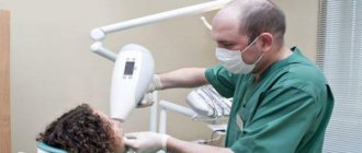
In many cases, dental treatment is simply unthinkable without X-ray examination. When you can't do without it:
- when filling thin and curved tooth canals;
- to detect hidden carious cavities, caries under fillings;
- when the question arises whether wisdom teeth need to be removed (for example, if they are incorrectly positioned);
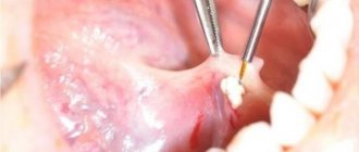
- to diagnose a tooth root fracture, cyst, determine the degree of inflammation of the tissues around the diseased tooth;
- when the issue of removing or restoring a tooth is being decided.
In the first three months, it is not advisable to take dental x-rays for pregnant women, since the fetus has not yet formed. Radiation can have an adverse effect on dividing cells, cause chromosome abnormalities and provoke intrauterine developmental defects. By the second trimester, at 16–20 weeks, the fetus is already fully formed - a tiny dose of radiation will not harm the baby in any way, so pictures can be taken without fear.
Dental X-rays during pregnancy are much safer than examinations of the pelvis or chest, because the mother’s belly is reliably protected by a lead apron, and dental examinations are always carried out using special devices with reduced radiation exposure. This applies to both old models and modern visiographs.
Photo of teeth at different times
In early pregnancy (1st trimester)
Why pregnant women should not have dental x-rays done in the first trimester of pregnancy was stated earlier. If a woman had an X-ray examination of her teeth at a time when she did not yet know about her situation, she should not worry too much about this, much less think about what pathologies this will cause in the fetus. In any case, a routine ultrasound will be performed in each trimester of pregnancy, which will show how the child is developing.
In the 2nd trimester
The second trimester has always been considered the safest period of pregnancy. The baby’s organs and internal systems have already formed, and now they are gradually developing and growing. It is during this period that women can be prescribed x-rays (but choose methods with minimal radiation) if there are certain indications.
Visiograph - an alternative to x-ray
A visiograph is a modern analogue of an x-ray machine. Its advantages:
- Allows you to take an image instantly, high-quality data is immediately displayed on your computer monitor.
- A narrow beam of rays is directed exclusively at the diseased tooth, does not affect adjacent tissues, and certainly cannot “touch” the abdomen.
- The duration of exposure (irradiation) is 10 times lower than when using old-style X-ray machines and is only 0.05–0.3 seconds. Radiation exposure is only 2 microsieverts (when using old devices - from 7 to 80 microsieverts).
For comparison, just sitting in front of the TV for three hours and enjoying your favorite series, a mother receives a radiation dose of 5 microsieverts. The safe radiation dose per year is 1,000 microsieverts (that’s 500 shots). Of course, you shouldn’t take the procedure lightly, but you shouldn’t panic and overestimate the danger to the baby either.
The need for x-rays before pregnancy
An X-ray examination performed during pregnancy planning may be needed in different cases. For example, in the absence of fluorography. It may be required at work if this period coincides with the next preventive medical examination. Or the expectant mother simply has not undergone such an examination for a long time, and now, in preparation for conception, she will have to do it.
Another option for x-ray examination is dental treatment. Before pregnancy, it is advisable to carry out a complete sanitation of the oral cavity to exclude foci of infection. Often during dental treatment, especially in cases of dental problems, it is necessary to take an x-ray.
Before planning a pregnancy, it is necessary to sanitize your teeth
There are situations when, in order to undergo a full examination before conception, the doctor, in addition to directions for tests, prescribes an X-ray examination of the abdominal organs. This procedure is especially necessary when you need to check the patency of the fallopian tubes.
How to minimize risks
What should an expectant mother do so as not to worry about the baby’s health?
- Not all clinics, especially municipal ones, have visiographs. It is better to inquire about this in advance.
- Be sure to use a protective apron and collar.
- You need to inform your doctor about the exact timing of your pregnancy. In case of a delay, even if the test shows a negative result, you must inform the specialist. In unclear situations, diagnosis (and treatment) are carried out as if pregnancy was definitely established.
- Severe pain, swelling, inflammation are good reasons not to delay a visit to the dentist, even in the first trimester. Unless absolutely necessary, the doctor will never prescribe dental x-rays during pregnancy and will try to do everything to postpone treatment to the 2nd, safe trimester or to the postpartum period. However, some acute conditions may require emergency intervention, such as purulent pulpitis or acute periodontitis. In this case, the mother needs to remember that a rapidly developing infection in the oral cavity will cause much more serious damage to the baby than 1-2 pictures on a visiograph.
- If there are concerns that the X-ray has somehow damaged the baby, you should consult with a geneticist and do an ultrasound. This will reassure those mothers who may have taken the photo without knowing that they were expecting a baby.
Since dental treatment during pregnancy is still associated with certain difficulties, the best thing an expectant mother can do is to go to the dentist and have all her teeth treated at least a month before the date of planned conception. And already in pregnancy, mommy should brush her teeth thoroughly and eat foods high in calcium.
Diagnostic results: interpretation of images
The contrast agent does not transmit x-rays, and the cavities filled with it are visible on the image as bright white spots. An indicator of good tube patency is the spreading of the contrast agent along the peritoneum to areas remote from the insertion site. If the contrast stops at any section of the fallopian tube, it means that there is an adhesion or other pathology in this place. Depending on the shape of the uterus and tubes, the doctor can determine which pathological process is preventing conception. Interpreting images of the uterus and fallopian tubes requires extensive knowledge and considerable practical experience. Self-diagnosis is a completely useless exercise.
If the results of the examination give reason to suspect the presence of uterine cancer, it is necessary to order additional examinations, including taking tissue for a biopsy. The diagnostic doctor's conclusion, along with the photographs, is transmitted to the attending physician who referred the woman for examination.
Interpreting images of the uterus and fallopian tubes requires extensive knowledge and considerable practical experience. Self-diagnosis is a completely useless exercise.
Risks of dental caries
A child is a key task for a pregnant woman’s body.
If he needs something, the body would rather give up resources to its detriment than allow a shortage for the future baby. A huge amount of calcium is required for the formation of bone and muscle structures of the fetus. Normally, if mom continuously chews cottage cheese pancakes, washing them down with whole milk, calcium will not be a problem. In the case of pathology of calcium metabolism or poor nutrition of the mother, the concentration of calcium in saliva may decrease. This leads to a decrease in its remineralizing properties, since normally saliva replenishes calcium ions washed out under the influence of bacterial acids. As a result, if nothing is done, during nine months of pregnancy you can not only get new foci of acute caries, but also have time to lose several teeth, where the carious process from chronic will turn into a rapid acute course.
In the first trimester, we try not to touch small carious lesions and deal only with remineralization. But in the future, caries must be treated.
Features of x-rays before pregnancy
A situation may arise when a woman, having undergone an X-ray examination, subsequently finds out that she was already pregnant at that time. Awareness of what happened, and fear of “what will happen?” give no rest. In this case, you need to know exactly the timing of the procedure and the date (at least approximately) of conception.
The fact is that if radiography was carried out in the first third of the menstrual cycle (days 1-8), when the egg has not yet matured or been released, then there is nothing to worry about. If the egg was irradiated after ovulation, that is, when it had already left the ovary (in the second third of the menstrual cycle) and conception had occurred, then the question of continuing the pregnancy should be entrusted to a doctor.
Everything will depend on how strong the radiation was, how often it had to be done, and what it was intended for. So, if, when planning a child, the expectant mother takes an X-ray of a tooth, then that’s one thing (there is no need to talk about any irradiation of the egg here). But if an X-ray examination of the pelvis was carried out, and more than once, then, of course, this can negatively affect both the pregnancy that has already occurred and the planned one. In the second case, it will be better if conception occurs a month after the procedure.
What's the result?
Pregnant women are very complex, fragile and emotionally vulnerable patients.
We always try to be especially attentive to their treatment. If a difficult situation arises that requires the prescription of some potent drug, we often arrange a consultation with the obstetrician who is caring for the patient’s pregnancy in order to jointly make the least risky decision for her and the baby. Pregnant women have other specific difficulties. For example, we cannot keep them in a chair in one position for a long time. Expectant mothers should not lie on their backs for a long time, as this position compresses the blood vessels supplying the uterus. After some time, the child usually begins to push indignantly and demand to change the position. It is necessary to give the opportunity to turn on one side or even stand up and walk around, if the treatment protocol allows.
In pregnant women, the volume of the bladder is greatly reduced, which is compressed by the growing uterus. This also has to be taken into account when planning long-term manipulations. In short, there are a lot of nuances, and they are all important. The most important thing is to remember that refusing dental treatment and support often carries greater risks than using local anesthesia or even x-rays.
Risks for mom
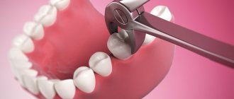
You can lose a tooth
We try not to use antibiotics, especially in the early stages of pregnancy.
This greatly changes the treatment tactics if we discover a chronic or aggravated purulent focus that has arisen due to an affected tooth. In a normal situation, we can try to save the patient's tooth under the additional cover of antibiotics that accumulate in the bone tissue. For example, this is a fairly common fluoroquinolone - ciprofloxacin. Everything is great, but it is strictly prohibited for use by pregnant women and children under three years of age due to the fact that it can cause defects in the formation of joints and cartilage. In a situation where the prospects for a tooth without antibiotic therapy in a pregnant woman are questionable, and the risks of developing an infection are high, we are usually inclined to remove such a tooth. It is much better to remove a potentially problematic tooth than to risk the health of the baby or the expectant mother. You cannot leave a chronic purulent lesion: it is dangerous for the child and for herself if it eventually worsens. If periostitis or, especially, phlegmon develops, the mother’s life will have to be saved. Then you will definitely have to prescribe antibiotics and other drugs.
Orthodontics
We stop the orthodontic treatment process if the patient reports pregnancy. First of all, we do this due to the impossibility of X-ray monitoring of the treatment process. Working with a bite blindly is a very bad idea and can end in even bigger problems. Most often, the patient remains at the stage when she found out about pregnancy. Either we continue to use the same aligner, or we simply fix the current condition in the case of braces.
Periodontal problems
The pregnant woman's body directs all efforts to pregnancy.
At the same time, he may ignore some of his own needs and problems. For example, large doses of sex hormones ensure good blood supply to the uterus and the desired condition of the mucous membranes. But in parallel, these same mechanisms lead to gum hypertrophy in the oral cavity. This is especially pronounced in the last trimester, when it becomes noticeable. According to various authors, gingivitis in pregnant women occurs in 25–100% of women, usually from the second to the eighth months of pregnancy. Moreover, in the region, 80% of women experience bleeding gums and have at least chronic catarrhal gingivitis. For the most part, all these symptoms will go away after childbirth and normalization of hormonal levels. However, without high-quality professional hygiene, there is a risk of developing more severe forms of periodontitis and damage to its structures. This also creates additional risks of accumulation of subgingival dental plaque and the development of multiple cervical caries.
To prevent such problems, we always recommend professional hygiene and give recommendations on what to do at home.
What is the relationship between a man's reproductive function and his age?
With age, hormonal changes occur in a man's body, the secretion of the gonads decreases, spermatogenesis decreases, and the consequences of environmental exposure accumulate. All this does not have the best effect on the ability to conceive. However, if a man leads a healthy lifestyle and monitors his physical condition, then his reproductive functions can be maintained until old age.
But in fairness, it is worth noting that in the sperm of older men, a larger number of cells with a broken DNA strand are found than in the sperm of younger men. Fertilization with such sperm leads to genetic abnormalities of the fetus and miscarriages (even if the woman is much younger than her partner). However, the sperm of an older man also contains sperm that can fertilize an egg.
What do doctors think?
Doctors do not have a consensus on the effect of X-ray radiation on the unborn child at the planning stage. Some claim that it is absolutely safe and will not cause any harm.
Others say that there is still a certain percentage of negative impact, but it depends on the frequency of exposure over any period of time.
What should you do if, at the stage of pregnancy planning, you have to visit an x-ray room? How long before you can think about conceiving? It is clear that you still need to undergo an examination, and there is no escape from it. Therefore, it is best to count the days of the menstrual cycle. It is not recommended to take x-rays in the second and last third. However, this applies to x-rays of the pelvis or lower spine. If you are a very suspicious person who avoids pills, microwaves and computers, then you can postpone conception until the next menstrual cycle, so as not to worry then for the entire nine months.
Is there a relationship between a man’s reproductive function and his potency?
More likely no than yes. A man may complain of poor erection, but he may have a normal spermogram. There are also cases when patients are completely satisfied with their intimate life, but suffer from severe infertility.
However, sometimes decreased potency and reproductive dysfunction are related. There are diseases that lead to disorders of potency and spermatogenesis. Another possibility is reduced fertility caused by weak ejaculation or sperm backing up into the bladder.
How much does the frequency of sexual contact affect the possibility of conception?
The frequency of sex is very important for conception. A healthy married couple who has sex 4-5 times a week has the best chance of conceiving. At the same time, repeated sex over several days, unfortunately, will not contribute to the growth of male fertility. In this case, the sperm concentration drops, and the likelihood of getting pregnant decreases. However, if a married couple is healthy, then they can choose any intimate schedule convenient for themselves. A healthy man's body produces enough healthy cells every day to conceive a child.