I. K. Lutskaya, Doctor of Medical Sciences, Professor BelMAPO (Minsk)
Modern endodontics in most cases guarantees high efficiency in the treatment of pulpitis and periodontitis. However, violation of the algorithm of interventions or clinical protocols can contribute to the development of errors and complications.
Regular clinical and radiological examinations are extremely important to assess the quality of endodontic treatment.
According to the European Society of Endodontics, assessment of the results of root canal treatment should be carried out within 1 year after treatment and thereafter as necessary. The high quality of therapy is evidenced by the following results: absence of pain, swelling and other symptoms, absence of changes in the sinuses, preservation of tooth function and x-ray confirmation of the presence of a normal periodontal gap around the root. Uneven expansion can be considered as the outcome of the disease - scar tissue changes.
The causes of complications after root canal filling may be errors made at the stages of endodontic treatment.
1. At the preparatory stage:
- Root canal infection.
- Lack of adequate access to the root canal orifice.
- Perforation of the bottom and walls of the tooth cavity.
2. During mechanical treatment of the root canal:
- Obturation of the root canal lumen with dentine filings.
- Formation of an apical ledge when the canal is bent (“Zipping”).
- Excessive lateral expansion of the middle third of the canal along the internal curvature of the root (“Stripping”).
- Perforation of the root walls.
- Destruction of anatomical (physiological) narrowing.
- Fracture of the instrument in the canal.
3. During the process of filling the root canal:
- Heterogeneous, insufficient filling of the canal lumen.
- Removal of filling material beyond the apical foramen.
- Longitudinal root fracture.
Root canal infection
Penetration of microorganisms into the root canal can occur due to merciless preparation with pressure on the coronal pulp, careless amputation and removal of tissue from the orifice. The development and proliferation of microbes is possible due to the repeated use of tools, including burs and excavators. Infection of the root canal increases the risk of post-filling complications such as painful percussion, lack of positive dynamics after treatment of pulpitis or periodontitis. In preventing this complication, great importance is attached to careful isolation of the surgical field, since microflora can penetrate into the canal along with oral fluid. It is optimal to use protective equipment such as rubber dam and its analogues (Fig. 1). Before instrumental treatment, it is advisable to completely excise carious dentin from the walls of the carious cavity in order to prevent infection from entering the root canal.
Rice. 1. Treatment of pulpitis using a rubber dam.
Errors in creating access to root canal orifices
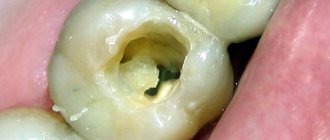
The reasons for this situation are insufficient preparation of the carious cavity, incomplete excision of the roof of the pulp chamber, and lack of control over the insertion of the endodontic instrument (Fig. 2). The consequence is the following complications. Overhanging edges of the cavity do not allow completely removing the remaining pulp from the tooth cavity, which inevitably leads to the appearance of pigmentation and worsens the aesthetic parameters of the tooth.
Rice. 2. Incomplete opening of the tooth cavity.
Due to poor visibility, it is not always possible to identify all existing root canal orifices, which precludes treatment and filling of undetected canals (Fig. 3).
Rice. 3. Poor quality treatment of the cavity walls.
The apparent “saving” of hard tooth tissues in the process of cavity formation can lead to poor-quality endodontic treatment.
At the same time, excessive, excessive tissue removal causes a decrease in the tooth’s resistance to mechanical stress.
A measure to prevent such an error is the formation of correct access, which is characterized by the absence of overhanging edges and the straightness of the cavity walls, which should be smooth, without roughness or notches.
Treatment
Small cracks are treated with remineralization therapy - the enamel is coated with products containing calcium and fluoride. Veneers are also used for treatment - thin ceramic or composite plates that cover the surface of the tooth. The technique is used on the front teeth; for cracks in the chewing teeth, artificial crowns are installed.
According to their location, cracks can be vertical, horizontal, or inclined; deep vertical ones can eventually open access to the pulp, which leads to its death. In this case, after cleaning and filling the canals, a crown is installed on the tooth. If there is a vertical crack running through the entire root, the tooth must be removed and replaced with a denture.
Injury to the root pulp
When treating pulpitis using the amputation method, injury to the root part of the pulp is possible in the absence of adequate access to the mouths of the canals (Fig. 4).
Rice. 4. Hypertrophied gums obstruct the view of the cavity.
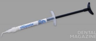
Excessive pressure on the bur or excavator will cause bleeding from the canal due to rupture of the neurovascular bundle. The application of a therapeutic pad over the mouth of the canal under pressure contributes to the disruption of blood circulation and the functioning of the root pulp (Fig. 5). In any case, trauma to the root pulp increases the risk of ineffective treatment of pulpitis with a biological method.
Rice. 5. Therapeutic lining over the mouths of the canals.
This complication can be avoided by careful preparation of the carious cavity with complete excision of the altered dentin and subsequent careful removal of the roof of the pulp chamber.
Perforation of the bottom and walls of the tooth cavity
May occur during the search for root canal orifices and their expansion; with poor visibility of the bottom of the tooth cavity as a result of inadequate formation of access to the root canals.
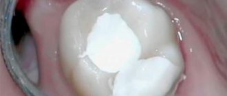
The presence of softened pigmented dentin, intense staining of hard tooth tissues after previous treatment (resorcinol-formalin method, silvering) also greatly complicate the search for root canal orifices (Fig. 6).
Rice. 6. Dentin pigmentation and paste residues at the bottom of the cavity.
In some cases, the causes of perforation are the following factors: insufficient or, on the contrary, excessive expansion of the tooth cavity; carrying out endodontic treatment through an artificial crown. Insufficient knowledge of anatomical features, such as displacement of the tooth axis and a decrease in the height of the crown due to its significant abrasion, contributes to the commission of errors.
Preventive measures for perforation of the walls of the tooth cavity are rational excision of hard tissues, adequate pressure on the bur during the preparation process, its correct direction and precise control of the depth of insertion of the rotating instrument.
Incomplete removal of the root pulp is allowed in cases where adequate access to the canal mouths is not provided or the latter are inaccessible due to the location of denticles in them. The reason may be insufficient expansion of the canal mouths or incorrect determination of the working length. The anatomical features of the structure of the roots can also become a factor in the poor patency of the canal for instruments. Violation of the work technique, for example, removal of tissue with a pulp extractor with rupture of the neurovascular bundle, incomplete removal of the root pulp, leads to bleeding from the canal, which prevents further endodontic interventions.
Obstruction of the canal lumen by dentine filings is manifested by the impossibility of reintroducing a small endodontic instrument to the entire working length. The reason is the accumulation of dentinal filings in the lumen of the canal and their compaction. An attempt to re-pass the canal with force may entail pushing the products of mechanical treatment of the root canal (endolubricants, dentinal filings, pulp residues, etc.) beyond the apical foramen, which can cause pain after endodontic treatment.
This complication is prevented by carefully passing the canal to the apical narrowing with small instruments after every second step, as well as washing the canal lumen with solutions.
The formation of apical enlargement (Zipping effect) most often occurs in curved canals. During canal treatment, slipping of the instrument tip during rotation leads to the so-called “Zipping” effect. The reason is the use of large, inflexible files, which cannot follow the shape of the canal. The canal lumen may be blocked by dentine filings. The risk of creating an apical expansion significantly increases when working with files that have an aggressive apex.
Excessive longitudinal expansion of the canal in the middle third along the internal curvature (Stripping) occurs during mechanical treatment of curved root canals. The reasons may be the following: the use of rigid, inflexible files; mechanical treatment without taking into account the thickness of the canal walls, as well as underestimation of the degree of root curvature.
Due to excessive removal of dentin in the area of the internal curvature of the root, not only does the tooth’s resistance to mechanical stress decrease, but there is also a real risk of longitudinal perforation of the root canal wall.
Destruction of the anatomical (physiological) narrowing occurs when the working length is incorrectly determined. Another reason is a slight decrease in the working length of the channel during straightening. If further processing of the canal is carried out to the previous working length, destruction of the physiological narrowing is inevitable.
Prevention of this complication consists in accurately determining the working length and its correction during mechanical treatment of a curved root canal.
Perforations of the root canal walls occur most often during instrumental treatment of curved roots.
Perforations of the mouth and middle thirds are formed mainly when the filling material is removed from the canal in the process of creating a bed for the anchor pin, as well as when the latter is screwed into the canal.
Apical perforations can occur when using insufficiently flexible rotating instruments in difficult, curved canals. A similar complication is possible from the application of excessive pressure during machining with hand tools, when trying to forcefully pass the canal. The cause of lateral perforation is the passage of a curved canal with an endodontic instrument with an aggressive tip without prior bending.
Measures to prevent various types of perforations are good access to the mouths of the root canals, analysis of the configuration of the root canals according to radiographs (Fig. 7). During mechanical treatment, obstruction of the canal lumen with dentinal filings should be avoided; pre-bend the tool; use an anticurvature technique for passing the canal.
Rice. 7. Excessive preparation and perforation of the wall of the first molar.
Crown fracture without opening the pulp chamber
This fracture affects not only the enamel, but also the dentin; the pulp is not exposed. Patients complain of pain when eating, caused by thermal, chemical and mechanical irritants. The closer the pulp is to the fracture plane, the more severe the pain.
A dental examination reveals:
- defect of part of the crown limited to dentin;
- pain on percussion;
- pain when probing the exposed dentin surface.
X-ray diagnostics are performed to exclude a root fracture, EDI and transillumination examination.
Treatment
To treat this type of fracture in baby teeth, a protective bandage is applied to the exposed dentin, covered with glass ionomer cement on top. The tooth is left in this condition until it is replaced with a permanent one. If the integrity of the bandage is damaged, the bandage is changed; X-ray monitoring of root formation is periodically carried out if it has not been formed.
Subsequently, thermal diagnostics and EDI are carried out to check the viability of the pulp: one, three and six months after the injury, then every six months until the root is formed.
When treating permanent teeth, sharp edges are ground off, teeth are restored with filling materials and crowns. If the non-viability of the pulp is detected due to a strong blow, it is removed.
Instrument fracture in the root canal
The risk of instrument fracture is very high in case of file deformation (bending, unwinding of turns) and most often occurs when passing and expanding narrow, curved, previously sealed canals (Fig. 8). The main reasons for this complication may be the lack of adequate access to the mouth of the root canal; violation of the sequence of use of endodontic instruments; use of instruments without taking into account indications; non-compliance with operating mode and rotation speed; application of significant force during manual or machine endodontic treatment; metal fatigue caused by repeated use of the tool.
Rice. 8a. Introduction of a curved file.
Rice. 8b. Broken instrument in the root canal.
Prevention of tool breakage consists in strict adherence to the operating mode and use of the tool according to indications. The sequence of use of tools must be taken into account. During machining, the use of endolubricants is recommended.
Incomplete and insufficient obturation of the root canal is mainly due to incorrect determination of the working length, incomplete passage of the canal (Fig. 9), the use of the technique of one gutta-percha or silver pin in canals that have an oval, dumbbell-shaped, slit-like (irregular) shape that does not correspond to the shape of the pin, and also using liquid-mixed paste for filling (using a channel filler). As a result, shrinkage is inevitable, as well as dissolution of the paste some time after filling.
Rice. 9a. Obturation of root canals: high quality.
Rice. 9b. Obturation of root canals: incomplete.
The removal of filling material beyond the apical foramen is often observed after excessive mechanical treatment of the root canal. The result is destruction of the physiological apical constriction. It can also be disrupted due to a chronic inflammatory process in the tissues of the apical periodontium. In addition, there is a real possibility of removing material beyond the apex when using a machine channel filler. The risk of complications increases sharply when filling a root canal without taking into account the working length (Fig. 10).
Rice. 10. Removal of a significant volume of sealer beyond the apex.
The removal of the filling material beyond the apical foramen is observed in the case of using a large amount of sealer, as well as as a result of excess pressure during the condensation of the filling material in the root canal.
Pushing the gutta-percha pin beyond the apex may be a consequence of incorrect determination of the working length and/or incorrect selection of the size of the main pin (Fig. 11).
Rice. 11. Removing the gutta-percha pin beyond the root apex.
Removal of gutta-percha beyond the root apex is possible during the process of lateral condensation of gutta-percha (Fig. 12).
Rice. 12. Lateral condensation of pins.
Preventive measures: control of working length at all stages of endodontic treatment; competent formation of the root canal; maintaining the integrity of the anatomical (physiological) narrowing.
If removing a small amount of sealer beyond the apical foramen may not cause problems, since it is quickly resorbed, then gutta-percha removed beyond the apex, which itself is biologically inert, can maintain inflammation in the apical periodontal tissues for a long time, being a mechanical irritant.
Longitudinal root is possible during the process of lateral condensation of gutta-percha pins and is a consequence of excessive thinning of the walls of the root canal during mechanical treatment. In addition, a longitudinal root fracture can be observed with strong lateral pressure on the sprider during the condensation of gutta-percha pins.
Prevention measures include assessing the condition of the hard tissues of the tooth root, their thickness, as well as improving manual skills and applying adequate efforts in the process of condensation of gutta-percha pins.
Crown fractures, methods of their treatment
Incomplete crown fracture
An incomplete crown fracture (fracture) is a tooth crack that appears as a result of acute (dislocation, bruise) and chronic (abnormal bite, bad habits) traumatic injuries, while the integrity of the tooth is not compromised. The cracks look like lines that run randomly across the enamel; they can appear on teeth located next to a tooth that has undergone more severe traumatic effects.
During a normal inspection, cracks may not be visible; they are detected using transillumination. Transillumination is an examination method that involves shining a cold light beam through the injured tooth. Transillumination lighting is used not only to detect cracks, but also to identify contact caries, subgingival dental plaque, and pulpitis.
When a patient with an incomplete crown fracture seeks help, an electroodontodiagnosis (EDD) is performed to monitor the condition of the pulp after injury.
Typically, cracks do not penetrate deeper than the enamel-dentin border of the tooth, and therefore do not cause complaints in patients, except for minor sensitivity to temperature and chemical stimuli, which sometimes occurs when eating. Over time, the enamel at the site of damage darkens, caries develops, and the tooth may split along the crack.