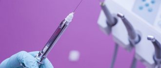
Most dental procedures are performed under local anesthesia. It allows for comfort for both the patient and the doctor.
Mandibular anesthesia is a conductive anesthesia that is performed for a mobile jaw and allows you to reduce the pain threshold of the lingual and inferior alveolar nerves. The technique is quite complex, and to obtain a high-quality result, a clear understanding of the anatomical structure of the dental system is necessary.
Using this anesthesia, the sensitivity of the following areas is blocked:
- half of the units of the dentition (from the insertion side);
- alveolar bone tissue;
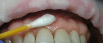
- gum mucosa;
- half lip;
- the area under the tongue and the tip of the tongue;
- chin skin
What is mandibular anesthesia
Dentistry today has a large selection of types of pain relief. This opens up wide opportunities for the dental surgeon, especially in terms of treating young patients. Mandibular anesthesia is a conduction-type local anesthesia. Provodnikova - temporary blocking of the transmission of nerve impulses through a large nerve trunk. It is used when there is a need for an absolute block of pain while keeping the patient fully conscious.
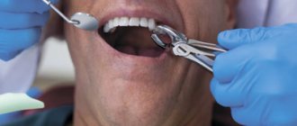
Mandibular anesthesia is required if surgical intervention on the lower jaw is planned. An unpleasant moment of injection is a short-term feeling of fullness, pain and burning in the injection area. This sensation lasts a few seconds and is comparable to a prick when blood is drawn from a vein.
Mandibular anesthesia by palpation
The target point is the mandibular foramen, through which the inferior alveolar nerve enters the canal, located on the inner surface of the ramus of the mandible. Location of the mandibular foramen .
The distance from the edges of the lower jaw: from the anterior edge of the lower jaw - 15 mm, from the posterior - 13 mm, from the semilunar notch - 22 mm and from the lower edge of the lower jaw - 27 mm (Fig. 93, A). Rice. 93. Lower jaw. A - lower jaw from the outside, slightly in front and above (V.P. Vorobyov, 1946): - articular process; 2— coronoid process; 3 - anterior edge of the lower jaw branch; 4 - mandibular eminence (torus); 5—temporal scallop; 6— retromolar fossa; 7 - mandibular foramen; 8 - tongue of the lower jaw; 9 - mental foramen. B - branch of the lower jaw: 1 - inferior alveolar nerve, 2 - internal pterygoid muscle.
The height of the hole in adults corresponds to the level of the chewing surface of the lower molars, in children and the elderly it is slightly lower. From below and in front, the mandibular foramen is covered by a bony protrusion (tongue of the lower jaw). In this regard, the anesthetic solution must be injected 7-10 mm above the level of the hole, where the nerve, before entering the mandibular canal, passes through a bone groove filled with loose fiber and in which the anesthetic spreads well. CONCLUSION : to approach the target point, the needle must be inserted 7-10 mm above the level of the chewing surface of the lower molars (Fig. 94, A).
Rice. 94. Needle position for mandibular anesthesia . (G.P. Ruzin, M.P. Burykh, 2000) A - on the mandibular bone. The needle passes 7-10 mm above the chewing surface of the lower molars, the end of the needle is located in the pterygo-maxillary space in the bone groove (sulcus n. mandibularis). 1 — level of the chewing surface of the lower molars; 2—tongue of the lower jaw; 3 - pterygomaxillary space. B - on a vertical section. B - on a horizontal section of the preparation.
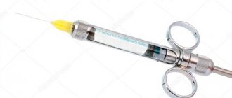
Injection equipment (for all types of intraoral mandibular and torusal anesthesia): carpule or disposable plastic syringe 2 ml, needle 41.5-50 mm long.
Methods of mandibular anesthesia
There are two common methods of performing mandibular anesthesia. The first method is intraoral and is carried out according to the modification of Gou-Gates and Akinosi. The second method is extraoral, carried out in three ways - mandibular, retromaxillary or subzygomatic.
From a technical point of view, there are the apodactyl (without palpation) method, the finger method, as well as modifications of Go-Gates and Akinosi.
Mandibular anesthesia using the apodactyl technique is performed most often. In the area of the required tooth in the lower jaw, infiltration anesthesia is first performed to anesthetize the buccal space. Then, with the maximum opening of the mouth, the patient feels the line between the lower and middle third of the pterygomandibular fold in its lateral slope. This is the place where the needle is inserted. First, it is inserted all the way into the bone, then it is turned in the opposite direction and one milliliter is injected at the level of the incisors. A total of 2 to 2.5 ml of anesthetic is injected.
With the finger method, an injection of an anesthetic solution in an amount of 3-4 ml is carried out in the area of the retromolar space and temporal ridge. The anesthetic is administered, as with the apodactylic technique, in two stages with a change in the position of the needle.
The Go-Gates modification involves anesthesia of three branches of the mandibular nerve at once. For this purpose, an injection of an anesthetic solution is administered in the area of the condylar process of the mandible.
If the patient has limited ability to open his mouth, mandibular anesthesia is performed using the Akinosi method. The patient does not open his mouth and closes his teeth. The injection is placed at the border of the buccal mucosa and the retromolar region of the upper jaw.
To perform mandibular anesthesia, the doctor must be highly qualified and have deep anatomical knowledge of the structure of the lower jaw.
Material and methods
A topographic-anatomical study of the pterygo-maxillary space was carried out on cadaveric material from the Department of Topographic Anatomy and Operative Surgery of Tver State Medical University. The work was carried out on 12 preparations taken from fixed corpses of people of different sexes and ages using the methods of macro- and micropreparation, morphometry, photography and sketching. Data obtained during the study were entered into the protocol manually. A clinical study of the use of the universal apodactylic method of mandibular anesthesia was carried out on the basis of the dental clinic of the Tver State Medical University. The study involved 20 patients (10 men and 10 women) aged from 20 to 52 years, who gave informed consent to participate in the study.
Possible complications
Mandibular anesthesia in rare cases leads to the development of complications. One of them is numbness of the tissues of the pharynx with subsequent limitation of movements of the lower jaw. Physiotherapy, mechanical therapy and administration of medicinal solutions are prescribed as treatment. A hematoma can occur as a result of damage to a vessel. If it does not resolve on its own, a puncture may be required. When a nerve is damaged, neuritis sometimes develops, the treatment of which requires physiotherapeutic procedures using heating elements or electric current. A very rare complication is temporary muscle paralysis as a result of medical error. If the injection technique is violated, the needle rarely breaks off and gets stuck in soft or tendon tissue. In such cases, surgical removal is used after an X-ray examination. If the needle does not cause concern to the patient and has grown into the tissue, it does not need to be removed.
Results and discussion
The conducted topographic-anatomical study made it possible to establish that on the inner surface of the branch of the lower jaw there is an area (triangle) free from the branches of the trigeminal nerve, through which safe access to the lower alveolar nerve can be provided when performing mandibular anesthesia. The boundaries of the mandibular triangle in front were the anterior edge of the ramus of the mandible and the tendon of the temporal muscle, the lingual nerve was located behind and medially, the buccal nerve and the lower edge of the lateral pterygoid muscle were located above. The average dimensions of this triangle were: on the lateral side - 18±0.5 mm, on the medial side - 20±1 mm, on the top - 15±0.5 mm. In a wide-open mouth position, this triangle was projected onto the oral mucosa behind the mandibular molars (Fig. 1).
Rice. 1. Topographic anatomy of the mandibular triangle located in the pterygomaxillary space.
Based on the topographic-anatomical study of the retromolar space, an “intermaxillary triangle” was identified on the oral mucosa, which was projected onto the mandibular triangle, identified on the inner surface of the mandibular ramus. The apex of the intermaxillary triangle was facing the retromolar fossa, the base was located at the level of the alveolar process of the maxilla. The medial border of the premaxillary triangle was the pterygomaxillary fold, and the lateral border was the groove of the oral mucosa, formed in the position of the mouth wide open by the anterior edge of the branch of the lower jaw.
The experimental study made it possible to topographically substantiate the universal method of apodactylic administration of mandibular anesthesia. To carry out mandibular anesthesia, the “intermaxillary triangle” was used as a landmark for the needle insertion site. The needle is inserted with a syringe into the oral mucosa directly in the center of the triangle indicated earlier. The bevel of the needle was directed to the branch of the lower jaw to facilitate its advancement. Inserting the needle within this triangle and advancing it 15 mm in depth was safe, since the needle was located between the inner surface of the mandibular ramus and the lateral surface of the medial pterygoid muscle along with the lingual nerve. The syringe was located in the corner of the mouth at the level of the canine on the opposite side of the lower jaw. An anesthetic depot was created in an amount of 1.7 ml. After the experimental study, the pterygomaxillary space of the deep region of the face was prepared using sectional material.
It was established that this method of mandibular anesthesia was carried out without damaging the internal pterygoid muscle, lingual, inferior alveolar nerves and blood vessels, and the anesthetic was located near the neurovascular formations (Fig. 2, 3).
Rice. 2. Performing mandibular anesthesia on a topographic-anatomical preparation of the lower jaw in the intermaxillary triangle.
Rice. 3. Method of performing mandibular anesthesia (diagram). A priority certificate for invention No. 2017141093 dated November 28, 2017 was received for this method of mandibular anesthesia.
In a clinical study, anesthesia was performed for the purpose of painless preparation of hard tissues of the mandibular molars and surgical manipulation of the mucous membrane of the lateral floor of the mouth and tongue. The technique of this apodactyl method of mandibular anesthesia: the anesthetic was administered, focusing on a number of anatomical formations. With the mouth wide open, the needle was inserted into the middle of the intermaxillary triangle. The direction of the needle was at an angle to the branch of the lower jaw. The syringe was located in the corner of the mouth, at the level of the canine of the lower jaw, on the side opposite to anesthesia (Fig. 4, a,
Rice. 4. Method of performing mandibular anesthesia. b). The needle was advanced until it made contact with the bone tissue. An anesthetic depot of 0.3 ml was created. Next, the needle was advanced parallel to the inner surface of the lower jaw branch deep into the pterygomaxillary space by 15-20 mm. After the aspiration test, an anesthetic depot of 1.7 ml was created. Anesthesia in the area of innervation of the lingual nerve occurred after 3-5 minutes, the inferior alveolar nerve - after 5-7 minutes and continued, depending on the local anesthetic solution used and the concentration of the vasoconstrictor, up to 1.5 hours. The onset of anesthesia was determined by the appearance of feeling in the patient numbness and tingling on the corresponding half of the tongue and lower lip. The manipulations performed were painless in all 20 (100%) patients. In 7 (35%) patients, there were signs of anesthesia in areas of the oral mucosa included in the zone of innervation of the buccal nerve on this side. When performing mandibular anesthesia using this method, complications associated with damage to the lingual, buccal, inferior alveolar nerves, and medial pterygoid muscle were not observed.
Recommendations after the event
Any type of anesthesia is stress for the body, which must be minimized. Immediately after applying anesthesia, you must rise from the dental chair carefully to avoid dizziness. After pain relief, you must avoid drinking hot and alcoholic drinks. Do not massage the injection area or eat hot food. It is necessary to rinse your mouth with soda-salt or any other solution as prescribed by your doctor several times a day. It is better to sleep on the side opposite to the injection.
Techniques
Mandibular conduction anesthesia can be performed intraorally and extraorally.
Intraoral administration techniques:
- palpation;
- apodactyl;
- appdactyl method according to A.E. Verlotsk.
Extraoral techniques:
- subzygomatic introduction in various modifications;
- insertion under the lower jaw;
- retromaxillary route of administration.
Next, we will consider the methods that are most often used in dental practice.
Contraindications for mandibular anesthesia
Mandibular anesthesia is not used for liver diseases, which is associated with a large load on it. Novocaine is the only anesthetic that is not subject to this contraindication. If a major operation is required, local anesthesia is not suitable due to the need to administer a large amount of it. Epilepsy and mental illness, as well as problems of the cardiovascular system are contraindications to mandibular anesthesia. During pregnancy, it is necessary to weigh the risk to the fetus when exposed to drugs. For blood diseases and bronchial asthma, mandibular anesthesia is not performed. Contraindications to anesthesia must be considered individually; sometimes their list can be shortened or, conversely, expanded at the discretion of the attending physician.
Anesthetic solutions
Various compounds are used to innervate the lower jaw. These include lidocaine and novocaine. These substances are time-tested, but are highly toxic. Along with them, articaine anesthetics are used, which give excellent results with less toxicity. These include ultracaine, articaine, ubistezin.
Novocaine is not used in dentistry, since there are more powerful and safer anesthetics of the articaine series. They are 4-5 times superior to novocaine in terms of the degree of analgesic effect.
Articaine drugs, in addition to anesthetic substances, contain vasoconstrictor components such as epinephrine or adrenaline. This ensures that the anesthetic substance remains in the tissues for a long time, since it is not carried by blood vessels outside the area of medical intervention.
Mepivacaine
This short-acting anesthetic is more attractive for patients who have simply had a tooth removed or a cavity filled. After instillation of a long-acting substance, the patient leaves the dentist with numbness in half of his face, and this causes great inconvenience. Loss of sensitivity for 5-6 hours is very unpleasant.
Preparations based on mepivacaine act on tissue for no more than 40-45 minutes, which is quite enough for short dental procedures. The numbness goes away quickly, and the patient can safely eat and drink drinks at the prescribed time without experiencing discomfort.
Medicines based on mepivacaine are recommended for patients with:
- cardiovascular pathologies;
- burdened with allergic anamnesis;
- small carious lesions of molars;
- periodontal pathologies.
Also, short-acting painkillers are indicated for removing surgical sutures, applying bandages, removing intact teeth and other simple manipulations. If the patient only needs to change the iodoform bandage, there is no reason to anesthetize half of the face for 4-6 hours.
Anesthesia during pregnancy
Pregnant women are prescribed anesthesia with drugs that do not cross the placental barrier. That is, they do not penetrate the placenta. These include articaine drugs - ultracaine and ubistezin. The listed solutions also do not pass into breast milk, so they can be used without fear during breastfeeding.
All dental procedures using anesthesia are recommended to be carried out no earlier than the second trimester; in earlier stages of pregnancy, unpredictable complications are possible.
Mandibular anesthesia in childhood
The child’s body is not yet sufficiently adapted to the environment, so the reaction to anesthetics is different. However, it is not recommended to treat children’s teeth without anesthesia due to the risk of severe psychological discomfort and fear. Therefore, anesthetics are used widely and successfully. The dose of administration of the medicinal solution depends on the age of the child and is calculated according to the established formula.
Complications after the use of anesthesia in children are the same as in adults:
- spasm of the masticatory muscles;
- loss of sensation;
- hematoma (bruise);
- swelling of the mucous membrane (allergies).
For children with a mobile nervous system and too excitable, a different type of anesthesia is used - sedation. The child is put into a state of superficial sleep.
Hypnosis as a method of anesthesia
A separate group is represented by verbal manipulation, which includes the method of suggestion - hypnosis. The doctor puts the patient into an altered state by instilling a certain idea. After this, the patient stops experiencing pain. This method is convenient for patients with intolerance to a large number of drugs, as well as with serious damage to the liver and internal organs.
Extraoral (tuberal) anesthesia
The method is practiced during long-term surgery or in case of jaw injuries, when the patient is unable to open the oral cavity.
Indications:
- extensive inflammatory process of oral tissues;
- long-term surgical intervention - 2 or more teeth;
- injury to the bones and muscles of the jaw.
The anesthetic anesthetizes a large area of the facial part of the skull, including 2/3 of the tongue, all teeth on the selected side, the alveolar process, gum tissue, and the skin of the lower lip.
Methods of administering anesthetic differ in the location of the needle insertion:
- submandibular;
- retromaxillary;
- anteriormaxillary.
This method of pain relief is especially indicated in the treatment of children, who may be difficult to force to open their mouths for therapy. Also, conduction anesthesia is indicated for severe infection of the oral mucosa, accompanied by copious secretion of saliva. In such conditions it is difficult to maintain sterility and it is easy to add a new type of infection to an existing one. Also, children do not always comply with the dentist’s request not to close their mouth, preventing the doctor from performing manipulations in compliance with sanitary standards.
Another problem when treating children: they do not understand the request not to touch the mucous membrane sanitized with antiseptics with their tongue and often change the position of their head, provoking contamination of the mucous membrane treated for medical manipulation. Therefore, the extraoral route of pain relief is the only possible method when dealing with young patients.
Postmaxillary route of mandibular anesthesia
This method was proposed by Peckert and Wustrow in 1937. The essence is to instill an anesthetic from a point at the posterior edge of the arch of the lower jaw to the pterygoid muscle. The advantage of the method is the accessibility of the inferior alveolar nerve, the path to which is not blocked by the uvula of the lower jaw. The nerve can be blocked from a great distance, so there are no obstacles to successful blocking.
However, the method is also characterized by disadvantages, among which there is a very significant one - puncture of the parotid gland. Also, for instillation, a needle of a special shape is required - with a curve. If the needle breaks, it will be difficult to remove the fragments. For patients, instillation at the specified point is felt as painful, and the proximity to the injection point of the carotid artery adds additional risk to the manipulation. Therefore, the retromaxillary method is practically not used in modern dentistry.
Submandibular path
This method is much safer than the mandibular method. During the puncture, the needle moves parallel to the jawbone. To find the correct place to insert the needle, you need to place your hand on your neck so that your index finger touches the lower edge of the auricle. Then the thumb will indicate the point through which instillation is carried out.
If it is necessary to block the nerves in the right facial part, the patient's left hand is used to determine the injection point. Accordingly, to determine the point of entry of the needle into the left part of the facial zone, use the right hand. This technique was proposed by German scientists Sicher and Klein in 1915.
The extraoral anesthesia technique should be mastered by every practicing dentist, as it is used in cases of inflammation in the mucous membranes of the gums and soft tissues. Thanks to it, it is possible to carry out surgical intervention, stopping pain impulses and gaining free access.
Bershe Dubov method
This is one of the types of extraoral anesthesia of the lower jaw, from the subzygomatic part of the skull. The needle is inserted under the zygomatic area of the face two centimeters from the tragus of the ear. Novocaine is used as an anesthetic, but other options are also possible. After instillation, the entire half of the jaw is frozen.
Dubov slightly modified Berche's technique, simply changing the depth of insertion of the needle into the tissue: it increased by 1 cm.
Uvarov also made his own adjustments to the technique of administering the anesthetic according to Berchet, proposing to insert the needle to a depth of 4.5 cm. Compared to the proposal of Berchet (2.5 cm) and Dubov (3.5 cm), this looks somehow bold. Berdyuk and Egorov proposed their own adjustments. Their innovations are associated with changing the angle of the needle.
Anteriormaxillary path
This anesthesia technique is not widely used due to the risk of puncture of the cheek and penetration of the needle into the oral cavity. Although, with a successful injection, three nerves can be immediately anesthetized - buccal, lingual and inferior alveolar.