Dental abnormalities are divided into malocclusion, configuration and location of specific teeth and rows.
A special pathology that can lead to severe complications:
- reducing the distance between the jaw arches and disrupting their configurations;
- narrowing of the dentition.
The configuration of a normal dentition is an ellipse (upper teeth) or a parabola (lower teeth). The remaining configurations are an anomaly. In mathematical terms, when the row decreases, the transverse diameter of the jaw arch decreases.
Description of the pathology
The normal shape of a row of permanent teeth resembles an elongated horseshoe. The lower jaw is naturally slightly narrower than the upper jaw - this is necessary for better closure of teeth and chewing food.
Narrowing of the jaw is called its reduction in diameter - the appearance of a “dent”. This happens more often with the top than with the bottom. It is formed by more porous bone and is more closely connected in the patterns of its development with the upper palate and other elements of the skull. But in fact, the process can affect both jaws, only the right or left side of one of them, or even both.
Another weak but obvious pattern is the increased frequency of curvature in the premolar area (4-5 teeth on each side), while “concave” anterior teeth are less common.
General overview
Within normal limits, the upper dentition has the shape of an ellipse, and the lower one resembles a parabola. Other forms of jaw arches, for example, narrowing of the dentition, are recognized as pathology.
The narrowing of the dentition is characterized by a reduced transverse size of the jaw arch. Pathology can be bilateral or unilateral. As a result of its development, a decrease in the width and deepening of the palate and a violation of closure are observed.
The anomaly is most often diagnosed in the upper jaw. This is due to the structural features and density of bone tissue. Its structure is looser than that of LF.
The flaw leads to the development of problems in the dental system and the occurrence of other malfunctions in the body:
- the reduced volume of the nasal cavity provokes breathing problems;
- limited space for the tongue causes diction defects;
- Poor chewing of food leads to dysfunction of the digestive system.
Varieties
This disease has several variants.
- Narrowing of the dentition along the entire length. Its typical sign is the appearance of a “horse” smile due to the fan-shaped incisors.
- Trapezoidal deformity. With it, the patient's jaw takes on the shape of a trapezoid with clearly visible visually defined angles in the area of growth of the 3rd incisor or 1st premolar.
- Narrowing in the anterior region. Here the incisors converge like a wedge and “climb” onto each other.
- Saddle defect. With it, the row is “dented” inward at the level of the premolars (4 and 5 units on each side) with 6 and/or 7 molars. And the incisors and “eights” are usually positioned correctly, or they may be moderately crowded and rotated.
- Asymmetry. This depression is observed only on one side, but, as a rule, affects both jaws. Often combined with normal occlusion in other areas.
All of them can be I, II or III degrees of severity. The pathology first appears in childhood, as temporary teeth are replaced with permanent ones. In adolescence and adulthood, consolidation of malocclusion leads to a change in the load on the mandibular joint, spreading secondary deformations to healthy areas.
The narrowing of the upper jaw is also accompanied by a deepening of the palatine vault and makes nasal breathing difficult. Plus, these processes provoke uneven wear of crowns and can lead to underdevelopment of units that do not have enough space.
Bad habits
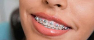
In dentistry, “bad” habits include sucking a pacifier, finger, lips, biting your lips or tongue, and habitually holding various objects in your mouth. If you discover bad habits in your child, a visit to the orthodontist is recommended, regardless of the child’s age and the stage of formation of his bite. What are the dangers of bad habits? They form an incorrect bite. The mechanism of malocclusion is associated with the prolonged presence of some object that should not be between the teeth - be it a finger or a tongue. For example, thumb sucking or pacifier sucking (if the habit persists in children for more than 2 years) leads to the formation of a narrow upper jaw, protrusion of the front teeth, and the formation of an open bite (when the teeth of the upper and lower jaws do not meet each other vertically). It can also lead to a distal overbite (where the upper and lower jaws do not fit together when viewed from the side).
In the future, the protrusion of the upper front teeth forward due to a bad habit leads to an increased risk of injury to the teeth, including their loss. If you discover some bad habit in your child, it is important to contact an orthodontist as early as possible to receive recommendations and exercises to eliminate the bad habit and correct the bite if problems have already occurred.
Causes
The most difficult reason in terms of prevention and treatment for a narrowing of the upper or lower jaw by 2 teeth or more (II degree and higher) is unfavorable heredity, since most of the structural features of the skull are determined at the genetic level. But other factors related to childhood also make a significant contribution:
- delay in introducing complementary foods;
- habit of thumb or pacifier sucking;
- early loss of baby teeth, especially molars or several units in a row on one side (the lack of chewing load in this area interferes with bone development);
- lack of solid food in the diet;
- metabolic disorders - rickets, type I diabetes mellitus, etc.;
- parafunction (unjustified increased activity of a nervous nature) of masticatory or facial muscles, including bruxism, involuntary movements of the tongue, etc.;
- rhinitis, sinusitis, tonsillitis, polyps and other diseases that force the child to breathe more often through the mouth than through the nose;
- injuries.
It can also be provoked by a cleft palate, caused by a delay in intrauterine development.
Diagnostics
It is not difficult to determine the narrowing of the jaw in a child or adult during an external examination. Its clear sign is the “pressed-in” dentition in certain areas or along the entire length with partially expanded, crowded units. Another characteristic feature is malocclusion.
At the same time, it is necessary to distinguish pathologies of jaw development from atypical orthodontic and dental situations such as supernumerary units, macrodentia, individual structural features of the skull, etc. They can also be combined. Therefore, a preliminary diagnosis made visually is confirmed with the help of one or more additional studies:
- According to Pon-Linder-Hunt , where the difference between the normal and existing width of the dental arch for a given patient is calculated using a special formula. The method requires taking several measurements directly in the oral cavity and taking into account different indicators for representatives of individual nations. But it gives the attending physician a fairly accurate answer to the question of whether several units will have to be removed to level the entire row.
- According to Howell-Gerber-Gerbst - another relatively complex mathematical scheme that is well suited for measuring the shape, length, width and other individual parameters of a row.
- Teleradiography is a type of X-ray examination that allows you to take a picture of the entire skull. It gives a detailed picture of the deviation itself and reveals its causes, if they are embedded in the structure of other bones.
- Anthropometry is an accurate three-dimensional cast, on which it is easier to take the necessary measurements, especially when the upper or lower jaw of a child is narrowed.
- X-ray - used to assess the condition of the upper palate and its suture, bone structure.
- Orthopantomography - a classic frontal image, is now increasingly being replaced by CT (computed tomography) due to its improved clarity and expanded capabilities - such as obtaining 3D images.
In combination, these methods allow the orthodontist to study the current condition of the bone, the degree and direction of deformation when narrowing the upper and/or lower jaw.
When choosing a correction method, it is also important that they provide an opportunity to evaluate the resource for moving teeth into the correct position and straightening the bite.
How is jaw expansion carried out in children and adults?
There are conservative methods that allow you to expand the dentition without surgical intervention. In most cases, the use of plates and fixed devices gives a stable and long-lasting result, but patients with complex pathologies require surgical correction.
The devices act systematically and delicately, so they do not cause pain to an adult or child. Under the influence of a slight force, the teeth move smoothly and the structure of the bone tissue is transformed. As a result, the narrowed dental arch increases.
How does a jaw expansion plate work?
Plates are recommended for bite correction at the age of 5-11 years, which makes it possible to cope with complex defects in the jaw relationship during the development and growth of bones. Plates for jaw expansion for children are made personally. After correction is completed, additional permanent structures may be required.
The orthodontic plate with screws consists of a steel spring and a part with a sectoral cut. To flatten the anterior part, the plate is equipped with vestibular arches. A removable appliance is fixed with clasps, a non-removable one is fixed with crowns installed on molars or premolars.
When is a jaw retraction device used?
A device for jaw expansion is used in cases where braces or plates will not give the desired result due to lack of space in the dental arch or congenital anomalies of the oral cavity. The device consists of rings that are installed on the lateral teeth and connected by arches. There is a screw in the central zone, the movement of which activates the operation of the device.
Under the influence of pressure, the median palatal suture opens and the jaw expands. The gap is quickly closed with restored bone tissue. In case of treatment of a child
Excellent results are possible, since as the patient grows older, the palatal suture becomes hard and more difficult to open. When treating adults, preliminary surgical dissection of the palatal suture will be required. During the correction process, the orthodontist often works together with the surgeon.
Main functions of the palatal expander
Palatal expanders are used to correct the upper jaw. The device has the shape of a cross, each edge of which is attached to the molars, and in the central zone there is a working sliding part.
Correcting the bite with a palatal expander in children will take about three weeks, but for adult patients this process may require a longer period. The cost of jaw expansion is offset by the result obtained: the gap is gradually filled with new bone tissue.
Mandibular distraction systems
Expansion of the mandible is performed using distractors. The devices ensure gradual divergence of the dentition with the simultaneous formation of new tissue. This method of correction gives a particularly pronounced result in childhood. In adult patients, dissection of the central zone of the lower jaw with an ultrasonic scalpel is required.
Devices of various types have unique designs, depending on the purposes and conditions of use. Many devices are suitable for treating adults and very young children. Such age-related indications make it possible to correct many congenital jaw anomalies in advance.
Based on: 7 votes
Treatment in children
The optimal age for correcting a narrowed jaw in children is from 6 to 15 years. For violations limited to the apical base and alveolar process (the area of root growth and placement of their holes), braces are recommended. They will align their row in conditions when the rest of the chewing apparatus is formed correctly.
If the pathology affects the entire jaw or palate, removable or permanent plates are prescribed. They consist of a plastic or silicone base with expansion screws in the center. They are fixed using a system of arms, brackets and locks to give the correct position to the displaced units. If it is necessary to correct a deep bite, cross bite, etc., two-jaw structures with cheek shields are used.
In each individual case, the elements of the plate are determined and adjusted individually, using casts. Only the rigid frame and screws remain unchanged, which must be expanded by approximately 0.25 mm every 4 days. The advantages of the plates are:
- wide frame color options;
- low visibility to others;
- mild impact on target structures.
Their low cost may also be an important argument. It rarely exceeds 10 thousand rubles, while the price of individual brace systems varies between 50-100,000 (ligatures made of plastic or precious metals, respectively). Plus, removable models are easy to care for and make it easier to maintain oral hygiene after eating. But when prescribing such plates to children, more control is required from parents.
The time of wearing them should be at least 20 hours a day, and the child is unlikely to comply with it himself due to the discomfort that persists even after the appearance of the habit.
Methods for expanding the upper and lower jaw
- Hardware method
It is used in relation to both removable and permanent dentition. Dental expansion is performed by gradually affecting the jaw tissue. For this purpose, special expanding devices are used.
Here are the most common expansion designs:
- The Derichsweiler apparatus is an orthodontic “device” with support rings and special arches. The supporting ring-shaped parts are placed on the molars, and the arches are fixed in the locks. The arches are also connected in the palate area, where there is a metal screw. When tightening the screw, pressure is applied to the jaw bones. Accordingly, the palatal suture gradually opens, and then the jaw itself expands. The free zone, which is formed when the palate opens, after a while is “overgrown” with hard tissue;
- plates that expand the jaw - are used to correct mixed dentition with pronounced deviations in the development of the dentition. They are used, among other things, for the gradual expansion of the arch in children under the age of ten years. In essence, this is a removable structure consisting of a base plate and a palatal screw. The plastic plate exactly matches the relief of the palatine bone;
- palatal expander – a special orthodontic device for expanding the upper jaw. The system includes support rings installed on the side crowns. One pair of expanders is placed on the premolar teeth, and the second is placed on the permanent molar teeth. Cross metal arcs are also soldered to the rings. There is a screw in the middle of the palatine vault, which is tightened and thereby exerts pressure on the lateral parts of the patient’s jaw;
- expansion springs improve the correction result and also shorten the expansion period. For this purpose, different types of springs are used in orthodontics: pin-shaped, Coffin, Koller and others;
- distractors - used to expand exclusively the lower jaw. In terms of its structure, the device differs from analogues in its flat body, which has a slider and a removable adjustment screw. Levers with ends in the form of wedges are needed for fastening in the tissues of the alveolar zone. The distractor helps expand the arch if the patient has a crossbite, eliminating the defect while gradually filling the bone tissue. This variability helps to correct pathology, including in young children.
- Surgical method
- the patient is given general anesthesia;
- the periosteum and mucosa are incised;
- soft tissues peel off, exposing the cortical bone.
- the selected expander is installed;
- the mucous membrane is returned to its original place and then sutured.
Surgical expansion of the upper jaw is prescribed if there is no proper result after hardware correction.
Stages of surgical correction:
Treatment options in adults
For patients over 18 years of age or with complex curvatures, more or less extensive surgical solutions are recommended. The simplest methods of treating narrowing of the upper jaw in adults:
- Installation of the Derichsweiler apparatus - a beam screw design for the upper jaw only. He breaks the palatal suture and sets its bones in a new, wider position. Then the teeth are straightened with braces.
- Normalization of the bite by removing several units in a row (in the vast majority of cases, the first premolars, 1 on each side). The method allows you to correct the bite and aesthetics of the smile without treating the narrowing of the jaw itself, if it is impossible or not necessary. Extracted premolars are not always redundant. But due to them, space is freed up for the remaining ones, and their location is again leveled with the help of braces.
In the case of the lower jaw, the layer of gum tissue is lifted, the bone is drilled in several places and the holes are connected with grooves. An expander is installed in them, with which the patient with a narrowing will need to go through several years, and the mucous membrane is returned to its previous position. This procedure is called jaw perforation.