A targeted dental photograph is a popular option for diagnosing most problems in the oral cavity. The method is based on the use of X-rays and a patch receiver for these rays (radiovisiograph), which makes it possible to obtain data on the internal tissues of the tooth, as well as the tissues surrounding the root. Taking into account the local direction, X-rays can be taken for 1-3 teeth. The purpose of the study is to supplement visual examination data, simplify the diagnosis of a number of diseases, and also control the quality of treatment provided by a specialist.
| For example, the image allows you to check the quality of cleaning and filling of the canals. If endodontic treatment has been carried out, a photograph of the teeth will allow you to find out how thoroughly the foci of infection were removed and how tightly the filling fits. |
Indications for prescribing a targeted photograph of a tooth
A modern targeted X-ray of the tooth (radiovisiography) should be performed before treating:
- caries. In the image, the doctor will be able to detect carious lesions under a filling or prosthesis;
- pulpitis, periodontitis. The doctor will be able to analyze the condition of the tooth roots, see the number of canals, and correctly carry out endodontic treatment;
- dental granuloma, cyst; The dentist must make sure that there are medical indications for tooth extraction. Otherwise, a “program to save” the tooth will be offered.
- pericoronitis. For example, a photograph of a wisdom tooth (“eight”) that has not yet erupted will allow you to clarify its position. In general, radiovisiography is rarely prescribed for wisdom teeth due to the difficulty of positioning the radiovisiograph sensor in the patient’s oral cavity. If radiovisiography is not possible, an OPTG (orthopantomogram) is prescribed.
- incorrect position of a growing tooth.
As for contraindications, traditionally, dental x-rays are not recommended in the first trimester of pregnancy. However, modern diagnostic equipment is extremely safe for the body. Therefore, if a correct diagnosis is needed for medical reasons, it is not prohibited to take a photo of the teeth while expecting a child.
Orthopantomogram (OPTG)
Orthopantomogram (OPTG, panoramic image) is the most common type of hardware diagnostics. Allows you to see the overall clinical picture.
The image is taken using an X-ray machine - an orthopantomograph. Metal accessories and jewelry should be removed before the procedure. The process lasts from 8 to 20 seconds:
- the patient stands in the center of the device;
- bites a special plate;
- The moving part of the apparatus rotates around the head.
OPTG shows the anatomical structure and hidden state of the roots of teeth and bone tissue. Based on the result, the doctor evaluates the bone tissue, identifies problems, and prescribes appropriate treatment for each clinical case. OPTG defines:
- relationship of the jaw with the maxillary sinus;
- location of the jaw nerves;
- hidden caries; violation of seal tightness;
- cysts, impacted teeth, periodontal pockets.
Advantages:
- obtaining a complete picture of the dental system
- focusing on individual areas of the jaw
OPTG and CT are not recommended for pregnant women in the first and third trimesters of pregnancy.
Types of targeted dental imaging
To take a single photograph of teeth, dental clinics use two types of equipment:
- an outdated analog X-ray machine: it can be used to take pictures on X-ray film. The process is not always comfortable, printing pictures takes time, and after 2 years the resulting image fades;
- modern digital radiovisiograph: a device that creates high-resolution electronic images. If necessary, the image can be enlarged and detailed information can be obtained. The images are permanently stored in the dental clinic’s database, and you can return to them at any time – even after several years. Plus, electronic equipment has 2-10 times less radiation exposure compared to analogue equipment.
How to do dental x-rays
The procedure is carried out by a doctor in a specially equipped room. The implementation mechanism takes place in 4 stages:
- The patient sits in a chair. The doctor examines the problem area.
- A special apron is placed on the patient to reduce the adverse effects of x-rays.
- The doctor fixes the head of the subject to obtain a clear image.
- On the affected area inside the oral cavity behind the teeth or on the front side, using a digital visiograph, the doctor directs the beam.
The whole process takes 3 minutes. After 15 minutes, you will be given a photograph of your teeth on film or electronically (if necessary).
There may be the following reasons for taking a photo:
- Clarification of the development process of pulpitis, periodontal disease and caries;
- Check after removal of damaged teeth;
- Checking the quality of treated teeth;
- Determining the specific structure of teeth before installing a crown or bridge;
- Checking neoplasms.
During endodontic treatment, at least 3 photographs of the tooth are taken: 1. Photograph of the teeth at the diagnostic stage. It is necessary to determine the condition of the tooth, the shape and number of roots, and the treatment method.
2. A photograph at the stage of tooth treatment with endodontic instruments inserted into the tooth canals. The picture is displayed on the computer screen.
3. Control image after completion of treatment to check the sealed tooth canals.
Permissible radiation dose for dental x-rays
The radiation dose for a targeted photograph for a person is 3-5 μ3v. During the year, an adult is allowed to receive up to 1000 microns. Consequently, a patient can take from 80 to 90 images per year using an X-ray machine. The radiovisiograph will make it possible to take up to 400 images. This indicates the harmlessness of this manipulation.
Advantages of a targeted tooth image
A dental image taken on a digital radiovisiograph has several advantages:
- a clear and detailed image of the tooth, as well as the tissues that surround it;
- safety of the study for the body and absence of harm to the body;
- you can prescribe repeated images in the required quantity without health consequences;
- organized and secure storage of images, the ability to return to previous results;
- results can be saved on digital media or sent by email;
- you can enlarge a certain area of the image, obtaining more information;
- There is no need to wait for the film to be developed - the results are available immediately.
The key advantage is high accuracy and information content, which makes it possible to detect problems that are invisible during visual examination, as well as pathologies and diseases in the early stages.
Varieties
To obtain targeted images in dentistry, two types of devices are used:
- A traditional X-ray machine that captures a planar image on film. It is not very convenient for the patient, and also requires some time to print the results, the period of maintaining sufficient brightness and contrast does not exceed 1.5-2 years.
- Digital radiovisiograph is a modern analogue that generates digital images of high resolution and quality. The results obtained are available for processing in special applications and scaling, and remain relevant over a long period. It is also worth noting that the radiation exposure associated with the use of electronic devices is three times lower than in the case of standard clinical equipment.
Current models of radiovisiographs are being developed to obtain digital images in two formats:
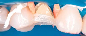
- Interproximal – assessment of the condition of the elements of the dentition, used in the case of planning the restoration of crowns with composite materials.
- Intraoral - an intraoral technique aimed at identifying carious formations adjacent to the affected units.
Stages of the procedure
The targeted image is formed in a clinical setting, in an office equipped in accordance with the operating requirements of the device used. The examination algorithm includes the following sequential steps:
- Placing the patient in the dentist's chair.
- Covering the body with a special protective apron.
- Fixing the head in a given position.
- An examination in which an x-ray beam is directed at the area of interest.
The duration of the entire cycle, including processing and printing the image, is no more than 15-20 minutes, after which the doctor gives the patient the results of the image.
Advantages and disadvantages
Positive aspects noted when using digital equipment to obtain targeted images include:
- Diagnostic safety.
- Clarity and high quality of the image.
- Option to enlarge the area under study.
- Convenient sequential storage.
- Possibility of printing a photo.
- Creating conditions for making a diagnosis.
- Minimum waiting time.
Radiovisiographic analysis is distinguished by its informativeness and accuracy of indications, which makes it possible to detect hidden pathologies and diseases at the primary stages of development. One of the disadvantages characteristic of the technology is the small coverage of the examination area, which limits the working space to 3-4 elements of the dentition.
Summary: important points
As a practicing dentist who knows the system from the inside, I want to draw your attention to the following points that are important for your safety. If the clinic has an X-ray machine, then a license must be obtained for it, the issuance of which presupposes the mandatory presence of a certified radiologist on the staff of the dental clinic. However, in reality, even in large clinics and public clinics, it is not always the case that x-rays will be taken by a trained specialist.
Even if he is, he may go on vacation or get sick, and a regular nurse (dental assistant) will take the pictures instead. This is a gross violation that leads to both the production of low-quality images and an increase in the radiation dose. In small clinics, the risks of receiving a poor-quality x-ray examination are much higher, and the first thing that makes you suspect a forgery is if the picture is taken not by a special employee, but by a nurse from the dentist you came to see.
The second very important point: if you see that the x-ray is not done in a special room, but the x-ray machine is located right next to the dentist’s chair, then you should change the clinic and the doctor. The fact is that all objects in this office (including the dental chair and doctor’s instruments) will have an increased background radiation, and this is no longer safe for health. We hope that our article on the topic: X-ray of the jaw and teeth was useful to you!
Safety of X-rays before implantation
When planning implantation and in the postoperative period, it is necessary to monitor the process of implant healing. Therefore, repeated orthopantomograms and CT scans are necessary. But is this amount of radiation harmful to the body?
| Type of examination | Exposure dose, m3v* |
| Sight shot | minimum radiation exposure - 0.01 |
| OPTG | minimum radiation exposure - 0.01 |
| CT (3D) | with partial CT - 0.02; with full CT - 0.05 |
*The permissible annual dose is 5 m3v. For comparison, the natural background of Moscow is 0.02 m3v.
Photo interpretation
The image can only be deciphered by a qualified doctor. First, he looks at the condition of the tooth in the picture or its parts. Then assesses the condition of bone tissue and periodontium. After this, you can examine the areas of interest in more detail or see pathology that is not related to the tooth, in which case the specialist will prescribe an additional examination.
Based on the image, a diagnosis can be made and a treatment plan determined. Only with the help of x-ray methods is it possible to objectively assess the condition of the teeth and oral cavity as a whole.
Contraindications for the study
Sight radiography has such a low radiation dose that there are practically no contraindications to its implementation. It is not recommended to conduct radiographic examinations in the first trimester of pregnancy, therefore preparation for bearing a child should begin with sanitation of the oral cavity. Also, the procedure may not be comfortable for patients with an increased gag reflex in cases where examination of large molars is necessary. However, specialists carry out it very carefully, which most often does not cause any discomfort.
Further detailed analysis of the image
A detailed analysis of the image is carried out by a doctor who determines a treatment regimen or prepares for surgery, or by a radiologist. The dentist examines the image and evaluates the color of various areas. He makes a conclusion about the rigidity, density, homogeneity of the bone structures of both jaws, and the placement of parts of the dentition. The analysis makes it possible to identify carious cavities, granulomas, cysts, periodontitis and the consequences of injuries. The image shows areas of inflammation, blood vessels and nerves.
The most common dental diseases on a dental photograph are manifested by the following signs.
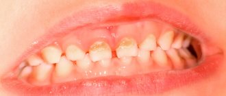
- Caries. This is the process of destruction of dental tissue. The picture shows areas of transparency of the enamel and the hard part of the tooth. The clearing zones have uneven, unclear contours.
- Pulpitis. Damage to the bone structure is characterized by heterogeneity of color in the interroot zone against the background of hypertrophy.
- Periodontitis. The progression of granulomatous periodontitis is accompanied by destruction of the hard part of the tooth and cement. Granulomas are observed in the area of tooth root formation, the interdental gap is enlarged, its edges are blurred.
- Periodontitis. The image shows signs of osteoporosis - the density of bone structures decreases, the height of the partitions in the dentition decreases, and pockets form.
To read the photo correctly, the image must be unblurred and in focus.