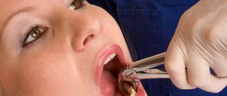
You can contact Dr. Granov's clinic for treatment of jaw injuries. This is one of the areas of treatment that we do.
The jaw is the bony structure that holds the teeth in place. The upper jaw does not move, only the lower jaw moves. Its movements are necessary for eating food and for producing speech. Both jaws connect at a point called the temporomandibular joint (TMJ).
Jaw injuries include cracks, dislocations and fractures. With a fracture or crack, the bone breaks; with a dislocation, the bone changes location and flies out of the joint. Such injuries occur after physical activity (bruise, blow, fall). After appropriate treatment, they heal safely, but the dislocation may appear again.
Jaw injuries are dangerous because they can lead to various complications. Among them:
- breathing disorder,
- bleeding,
- blood entering the lungs
- problems with chewing food,
- problems with diction,
- infection of the jaw or facial structures,
- joint pain,
- numbness of the jaw or face,
- tooth displacement,
- edema.
Symptoms of a jaw fracture:
- pain in the cheek next to the ear, which increases with jaw movement;
- swelling of the face, blood in the mouth;
- immobility of the jaw, inability to open or close the mouth;
- when opening the mouth, only one side of the jaw moves;
- jaw pain that gets worse when chewing;
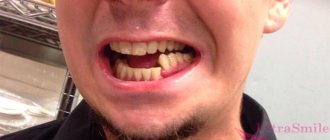
- dental injuries;
- swelling of the face or changes in the contours of the jaw;
- numbness of the face in the lower lip area.
Symptoms of a dislocated jaw:
- pain in the cheek next to the ear, which increases with jaw movement;
- feeling that the jaw is not positioned correctly in the joint;
- violation of diction;
- inability to close your mouth;
- drooling due to open mouth;
- spasm of the masticatory muscles or protrusion of the jaw forward;
- dental injuries.
First aid for jaw injuries
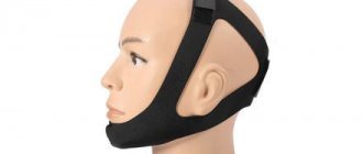
Jaw injuries require immediate medical attention. Call an ambulance or go to the nearest hospital. Before doctors arrive, you can put a bandage on your jaw to keep it immobilized. The bandage should be easy to remove.
Do not try to set a dislocation yourself or wait for the fracture to heal on its own. Be sure to contact a traumatologist.
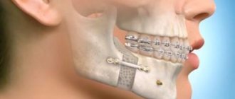
Treatment of a jaw fracture
Treatment depends on the extent of the fracture. If the patient has a small crack in the bone, it will heal on its own. The doctor will only prescribe a painkiller.
Severe fractures require surgery to allow the bones to heal properly. After surgery, a bandage is applied to the jaw to allow it to heal quietly. The jaw takes 6 to 8 weeks to heal. At first, elastic bandages are additionally applied, which are then removed to allow the muscles to be developed and their mobility restored.
During treatment you can eat only liquid food. You should always have scissors with you to cut bandages in case the patient suddenly chokes on something or starts vomiting. Then go to the hospital to have a new bandage applied.
Instruction No. 1 (from 2 to 5 weeks after surgery).
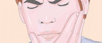
Basic exercise to restore sensitivity / Do this exercise using a small brush (for example, a toothbrush or cosmetic brush) at least 3 times a day for 5 minutes each time (morning, afternoon and evening).
First you will need a mirror. 1. Alternately touch and stroke the entire surface of the face with the brush. 2. Then make a series of light touches at a relatively large distance from each other, first on one side of the face and then on the other. The frequency of movements with the brush should be as if you were trying to touch a piece of fabric. 3. As the exercise progresses, remember the place where you made the touches and then repeat the next series in a similar place, but on the other side of the face. 4. Be careful and go over the entire surface of the face (forehead, cheeks, chin, upper and lower lip). Don't get stuck in one place. Additional exercises to restore sensitivity / You can perform exercises to restore sensitivity an unlimited number of times a day. At the initial stage, it is important to look in the mirror during the exercise. If you don't have a brush, you can do this exercise with your fingertip. 1. Close your eyes and continue alternately touching and stroking your face with the brush. Tell yourself before you do this what will happen next: a series of touches or stroking. It is important to feel this difference. 2. You can ask someone to do this exercise for you. The first few times, the assistant first tells you what movement he is going to do: a stroke or a series of touches. Remember that your eyes are closed. The assistant continues to do the exercise and then stops telling what movements will follow, and you must determine them yourself. If you can do this with ease, this is a good sign. It is also important to weaken the force of the touches and make them closer to each other, which will complicate the task. 3. Remember to make a series of light touches both horizontally and vertically. If you have difficulty feeling touch even at a fairly large distance, do not despair. For most people, sensitivity returns slowly and you need to be patient. We remind you that the full return of sensitivity in some cases can take up to 12 months.
Instruction No. 2 (from 5 to 12 weeks after surgery)
Basic exercise to restore sensitivity / Do this exercise using a brush in the morning, afternoon and evening for 5-15 minutes each time. First, monitor your actions in the mirror. 1. Use light tapping on one side of the face and then on the other. 2. As you complete the exercise, remember where you tap. 3. As you begin the side-to-side exercise, start on one side of your face and tap to a similar area on the other side. For example, start at the base of the ear on one side and work your way through the upper lip area to the base of the ear on the other side. 4. When you do the exercise in the vertical direction, also go over the entire face from above and below. Start, for example, at the neck and work all the way up the side of the face to the forehead, and then back to the neck. You can also do the exercise from the infraorbital region down to the neck and back. 5. As you complete the exercise, note your sensations, whether there is a difference and what its magnitude is. 6. Be careful and do the exercise over the entire surface of the face (forehead, cheeks, chin, upper and lower lip). Don't get stuck in one place.
Additional exercises to restore sensitivity / You can perform exercises to restore sensitivity an unlimited number of times a day. At the initial stage, it is important to look in the mirror during the exercise. Also, if you don't have a brush, this exercise can be done with your fingertip. 1. Close your eyes. Before you start lightly tapping your face, tell yourself exactly how you will start doing it and in what direction. 2. If there is someone who can help you do this exercise, then the first few times the assistant says where he is going to start and in what direction he will proceed: from side to side or from bottom to top. Next, without opening your eyes, ask your assistant to continue the exercise without notifying the next places and direction of touch. If you can easily identify the differences, this is a good sign. 3. Remember to try every day to make the touches softer and closer and closer together to make the task more difficult. If you have difficulty feeling touch even at a fairly large distance, don’t worry. For most people, sensation returns slowly and you will need to be patient.
Treatment of jaw dislocation
A dislocated jaw is treated simply: the doctor realigns the joint, placing the bone in the right place. Muscle relaxants or painkillers may be needed to relax tight muscles.
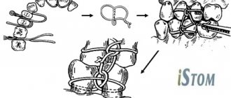
Then the jaw must be secured in a stationary state. To do this, the doctor applies a bandage. If the dislocation does not appear for the first time, then surgery will be needed to straighten it.
Recovery after a dislocation lasts 6 weeks. At this time, you should not open your mouth wide. When yawning and sneezing, the lower jaw should be supported with your hands.
Malunited mandibular fractures
Dentistry / Articles / Incorrectly healed fractures of the lower jaw
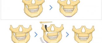
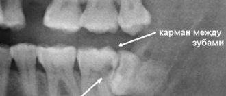
In case of jaw fractures, it is necessary to provide timely and qualified specialized medical care, timely treatment of the wound and fixation of fragments. Under such conditions, the jaw fragments grow together without complications and in the correct position. The consequence of violation of one of these conditions is that the jaws often heal with displacement of the fragments, which is why fractures are formed that do not heal properly, and the wound heals in the form of rough scars that limit the movements of the jaw, lips, and tongue.
The nature of such deformations is very different. Incorrectly healed fractures of the lower jaw are characterized by disorders of the dentoalveolar system and depend on the location of the fracture, the degree of discrepancy between the fragments, and the severity of the deformation. Of course, the patient's appearance changes. There is an elongation of the face and tension in the soft tissues of the area near the mouth. Facial asymmetry often occurs. Displacement of fragments of the lower jaw leads to displacement of the articular heads, which leads to disruption of the functioning of the temporomandibular joint, as well as the masticatory muscles. Changing the position of jaw fragments leads to speech impairment mainly due to a decrease in the volume of the oral cavity. The clinical picture is characterized by the degree of occlusal disturbances.
Surgical treatment for fractures that have healed incorrectly with minor functional impairment does not cause any particular difficulties.
Patients are divided into three groups. In patients of the first group, there is a tubercular appearance of occlusal contacts, in the second, the teeth contact only the lateral surfaces, in patients of the third group, the closure of the teeth is completely absent.
The method of treatment for fractures of the lower jaw that have not healed properly is, of course, surgical, i.e. The fragments are repositioned and immobilized. If patients refuse treatment, prosthetic, orthopedic, and hardware-surgical treatment methods are used.
The tactics of treatment depends on the presence of teeth in the jaw.
Treatment of improperly healed jaw fractures with preserved dentition has its own peculiarities. First of all, you need to pay attention to the age of the patient. Thus, if there are signs of an open bite, then at a young age orthopedic methods give good results, while in elderly patients with the same pathology, grinding down the teeth on which the bite is fixed, or even removing them, will be effective. This tactic must also be followed in the presence of other pathological bites, paying attention to the age of the patients.
Among orthopedic structures, plastic mouthguards are widely used - crowns, solid crowns, metal-ceramic and metal-plastic mouthguards or crowns, solid-cast arch prostheses, and in very complex cases - collapsible prostheses.
An orthopedic surgeon finds himself in difficult conditions when treating patients with fractures that have not healed properly and have defects in the dentition. The main task here is to restore the dentition, normalize occlusal relationships and restore the patient’s appearance. Among the most important orthopedic structures are fixed bridges and removable types of dentures.
Particular attention should be paid to the condition of periodontal tissues, from the point of view of the possibility of their use as a support for orthopedic structures. Depending on the location of the defect, the number of lost teeth, and pronounced occlusal changes after improperly healed fractures of the lower jaw, orthopedic denture designs are used. Defects in the anterior section and lateral section are restored with solid-cast bridges. In the presence of crossbite, removable types of dentures are used, including, if necessary, cast occlusal overlays.
Significant difficulties often arise in removing impressions from the jaws. Then it is necessary to make individual or collapsible spoons. The ways of introducing and removing prostheses are studied using parallelometry.
Based on materials from the medical website surgeryzone.net
Indications for removal of fixing structures
On the one hand, removal is an operation, but on the other hand, a foreign body can cause unwanted reactions in the body. The structure is installed for the time during which the bones are fused. The fact of fusion is confirmed by an x-ray. The treatment method involves removing metal structures after a fracture so that the likelihood of developing deforming osteoarthritis is minimal.
Removing a splint from a jaw in case of a fracture
Long-term wearing of a splint leads to diseases of the jaw joint, which can cause discomfort when chewing or speaking. In addition, it is imperative to sanitize the oral cavity and examine it to detect dental diseases after removing the jaw splint. The team of dentists at the KRH-Medical clinic can remove the splinting of the lower jaw, conduct a dental examination and prescribe the necessary procedures. Make an appointment in Lyubertsy and you will receive highly qualified assistance!
Signs, first aid for a fracture of the upper jaw
A fracture of the upper jaw is much less common - only 30% of all facial injuries. This is a dangerous injury, because in most cases it is accompanied by severe complications - meningitis, osteomyelitis. Moreover, the higher the crack (fracture line) is formed, the more severe the consequences will be.
Symptoms of a jaw fracture (upper) can vary and depend on which part of it is damaged:
- trauma above the palatine vault + nasal fracture + fracture of the maxillary sinus bone is accompanied by severe bleeding between the upper lip and upper teeth, extensive swelling of the upper part of the face;
- separation of the upper jaw from the calvarium + a crack running through the orbit and bridge of the nose, characterized by severe swelling and rapid spread of hematoma under both eyes, numbness in this area, nosebleeds and uncontrollable salivation;
- jaw avulsion + fracture of the base of the jaw can be diagnosed by drooping eyeballs and blurred vision.
First aid consists of completely immobilizing the upper part of the face and clearing the mouth of fragments of teeth/bones. In each case, the victim will experience nausea and may develop vomiting.
Activities and auxiliary things
If the fracture of the limb was uncomplicated, it is enough to wear insoles. They will help not to overload the injured limb. If a person breaks a hip, the recovery time will be more difficult and lengthy. You can’t do it without a special program and devices.
Even in the clinic, the patient’s bed is equipped with a belt. A person grabs it with his hands in order to sit or rise comfortably. Doctors prescribe physical therapy in the first week after an injury, when canes and crutches are still used.
First, breathing exercises are prescribed for the patient. Balloons are used for this. Afterwards, exercises are prescribed, aimed at developing the mobility of the torso. After about a month, the doctor prescribes the following complexes:
- General. Designed for general human health.
- Special. To restore a diseased limb from the day the cast is removed.
Well-thought-out treatment and rehabilitation based on physical education can achieve the following goals:
- Restoring blood circulation through massage;
- Prevention of dangerous complications;
- Serious strengthening of the muscles at the fracture site;
- Restoration of motor ability.
To achieve the above goals, it is important to start exercising while lying down. After this period, quitting classes is not recommended. The features of each gymnastics option should be studied in more detail.
Technical features of installing and removing fasteners
If the structure with an external location is installed correctly, removing the spokes after a fracture is not difficult - the spokes are simply removed. If the fixation is intraosseous, inside the joint, using nails and screws, then a full-fledged operation is required.
Fixation with knitting needles is used to connect the joints of the limbs and fingers. It can be performed externally, when the end of the needle rises above the surface, or internally, when the entire structure is under the skin. The technique is used as a temporary measure. In some cases, if patellar fractures, clavicular or elbow injuries are fixed, stable fixation and a longer time for fusion are required.
When a hand is broken, osteosynthesis techniques are used - metal structures, knitting needles and an Ilizarov apparatus is installed. They are removed only after complete healing.
Using plates you can fix any bone. This is a convenient and reliable method. Today, many variants of such plates are used, varying in size, shape, and functionality. The plate is used for fractures of the tibia and ankle, when there is a need to fix bone fragments. Its removal occurs as planned. The time of the operation is determined by the doctor.
Intraosseous rods (pins) are used to fix tubular bones - for example, in case of a fracture of the collarbone or leg. Such operations are performed quickly and are characterized by minimal trauma. After fixation, loading is allowed in just a few days.
Removal of the structure is carried out in half an hour if the installation was carried out correctly. If there is damage to the threads or screws, it becomes necessary to drill out the elements.
Working with gait
Along with the listed exercises, you need to add a complex for quick gait restoration and rehabilitation. Here are some effective options:
- Use your toes to grab and hold a small object;
- Roll a ball with the foot of the sore limb;
- Standing on your toes, on your heels;
- Walking sideways and backwards.
If possible, you can exercise on exercise bikes. The listed exercises and prescribed treatment after an injury cannot be neglected. Physical education during rehabilitation quickly restores and heals the body from the day the cast is removed.
Recovery after a fracture should continue until the body is completely restored. You can stop exercising after you have regained mobility, swelling has subsided, and there is no pain. The success of rehabilitation depends not only on the will of the patient and the treatment program.