Microabrasion is a modern method of combating discoloration on the surface of teeth, which involves carefully grinding off the thinnest layer of damaged enamel using a specialized abrasive composition. The thickness of the ground layer rarely exceeds 60 microns, which avoids the development of dentin hyperesthesia and tooth hypersensitivity. Competent, professional microabrasion helps to achieve the highest aesthetic results and completely eliminate pigmented spots and areas of demineralization on the surface of the enamel layer.
Indications for the procedure
If you are offered microabrasion, then there are probably the following indications for this:
- unsightly and uneven enamel color, stains that could not be removed with the help of professional oral hygiene and for which whitening alone is not enough,
- spotted hypoplasia,
- the initial stage of fluorosis in streaked and spotted forms: for erosive and destructive types of the disease, the procedure is not performed,
- the presence of areas of enamel demineralization: the procedure can be carried out as a caries prevention, as an addition to remineralization or fluoridation,
- white spots and unevenness on the enamel after removing braces.
How does the procedure work?
Before whitening stains on enamel using microabrasion, it is recommended to carry out a complete sanitation of the oral cavity. The best results can be obtained with professional dental cleaning. Further stages of the procedure:
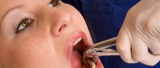
- The dentist examines the affected areas, determines the parameters of the enamel, then isolates the mucous membranes located near the treated area and takes photographs.
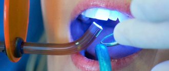
- Treatment of the modified surface with a micro-grained diamond bur for 10 s.
- Applying a composition consisting of abrasive silicon gel, hydrochloric acid and corbund to the treated surface.
- Next, each segment is ground again for 1 minute.
- If the initial changes are practically unnoticeable, the doctor chooses a more gentle method - coating with a suspension using a wooden stick without sanding.
- The area is washed, dried and then a gel is applied to help reduce the sensitivity of the incisors.
- Finally, control photographs are taken.
- After the manipulations are completed, they move on to bleaching hard tissues (if provided for in the treatment plan).
This procedure is painless and does not have a harmful effect on the enamel.
What are the contraindications
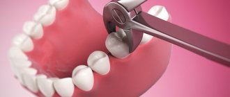
Removal of a thin layer of enamel is contraindicated in the following pathologies and conditions:
- severe thinning or destruction of hard tissues (including cracks and chips),
- multiple caries,
- erosion and wedge-shaped defect,
- advanced stages of fluorosis and hypoplasia (including erosive, grooved, mixed),
- bleeding gums and exposure of tooth roots.
The procedure is not performed on baby teeth or on pregnant and lactating women. Microabrasion is not performed on pulpless, that is, nerveless, elements. Its use is inappropriate for such a defect as tetracycline staining, as well as after treatment with resorcinol-formalin compounds.
The microabrasion procedure may have contraindications
Important! The success of microabrasion largely depends on the correct definition of the clinical case. If the treatment method is chosen incorrectly, it can disappoint the patient. After all, the procedure can only eliminate superficial discoloration of the enamel.
Efficiency and safety
Microabrasion allows you to get rid of enamel discoloration. As a result, not only the appearance of the tooth improves, but its further destruction is stopped. In addition, micro-abrasive treatment allows you to avoid tooth grinding and restoration using expensive composites. After the procedure, age spots do not return.
Efficiency depends on the degree of damage: microabrasion helps either completely eliminate surface stains on the enamel or mask discolored areas. Provides a natural glossy shine to the enamel surface. The result is noticeable immediately after the procedure.
The main complication after the procedure is increased tooth sensitivity. To eliminate it, it is recommended to use special toothpastes and rinses for sensitive teeth, for example, low-abrasive ASEPTA Sensitive toothpaste. Additionally, it is necessary to use remotherapy products as an opportunity to mineralize damaged areas, for example, ASEPTA Plus Remineralization. This professional toothpaste restores weakened and damaged enamel, saturates it with essential macro- and microelements.
Features of the method
- Enamel microabrasion is considered a minimally invasive procedure. It is painless and does not require anesthesia. It feels like an Air-flow procedure, polishing a filling or, for example, facial peeling. The result is achieved in 1 session.
- When enamel microabrasion is performed, according to various sources, on average, from 15 to 60 microns of hard tissue are removed, depending on the clinical situation.
- To remove the required thickness, doctors use various gels and suspensions, which may include pumice, hydrochloric acid, and silicon carbide microparticles. The gel is rubbed into the enamel using special brushes and rubber cups.
- After microabrasion, a smooth surface is formed on the enamel that reflects light well, and therefore visually the tooth looks whiter and healthier.
Expert opinion
Elina Ruslanovna Dzagurova
Specializations: Dentist-therapist
Experience: 11+
“To achieve a better effect, microabrasion is often used in the treatment of hypoplasia and other forms of discoloration in addition to other measures: bleaching, remineralization and fluoridation courses, artistic restoration with adhesive materials.”
Clinical researches
ASEPTA products are clinically proven effective. For example, repeated laboratory tests have proven that regular use of preventive toothpaste ASEPTA SENSITIVE for a month can reduce bleeding gums by 62%, reduce sensitivity of teeth and gums by 48% and reduce inflammation by 66%.
Clinical studies have proven that regular use of professional toothpaste ASEPTA REMINERALIZATION improved the condition of the enamel by 64% and reduced tooth sensitivity by 66% after just 4 weeks.
Sources:
- Report on determining/confirming the preventive properties of toothpaste “ASEPTA PLUS” GENTLE WHITENING” Author: doctor-researcher A.A. Leontyev, head Department of Preventive Dentistry, Doctor of Medical Sciences, Professor S.B. Ulitovsky First St. Petersburg State Medical University named after. acad. I.P. Pavlova, Department of Preventive Dentistry
- Clinical and laboratory assessment of the influence of domestic therapeutic and prophylactic toothpaste based on plant extracts on the condition of the oral cavity in patients with simple marginal gingivitis. Doctor of Medical Sciences, Professor Elovikova T.M.1, Candidate of Chemical Sciences, Associate Professor Ermishina E.Yu. 2, Doctor of Technical Sciences Associate Professor Belokonova N.A. 2 Department of Therapeutic Dentistry USMU1, Department of General Chemistry USMU2
- Clinical studies of antisensitive toothpaste “Asepta Sensitive” (A.A. Leontyev, O.V. Kalinina, S.B. Ulitovsky) A.A. LEONTIEV, dentist O.V. KALININA, dentist S.B. ULITOVSKY, Doctor of Medical Sciences, Prof. Department of Therapeutic Dentistry, St. Petersburg State Medical University named after. acad. I.P. Pavlova
- The role of anti-inflammatory rinse in the treatment of periodontal diseases (L.Yu. Orekhova, A.A. Leontyev, S.B. Ulitovsky) L.Yu. OREKHOVA, Doctor of Medical Sciences, Prof., Head of Department; A.A. LEONTIEV, dentist; S.B. ULITOVSKY, Doctor of Medical Sciences, Prof. Department of Therapeutic Dentistry of St. Petersburg State Medical University named after. acad. I. P. Pavlova
- Report on determining/confirming the preventive properties of toothpaste “ASEPTA PLUS” COFFEE and TOBACCO Author: doctor-researcher A.A. Leontyev, head Department of Preventive Dentistry, Doctor of Medical Sciences, Professor S.B. Ulitovsky. First St. Petersburg State Medical University named after. acad. I.P. Pavlova, Department of Preventive Dentistry
- Report on determining/confirming the preventive properties of commercially produced personal oral hygiene products: Asepta toothpaste used in combination with Asepta mouthwash and Asepta gum balm Head. Department of PFS Doctor of Medical Sciences Professor S.B. Ulitovsky St. Petersburg State Medical University named after Academician I.P. Pavlova. Faculty of Dentistry. Department of Preventive Dentistry.
Stages of the procedure
Regardless of the pathologies for which microabrasion is performed: fluorosis, hypoplasia, or simply pigmentation of the enamel, the essence and order of the procedure does not change.
| Stages | Description |
| First stage | The doctor assesses the condition of hard tissues, visually examines them, and identifies boundaries and areas of discoloration. |
| Second phase | Removing soft and hard plaque from the surface of teeth in order to best prepare them for exposure to abrasive compounds. |
| Third stage | Protection and isolation of the soft tissues of the oral cavity using a rubber dam or rubber dam. |
| Fourth stage | Applying an abrasive mass, rubbing it into the enamel using rubber cups, which are set to rotate slowly. The procedure lasts about 30-60 seconds. |
| Fifth stage | Rinsing the tooth with a water-air spray. The decision to re-use abrasives (for better effect, the procedure can be repeated 2-3 times at intervals of 60 seconds). |
| Sixth stage | Strengthening enamel using a remineralizing gel or fluoridated prophylactic composition. |
“While wearing braces, white spots appeared on my teeth. This became especially noticeable after removing the plates. To be honest, at first I was very upset, because even though my teeth were straight, these stains were not at all what I expected to see as a result of long-term treatment. I thought I'd have to bleach it. But the doctor suggested a better solution - microabrasion. In principle, after this, the teeth began to look very good, and those around them generally thought that they were perfect. So I recommend it!”
Orange, review from woman.ru
Complications after microabrasion
One of the most common complications of the microabrasion procedure is hyperesthesia, caused by a high degree of demineralization of tooth enamel. Treatment of this pathology comes down to rinsing the mouth with a 10% solution of calcium gluconate and excluding sour, spicy foods and too hot or cold dishes from the menu.
An illiterate approach to the selection and application of an abrasive mixture can cause erosion on tooth enamel or lesions on gum tissue. Such complications can be dealt with by regularly applying antiseptic and anti-inflammatory agents to the affected areas, as well as drugs that accelerate regeneration processes.
Advantages of the method
The procedure is simple to perform and can be used on permanent teeth in adults and adolescents (which cannot be said about whitening). In case of superficial enamel discoloration, microabrasion allows you to quickly and painlessly achieve good results, making your teeth aesthetically more attractive, white and shiny. Another advantage is that the procedure, if the tooth is intact and has not undergone severe destruction, helps to avoid more invasive intervention and refuse the installation of orthopedic structures. For example, crowns.
Before and after the operation
A number of studies suggest that hard tissues subjected to microabrasion are less susceptible to demineralization processes than those that were not treated. They show less colonization of carious bacteria, the main causative agents of caries - Streptococcus mutans.
What are the pros and cons?
The technique has quite a few advantages, among which specialists and patients often highlight the lack of need for any preparation. In addition, the procedure is completely painless and does not cause any discomfort. If microabrasion is used to prevent further tissue demineralization, then after its successful implementation it is possible to avoid large-scale tooth preparation for its further restoration. In general, the technique saves in cases where other options for removing plaque and pigmentation do not help.
“They performed microabrasion on me. I found white spots on my front teeth, the doctor said this is how caries begins. After the examination, the dentist suggested grinding off a thin layer to ensure that the lesions were removed. I agreed. There was no pain at all during the procedure. Only then did sensitivity to cold and hot appear. Personally, everything went away for me literally in 3-4 days.”
Evgenia S.S., Moscow, from correspondence on the woman.ru forum
Among the disadvantages, one can highlight a fairly impressive list of restrictions on the use of the method. In addition, after the procedure, almost all patients experience increased sensitivity of the enamel layer - hyperesthesia goes away on its own within a few days, but for this you need to strictly follow the instructions of your doctor.
Microabrasion results
At the end of the procedure, the patient receives a uniform shade of enamel and lightens the teeth by a couple of tones. It has been clinically confirmed that such manipulation is absolutely harmless to the body and does not lead to complications. The only point: 8% of patients may experience increased sensitivity of the enamel. But don’t panic, this is considered to be the norm. The doctor gives the necessary recommendations and soon this symptom disappears without a trace. As a result, you can get not only an aesthetic result, but also protect the tooth from further destruction due to caries.
Dental fluorosis - symptoms and treatment
It was possible to find out that fluorosis is associated with exposure to fluoride in 1931, although even at the beginning of the 20th century, Italian scientists thought that the cause of such damage was volcanic dust or other chemical compounds.
Fluorine (from the Greek “fluoros” - destructive) is an element of Mendeleev’s periodic table; it is quite widespread in nature and makes up approximately 0.081% of the earth’s crust. The fluorine content in natural water sources is due to its ability to easily dissolve in water. In the Russian Federation, open reservoirs have a fluorine concentration of 0.5 ml in 1 liter of water, with an acceptable value of 1.5 ml/l.
Fluoride is essential for the body; its main role is participation in bone formation and the formation of dentin and tooth enamel. In moderate quantities, this element prevents the development of caries [13]. 99% of fluoride occurs in bone tissue and tooth enamel, therefore, with a low fluoride content in children, the ossification process slows down, defects in bone mineralization appear, and in adults - osteoporosis.
However, an excess of fluoride, like its deficiency, is harmful to humans. The reactivity of fluorine is high: penetrating the body's protective barriers, it causes metabolic disorders. In large quantities, fluoride is a potent neurotoxin because it promotes the accumulation of aluminum in the brain, which can lead to Alzheimer's disease and other mental disorders. One of the first manifestations of excess fluoride intake is dental damage.
To understand the mechanism of fluorosis development, you need to know what enamel is and how it is formed.
Enamel is the outer protective layer of the crown of human teeth. It is the hardest tissue in our body due to the high content of inorganic substances - up to 97%. Enamel consists of enamel prisms and an interprismatic substance that glues them together. Enamel prisms, in turn, consist of the smallest structural units - apatite crystals, which are tightly packed together.
The process of enamel formation is called amelogenesis. It occurs in three stages.
1. Secretory stage, or stage of primary mineralization of enamel. Enameloblasts (enamel-forming cells) produce the organic base of enamel. Almost immediately, primary mineralization of the enamel occurs - its saturation with mineral components, leaching of organic substances (mainly proteins) and water.
2. Stage of maturation, or secondary mineralization. The enamel undergoes further calcification. The first two phases of amelogenesis occur before tooth eruption.
3. Stage of final maturation, or tertiary mineralization. Completion of enamel mineralization mainly through the entry of inorganic ions from saliva into it. It occurs after teething.
To date, there is no consensus on the mechanism of dental fluorosis. A number of foreign authors describe the effect of fluoride on ameloblasts. High concentrations of fluoride destroy ameloblasts, which stops the development of enamel prisms. At the same time, the composition of young enamel changes and the transport mechanism of calcium, phosphate, and bicarbonate ions is disrupted. Fluoride also reduces the activity of phosphatase (an enzyme that maintains phosphate levels in the body) and causes disturbances in the mineralization of tooth enamel.
According to Bykov L.V. (1998) [3], excess fluoride during the period of enamel maturation causes “abnormal calcification” (hypermineralization) of the enamel. This researcher also discovered the presence of multiple areas of hypomineralization in the enamel of fluorous teeth.