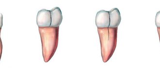
Fused teeth are one of the rarest anomalies in the development of the dental-jaw system, which consists in connecting the sides of the rudiments during the development of the embryo. It occurs in only 1% of Patients.
Most often, fused teeth are observed during a primary dentition; with a permanent dentition, such an anomaly is extremely rare.
The reasons for the formation of fused teeth can be:
- hereditary factor;
- too much pressure on the buds of neighboring teeth;
- jaw injuries;
- inflammatory and infectious diseases that cause metabolic disorders in the human body.
Often, the fusion of teeth is confused with their fusion, although these pathologies are completely different in nature. Teeth fusion occurs during the period after their growth and development, and fusion occurs during the formation of tooth buds.
What is ankylosis
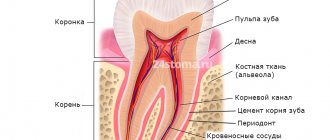
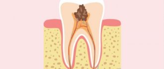
Tooth ankylosis is a pathology characterized by fusion of the cement of the tooth with the jaw bone. The tooth root is deprived of part of the ligamentous apparatus, which often leads to adhesion or germination of bone tissue. The aesthetics of the teeth are disrupted, they look shorter and change their angle. Ankylosis usually occurs during tooth eruption. Pathology can also form at a late stage in the formation of the dental arch.
It is important to know. The process is accompanied by periodontal necrosis and the incorporation of root cement into the alveolar bone.
Ankylosis of a primary tooth is caused by loss of the periodontal ligament. It is observed in cases where a primary molar does not fall out due to the absence of a permanent dental unit. Ankylosed teeth are different in size from their neighbors, so bite correction will be required.
Types of Teeth Fusion
In orthodontics, there are several types of dental fusion:
- first: supernumerary fusion. In this case, the second tooth is much smaller than the first. It is characterized by a spiky shape and a bumpy surface;
- second: non-gingival, observed only in the supragingival part involving a certain area of enamel;
- third type: root. Occurs in the root part at the level of cement and is characterized by deep damage to dentin. At the same time, both teeth have the same size;
- fourth: observed along the entire height of the tooth with damage to the crown and root parts.
Symptoms
Parents should be wary of the fact that their child’s baby teeth are not falling out. It is fused to the bone and cannot easily separate from the alveoli, as is normal. If the pathology is not detected and treated in a timely manner, the child’s jaw begins to develop incorrectly. The diseased tooth is located lower than the others, and the neighboring teeth are placed at an angle in relation to it. Problems with bite occur.
Another scenario is also possible. The baby tooth does not erupt completely and is delayed in its development. Because of this, the permanent tooth also cannot develop, and after the milk tooth falls out, it becomes fused with the jaw. This is how ankylosis of permanent teeth occurs. The final diagnosis is made after analyzing the results of a computed tomography scan.
Good to know. The service life of ankylosed teeth is completely different. They can last for decades or fall out, thinning out the dentition.
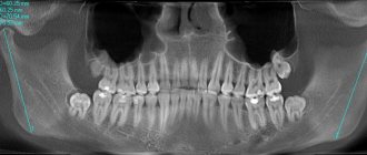
Impacted supernumerary teeth
Often, supernumerary teeth stop growing and remain impacted, that is, unerupted. Such teeth may not appear for a long time; they are discovered by chance during an X-ray examination of the oral cavity. The presence of an impacted supernumerary tooth may be indicated by the following symptoms:
- The appearance of mobility of complete teeth;
- periodic aching pain in the area where the tooth is located;
- tissue compaction if the tooth is located close to the edge of the jaw.
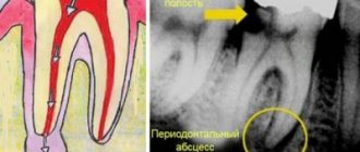
Cysts sometimes develop on the root of the tooth, which may not cause pain for some time; they are also detected during x-ray examination.
Treatment of ankylosis
Treatment of ankylosis with traditional orthodontic methods (braces) is impossible. The tooth grows into the bone and it is impossible to return it to its correct position. But restoring the functionality of the dentition is quite possible. In dental clinics, the method of treatment is selected individually in each individual case.
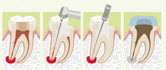
First of all, the patient is referred for a dental computed tomography scan. The dental surgeon studies panoramic images and draws up a treatment plan. Most often, surgical treatment methods are used: excess fibrous tissue is removed and joint mobility is restored. Crowns made of metal-ceramics, ceramics and zirconium dioxide are also used to treat dental ankylosis. With their help, the bite is corrected.
Correction methods
Several decades ago, a person with such an anomaly had two options: remove the merged specimens, or walk with them all his life, building up an inferiority complex.
Today, dentists, using modern techniques and materials, can successfully correct this problem while the main tooth remains intact.
What problems need to be solved?
When choosing a treatment method for this anomaly, the doctor is primarily guided by the tasks that must be solved through therapy.
The main tasks include:
- Correction of pathology while preserving the more significant half.
- Freeing up the necessary space to correct its position.
- Creating favorable conditions for the formation of normal occlusion and dentition.
- Preservation of the pulp chamber and the pulp of the main half to extend the life of the tooth.
What determines the choice of method?
Unlike the treatment of other dental anomalies, in which the fundamental factor in choosing a method is the patient’s age, in this case they rely only on the degree of pathology :
- To eliminate the problem in the first degree of anomaly, they resort to gentle treatment, which involves grinding off the adjacent part.
This method allows you to completely preserve the main one, without the use of additional equipment and special filling materials, since the affected units do not have a common pulp chamber.If necessary, the procedure ends with restoration.
- For the second type hemiresection , in which the supernumerary part is disconnected and removed. In this case, periodontal tissues are not injured.
- With the third type of anomaly, hemisection , which involves exfoliation of the gum tissue and dissection of the root part.
- Treatment for the fourth degree is prescribed exactly the same as for the third, but the dissection is carried out along the entire length .
Sequence of procedure
The treatment takes place in several stages, the first of which involves the immediate removal of the secondary part.
If it is necessary to carry out removal with dissection of only the upper part, then conduction anesthesia is used. In case of complete dissection, general anesthesia may be administered.
Depending on the technique, the main points of the procedure may differ.
Hemiresection involves several steps:
- The dentist uses a diamond disc to cut off a smaller portion of the tooth.
- Performs extraction by rupturing the periodontal ligaments and dislocation.
- Grinding the cut areas with constant irrigation with an aseptic preparation.
- Washing the wound and applying a medicinal application with biologically active components that restore damaged tissue. The bandage is fixed for 2 weeks, after which it is removed.
How to recognize macrodentia, characteristic signs and treatment methods.
In this material we will look at why wisdom teeth are characterized by manifestations of dystopia.
Here https://orto-info.ru/zubocheliustnye-anomalii/zubov/polozheniya/krivyie.html you will see in the photo how effective the fixation of veneers is on crooked teeth.
Hemisection , which is a more traumatic operation, proceeds as follows :
- To gain access to the root part of the tooth, the dentist performs an excision of soft tissue by making an angular incision on the vestibular side of the jaw, plunging the scalpel all the way to the alveolar bone.
In order for the excised tissue to move to the side without any problems, it is cut on both sides of the problem tooth and at its cervical part.The result is a trapezoidal flap that is easily separated from the jawbone.
- It is then peeled off, exposing the cortical bone, from which a limited area is removed. This allows access to the roots.
- Using a special fissure-type bur and diamond discs, the root and then the crown part of the tooth are cut off. The cut off parts are removed.
- The disconnection site is polished, while the wound is constantly treated with saline solution, which ensures the safety of the main part of the pulp left in the alveolar arch.
- The cleaned area is covered with a special composite that restores the tooth wall.
- The mucosal and periosteal flaps are placed in place and sutured with simple surgical sutures, which are removed after 7 days.
- Next, the tooth is splinted for 2 weeks.
2 weeks after removal of the complementary half, the second stage of treatment begins - orthodontic. This type of treatment allows you to completely restore the normal position of your teeth and correct your bite.
Most often, braces with arches of varying degrees of rigidity are used for this, regular replacement of which leads to complete restoration of the row after 8–15 months .
To prevent the tooth from returning to its previous position after correction, retainers are additionally prescribed. The retention period, as a rule, lasts almost as long as the main treatment.
How to remove a tooth with root separation, watch the video:
Primary and secondary edentulous teeth
Experts distinguish several types and subtypes of adentia. For greater convenience, below are tables showing the types of the disease, combined by certain characteristics.
Primary adentia
Caused by hereditary factors and diseases at the stage of fetal formation. With primary adentia, there is an absence of tooth germs.
Varies:
- partial primary adentia - the absence of one or more rudiments (for example, primary adentia of two incisors);
- complete primary adentia – absence of all tooth buds. It is extremely rare;
- true adentia – absence of tooth buds (without their destruction by diseases and infections);
- false edentia, when different teeth merge into one.
Secondary adentia
Secondary adentia occurs as a result of illness and injury during life.
Classification of edentulism by the number of missing teeth
| Number of missing teeth | Classes | general description |
| Partial edentia (ICD K00.00) | Partial primary adentia. Partial secondary adentia. | With partial edentia, from one to 5 teeth or their rudiments are missing on one jaw. Most often, three teeth are missing. |
| Multiple edentia | Multiple primary adentia. Multiple secondary adentia. | Many experts combine the concepts of partial and multiple edentulism. Criteria:
And so on up to 15 units on one jaw. |
| Complete edentia (ICD K00.01) | Complete primary adentia. Complete secondary edentia. | Complete absence of teeth or missing teeth |
Deformations of the dentition
The pressure exerted by the erupting wisdom tooth on the dentition when there is not enough space causes crowding of the teeth in the dentition and the development of malocclusion. Aesthetics and smiles are affected mainly in the anterior region, and oral hygiene becomes difficult.
Growing and expanding wisdom teeth are often the cause of reoccurrence of malocclusion after long-term orthodontic treatment. Therefore, it is strongly recommended to remove wisdom teeth before starting orthodontic treatment!
Indications for wisdom teeth removal can be formulated as follows: ANY DISEASE, COMPLICATION ASSOCIATED WITH WISDOM TEETH, AS WELL AS THE RISK OF THESE DISEASES OR COMPLICATIONS IS A DIRECT INDICATION FOR WISDOM TEETH REMOVAL.
Classification of missing teeth according to the location of the disease
| By localization | Description |
| Missing upper teeth | The absence of teeth in the upper jaw can be either partial or complete, as well as single. |
| Missing lower teeth | A general designation for a disease indicating the absence of teeth in the lower jaw. |
| Lack of chewing teeth | May be referred to as missing lateral teeth. Lack or complete edentulism in the chewing region. In particular, we are talking about the absence of small molars (premolars) and molars. |
| Missing front teeth | Edentia of incisors or canines in the frontal zone. |
In addition to the classification described above, the type of teeth itself is also taken into account. Based on this, the absence of molars and edentulous primary teeth (not associated with the normal timing of their replacement) are distinguished.