Find out more about nervous diseases starting with the letter “N”: Sleep disturbance; Narcolepsy; Hereditary cerebellar ataxia of Pierre-Marie; Spinal circulatory disorders; Trigeminal neuralgia; Neuralgia of the submandibular and sublingual nodes; Neuralgia of the glossopharyngeal node; Neuralgia of the ear ganglion; Neurasthenia; Neural amyotrophy of Charcot-Marie-Tooth; Acoustic neuroma; Neuroma; Optic neuritis; Pharyngeal neuritis; Neuritis of the facial nerve; Neuritis; Obsessive-compulsive neurosis; Pharyngeal neurosis; Neuroses; Neurosis-like stuttering; Femoral nerve neuropathy.
What is glossopharyngeal neuralgia?
If the cranial nerve is affected by a quarter on one side, and paroxysms of pain appear in the tonsils, soft palate, pharynx and ear, then the patient may have neuralgia of the glossopharyngeal nerve. With this pathology, a violation of the taste perception of the posterior third of the tongue, on the affected side, is detected. Hypersalivation and decreased reflex of the palate and pharynx are also noted. To make a diagnosis, an examination by a neurologist or dentist is required. CT and MRI of the brain are indicated as hardware diagnostic methods. In most cases, treatment is conservative, including:
- Anticonvulsants;
- Analgesics;
- Sleeping pills and sedatives;
- Vitamin therapy;
- Physiotherapy;
- General strengthening procedures.
4.Treatment
Treatment for glossopharyngeal neuralgia is aimed primarily at relieving pain. Given its severity and specificity, in some cases it is necessary to prescribe both non-narcotic and narcotic analgesics; Antiparoxysmal and anti-inflammatory drugs, vitamin complexes, restorative therapy, and electrophysiotherapeutic methods are also used. In situations where the established cause of neuralgia is mechanical compression (compression) of the nerve, for example, with hyperplasia of structures adjacent to the nerve, neurosurgical intervention is performed.
Conservative treatment usually lasts quite a long time (up to several years), but in most cases it leads to the elimination or significant reduction of neuralgia.
Structure of the glossopharyngeal nerve
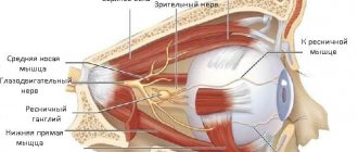
N. glossopharyngeus or glossopharyngeus nerve begins in the nuclei of the medulla oblongata. It is based on motor, sensory and autonomic parasympathetic fibers. Moreover, the sensory fibers begin in the sensory nucleus common to the vagus and glossopharyngeal nerves. They innervate the mucous membrane of the pharynx, soft palate, tongue, tonsils, and Eustachian tube. The sensation of taste in the anterior two-thirds of the tongue is provided by taste fibers emerging from the nucleus of the tractus solitarius. The taste fibers of the glossopharyngeal nerve are responsible for the taste sensations of the posterior third of the tongue and epiglottis.
The motor fibers originate in the nucleus ambiguus and innervate the stylopharyngeal muscle, which is necessary for raising the pharynx. Together with the vagus nerve, they form the reflex arcs of the pharyngeal and palatal reflexes.
Parasympathetic fibers originate in the salivary nucleus. Being part of the tympanic and lesser petrosal nuclei, they reach the autonomic ganglion, and with the branch of the trigeminal nerve they regulate salivation of the parotid gland.
Since the pathways and nuclei of the glossopharyngeal and vagus nerves are common, isolated pathology n. Glossopharyngeus. But more often symptoms of combined lesions are observed.
How to protect yourself from lingual nerve damage
In order not to encounter this problem, you should contact a good specialist who can carefully plan and ensure qualified execution of the manipulations.
The lingual nerve provides sensation to the tongue. If unfavorable circumstances occur during the provision of dental care, it may be damaged. The nature and degree of the complication may be different - these indicators determine whether the functionality of the lingual nerve will be restored. Today, dentistry can offer complex therapy for successful but long-term restoration of lingual nerve function in more than half of cases.
Pathogenesis
There are cases of idiopathic Sicard syndrome. At the same time, it is almost impossible to establish the etiology of the disease. Provoking factors may be:
- Acute or chronic intoxication;
- Otitis;
- Pharyngitis;
- Tonsillitis;
- Sinusitis;
- Atherosclerosis;
- Viral infections incl. flu.
Secondary neuralgia occurs due to:
- Arachnoiditis, encephalitis and other infectious lesions of the posterior cranial fossa.
- TBI.
- Hyperthyroidism.
- Diabetes mellitus.
- Meningiomas, gliomas, medulloblastomas and other intracerebral tumors of the cerebellopontine ganglion.
- Compression and irritation of any part of the glossopharyngeal nerve.
- Nasopharyngeal tumors.
- Intracerebral hematomas;
- Carotid artery aneurysm;
- Hypertrophy of the styloid process.
- Overgrowth of osteophytes of the jugular foramen.
- Ossification of the styloid ligament.
In some cases, pathology may be the first symptom of cancer of the larynx or pharynx.
Diagnosis of neuralgia of the submandibular and sublingual nodes
A neurologist can detect and diagnose diseases of the vegetative nodes during a personal examination of the patient. To establish an accurate diagnosis, topical diagnostics is used, which includes:
- patient interview;
- analysis of complaints;
- examination using palpation and additional techniques;
- as well as identifying objective signs of pathology.
Since there are several vegetative nodes in the head and face area, and the symptoms of their diseases are very similar, the main task of the neurologist during the initial examination will be to identify the location of the disease.
Using sliding and fixing palpation, the specialist identifies the localization of the pathological process. Sometimes pinching or superficial pain stimulation methods are used for accurate diagnosis.
If the doctor has doubts about the results of topical diagnostics, a diagnostic blockade technique is used, which makes it possible to absolutely accurately establish the source and localization of neuralgia. The blockade is carried out on an outpatient basis by injecting local anesthetics (novocaine, lidocaine, trimecaine) into the painful area. Pain relief for a while allows the doctor to make an accurate diagnosis.
Symptoms of neuralgia of the submandibular and sublingual nodes often have similar symptoms to inflammation of the gums and internal structure of the teeth. To rule out dental diseases, you may need to consult a specialist in this field.
Symptomatic picture
Clinical signs include painful paroxysms lasting from a few seconds to three minutes. A sharp acute pain originates at the root of the tongue and instantly spreads to the tonsils, soft palate, ear and pharynx. May radiate to the eye, neck and lower jaw. Pain can be triggered by coughing, chewing food, its temperature, yawning, swallowing and even talking. During paroxysm, dry mouth is noted, immediately after hypersalivation. But dryness is not a mandatory sign of diagnosis, because the secretory insufficiency of the parotid gland can be compensated by the remaining salivary glands.
Paresis of the levator pharyngeal muscle does not cause swallowing disorders. But difficulties in chewing and swallowing associated with impaired proprioceptive sensitivity, which is responsible for the position of the tongue, may be noted by patients.
The disease has a wave-like course, worsening in autumn and winter.
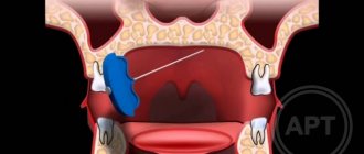
Causes of lingual nerve damage
The etiology of this phenomenon lies in the peculiarities of the topographic location of the lingual nerve and its adjacency to the surgical area, as well as the sensitivity of the nervous tissue to ischemia. Post-traumatic neuropathy of the lingual nerve, after complex extractions of impacted and dystopic third molars of the mandible, accompanied by complications, occurs in 2-7% of clinical cases [5].
A damaged lingual nerve is often caused by the following factors:
- Failure to observe anatomical landmarks by a doctor.
- Direct injury to the nerve with a needle at the time of partial or complete anesthesia, provided that it has low mobility in the tissues and is adjacent to bone structures.
- Injury from a deformed needle point.
- Neurotoxic effects of the anesthetic and hematoma: injury to the lingual nerve can occur during anesthesia due to the formation of a hematoma, as well as the chemical action of the components of the anesthetic solution.
- Endoneural administration of anesthetic [2].
Diagnostic methods
The diagnosis is made by a neurologist, who, if necessary, can involve an otolaryngologist and dentist. During the examination, the doctor determines analgesia, that is, the absence of pain sensitivity at the base of the tongue, upper parts of the pharynx, and tonsils. Taste sensitivity is also studied, for which a taste solution is applied to symmetrical areas of the tongue. The diagnosis is confirmed by isolated unilateral taste disorder in the posterior third of the tongue. Bilateral disruption is typical for diseases of the oral mucosa, such as chronic stomatitis.
The pharyngeal reflex needs to be checked. To do this, touch the back wall of the throat with a paper tube, which provokes a swallowing movement and occasionally a cough. Normally, touching the soft palate should be accompanied by a lifting of the palate and uvula. Sicard syndrome is characterized by the absence of reflexes on one side. But a similar symptomatic picture can occur with damage to the vagus nerve. If the pharynx and pharynx are strewn with herpetic rashes, the doctor can diagnose ganglionitis of the ganglionitis of the glossopharyngeal nerve nodes, which differs in almost identical n. Glossopharyngeus clinical picture.
To establish the root cause of symptomatic neuritis, neuroimaging diagnostics is needed:
- MRI or CT scan of the brain;
- Electroencephalogram;
- Echo-EG;
- Ophthalmoscopy with consultation with an ophthalmologist.
It is necessary to distinguish between neuralgia of the glossopharyngeal nerve and diseases that cause painful paroxysms of the face and head. These include:
- Neuralgia of the ear ganglion;
- Trigeminal neuralgia;
- Oppenheimer's syndrome;
- Glossalgia;
- Ganglionitis of the pterygopalatine ganglion;
- Retropharyngeal abscess;
- Tumors of the pharynx.
Symptoms of throat neurosis in adults and children
Throat neurosis is characterized by stable symptoms that differ depending on the type of disease. The main manifestations of the disease are tissue numbness and weakening of the swallowing reflex. The disease is accompanied by severe pain throughout the body. Patients may experience increased sensitivity of the hard tissues of teeth.
Throat neurosis is manifested by the following main symptoms:
- Feeling of lack of oxygen even after taking a deep breath;
- Feeling of a lump in the throat that does not go away after drinking liquid;
- Dryness in the larynx;
- Intense thirst;
- Bronchial spasm, which is accompanied by a gag reflex and is not relieved by bronchodilators;
- Sore throat, itching, sore throat, worsening at the moment of greatest irritation;
- Pain in the neck;
- Shooting pain in the ear.
Symptoms of throat neurosis are aggravated when a person is in a dusty or smoky place. An increase in reflex reactions leads to a gag spasm during a hacking dry cough.
Symptoms of throat neurosis intensify with excitement, irritability, psycho-emotional lability, and a migraine attack. Patients' mental and physical activity decreases.
A lump in the throat with neurosis provokes the occurrence of additional symptoms:
- Mood swings;
- Disturbances in sleep and wakefulness;
- Decreased appetite;
- Hypochondria;
- Manifestations of depression;
- Fear of suffocation.
It is especially difficult to diagnose throat neurosis in children, since they are susceptible to colds, the symptoms of which are similar in symptoms to those of pharynx and larynx neurosis.
Under the influence of provoking factors, the function of the child’s immature nervous system is disrupted, which leads to the loss of neural connections between active cells of the mucous membrane of the upper respiratory tract. Children are psycho-emotionally immature. They react sharply to all irritating factors of a psychogenic nature.
For this reason, throat neurosis in children is often accompanied by somatic pathology. The child's sleep may be disturbed and emotionality may increase. Children can swallow whole pieces of food, which leads to disruption of the digestive system. Sometimes children suffering from throat neurosis develop hoarseness or aphonia (complete absence of voice while maintaining whispered speech).
Therapy
To treat neuralgia of the glossopharyngeal nerve, conservative therapy is prescribed. The only exception is if the underlying cause is tumors and hypertrophy of the styloid process, leading to compression of the nerve. In these cases, surgery is indicated.
To relieve pain, a 10% solution of cocaine is prescribed, which is applied to the root of the tongue and pharynx. This allows you to eliminate paroxysms for 6-7 hours. If this remedy turns out to be ineffective, a 1-2% novocaine solution is injected into the root of the tongue. Non-narcotic analgesics and anticonvulsants are also prescribed internally. If the pain syndrome is pronounced, it is advisable to prescribe sedatives, hypnotics, antipsychotic medications and antidepressants. AFT, multivitamin complexes and FiBS are used as general strengthening agents.
Among the physiotherapeutic procedures that have proven effectiveness are:
- SMT on the area of the larynx and tonsils;
- Diadynamic therapy;
- Galvanization.
1.General information
One of the paired cranial nerves is called the glossopharyngeal. This (IX) pair received its name in connection with the functions that it controls: the activity of the stylopharyngeal muscle, the sensitivity of the taste pimples of the basal part of the tongue, the secretory activity of the parotid gland, as well as the tactile sensitivity of a number of structures of the hearing organs and the oral cavity.
The term "neuralgia" is translated as "nerve pain" or "nerve pain." Neurogenic pain syndromes are distinguished by their specific nature (the pain is usually piercing, the sensations are similar to an electric shock extended over time), significant severity and persistence: it is impossible to find a forced position, conventional analgesics have little effect, etc.
A similar syndrome, caused by an isolated lesion of the glossopharyngeal nerve, is very rare in clinical practice - its statistical frequency is one case in approximately 620,000 people. Among the patients, men predominate, and in the age group - people over 40 years old.
A must read! Help with treatment and hospitalization!