How many canals are there in the teeth of the upper and lower jaws?
The main parts of the tooth are the crown (rising above the gum) and the root part.
Each tooth has its own shape and structural features. One of them is the number of roots and canals. The more a tooth has to withstand the load, the stronger it needs to “hold” in the gum. Therefore, it requires more roots. If you can figure out the number of roots of the teeth, then with the canals the situation is much more complicated. How many canals a tooth has has nothing to do with the number of roots. For example, the top “five” has only one root. At the same time, 75% of “fives” will have 1 channel, and 24% may have 2. In addition, there are another 1-2% of “fives” in whom the doctor detects 3 channels.
The root canal is the space inside the root . Complex anatomical branches can connect different channels to each other. At the tip of the horse there is an apical foramen. A feature of the tooth root system is that one hole can serve as an exit for several canals. Also, the channel sometimes bifurcates, ending in two exits. No two root systems are alike. Therefore, the doctor cannot do without an X-ray examination.
What are channels?
Each tooth has a certain number of roots located under the gum.
Read also: What is the difference between pulpitis and periodontitis?
How many roots do teeth have? The answer to this question depends on several factors - the position of the unit, the person’s age, heredity, even race. It is known that Mongoloids have more roots than Caucasians.
The standard quantity is as follows:
- Incisors, canines – 1.
- Premolars – 1-3.
- Upper molars – 3-4.
- Lower molars – 2.
- Third molars – 3-5.
Inside the crown is the pulp, a tissue consisting of blood vessels and nerve endings. They pass into the pulp through the apical foramen, located at the apex of the root, and through canals, narrow cavities inside the root. Their number is not always equal to the number of roots.
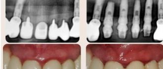
The photo shows the beginning of the root canals.
Why is there no accurate data on the number of canals in each specific tooth?
This situation is explained by the individual characteristics of the structure of each person’s teeth. Therefore, the dentist relies on statistical data and, if necessary, receives accurate information after appropriate research.
How do doctors figure out how many canals are in each specific tooth? A certain pattern does exist in this matter: the deeper a tooth is located in the mouth, the more complex its canal system is. This feature is due to the load that the tooth has to withstand.
In addition, statistics show that there are more canals in the upper teeth than in the lower ones, but not always. Therefore, it is possible to say about the exact number of canals only after opening the tooth or based on radiography. To preliminary determine the number of canals in teeth, statistical data are used, and to obtain accurate information, radiography results are used.
Why do you need to know about the number of channels? This question is very important during treatment and filling. If the doctor has not treated one of the canals, then an infection will remain in it, which will negate the result of the treatment.
Patency of dental canals
In addition to the number and length, important information is the patency of the root canals, which depends on the degree and location of the curvature. If the curvature is less than 25 degrees, then the canal is instrumentally accessible, from 25 to 50 degrees it is difficult to access (the so-called difficult tooth canals), and over 50 degrees it is inaccessible. When the curvature is localized near the mouth of the canal, it is possible to expand the latter and improve patency.
If the examination reveals a too narrow, deep canal in the tooth, a CT scan may be required to clarify its configuration. Treatment of complex teeth requires particularly painstaking work, which can be made easier with the help of a microscope.
Sometimes the doctor cannot find the canal in the tooth. This situation is usually associated with obliteration (narrowing or overgrowing) of the canals due to an inflammatory or tumor process, incorrect treatment in the past, or age-related changes.
Remember that only a specialist can assess the condition of the root canals and, depending on their structural features, determine treatment tactics.
Accurate determination of the number of channels using an x-ray image
An x-ray allows you to determine exactly how many canals there are in a tooth. This method allows the doctor to see a complete picture of the condition of the teeth: the location of the roots, the presence of a cyst. The image helps to evaluate the quality of the filling performed, as well as to calculate how many canals there are in a particular tooth.
Some patients associate the word “X-ray” with something dangerous. However, modern devices do absolutely no harm to humans. The procedure does not require preliminary preparation; it is often carried out directly in the dentist’s office. The whole process takes no more than 5 minutes.
As a result of radiography, the doctor has a complete, and most importantly, clear picture of what is happening to the teeth. Where crowns or fillings are installed, white areas appear in the image, cavities appear black, and tissues and fluids acquire gray shades. The information obtained allows the doctor to determine the number of channels as accurately as possible and perform treatment while minimizing negative consequences.
Dental X-rays can be done even for nursing and pregnant women. Of course, there must be serious reasons for this, but in general the procedure is absolutely safe. The only unpleasant moment may be the appearance of a gag reflex. It occurs when the film is fixed to the gum. Deep breathing through the nose helps reduce the urge to vomit.
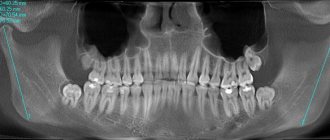
The images that can be obtained using x-rays are divided into two types:
- Orthopantogram - displays a complete picture of the condition of the teeth of the upper and lower rows. Such photographs are needed at the initial stage of treatment in order to draw up a general plan of the necessary procedures, identify pathologies, structural features, and the relative position of the teeth.
- Targeted – allow you to obtain complete information about a specific tooth. The image gives a clear idea of the internal structure, number and location of the channels, and helps make a final decision on the treatment method.
- Sight shots are sometimes called control shots. Their implementation after treatment allows us to evaluate their effectiveness and quality of implementation.
Approximate number of canals in the teeth of the upper jaw
The number of dental canals is one of the few points on which dentistry cannot give a clear answer. Such a concept as “norm” does not apply here. This is due to the individual characteristics inherent in the teeth of each person. They are determined by the location of the tooth in the row and on the jaw.
In general, you can rely on the following data:
- canines and incisors – usually 1 root;
- premolars 1-2 roots;
- molars have 3-4 roots;
- Wisdom teeth have up to 5 roots.
The main purpose of molars is to grind food. Therefore, they have a wide flat surface, and their root part is firmly fixed in the gum. The chewing teeth of the upper jaw usually have 3 roots, but with 4 canals. For comparison, the same teeth in the lower row often have 2 roots, but 3 canals.
Statistics tell you best about the number of channels:
The teeth located on the upper jaw are very different in the number of canals from the lower ones. The situation is simpler with incisors. The 1st, 2nd and 3rd incisors usually have only 1 canal. The fourth tooth is a little more complicated: in 85% of cases it has 2 canals, in 9% 1, and in only 6% 3 canals. In the fifth tooth, statistics give the following result: most often (75% of cases) there is 1 channel, less often (24%) - 2 and only 1% of cases - 3 channels.
In the sixth tooth, dentists find 3 and 4 canals, respectively, in 57 and 43% of cases. Among the “seven”, 3 channels are more common (57%), less often – 4 (43% in total). The doctor obtains the exact result in each specific case through direct examination or with the help of an image.
Prevention of root canal diseases
For ideal “order” in the oral cavity you need:
- take proper care of it;
- use high-quality instruments and oral hygiene products;
- visit the dentist twice a year;
- after each meal, rinse your mouth with water;
- quit smoking and alcohol;
- reduce the amount of coffee and tea consumed;
- Healthy food.
Once I went to the dentist to have a tooth treated, in the end they pulled it out and said that the dentition was incorrect. That there should be 3 roots in a tooth, but I have 2. The doctors were mistaken, the root remained in the gum. I got it with my own strength. And they didn’t even deign to double-check everything. Just like that...
Approximate number of canals in lower jaw teeth
The situation with the teeth of the lower jaw is somewhat different from the upper one. Statistics show that in the first tooth, 1 canal is most often found (70%), 2 canals - respectively, in 30% of cases. In the second tooth, we can say it’s 50/50: a little more than half (56%) for there to be 1 canal, and 44% for two. In the “troika,” 1 channel is predominantly found (94%), the remaining 6% are 2. Slightly more often, 2 channels are found in the “four” (26%). The probability of meeting them in the “five” is even greater: 89%, the remaining 11% for 1 channel.
The “six” and “seven” have a more branched structure. In the sixth tooth, 3 (65%) or 4 (29%) canals are most common, 1 canal is least common - 6%. In the “seven” the dentist usually finds 3 canals (77%) and 2 canals (13%). It is very rare, but even in it you can find only 1 channel. This result shows only 1% of cases.
How many canals are there in a wisdom tooth?
Wisdom teeth are considered extraordinary in all respects. Even when they start to cut themselves, they cause a person a lot of unpleasant moments. Then they most often occupy the wrong position, and cleaning them is another almost impossible task. There are even special brushes available that help you reach hard-to-reach places.
The number of G8 channels is also difficult to quantify. They are unique for each person. The number of channels in the upper eight can be 1-5, and in the lower eight most often it is 3. Although often opening a tooth reveals the presence of additional branches. Sometimes the dentist says that the patient has a single-channel “eight”. This usually means that there are actually more roots, but they have grown together.
A special feature of wisdom teeth canals is their non-standard shape. They are usually strongly curved, with narrow passages. This greatly complicates the dentist’s work during treatment and filling.
Wisdom tooth and features of its anatomical structure
The outer eighth units of the lower and upper jaw are called wisdom teeth.
The cavity of these teeth is often affected by pulpitis, since they erupt very fragile. These curved wisdom units have a unique anatomical structure of the tooth cavity. They appear later than everyone else: at 20, and at 30, and even at 40 years old. The difference in their anatomical structure lies in the number of roots, which can be from two to five. These roots are quite crooked (see photo), therefore they cause many problems during treatment procedures, and especially during determination of the working length, expansion of canals and filling. The number of channels for “eights” can reach up to five.
Table and diagram of the location of canals in teeth
Often in dental clinics you can find useful information on educational posters: find out how many canals are in the teeth, look at the table and diagram of their location, study the structure of the tooth. These are questions that do not interest us in everyday life, but become especially relevant during a visit to the dentist. In order to come to your appointment fully armed and understand exactly what manipulations the doctor is performing and why, it is better to study the theoretical aspects in advance.
Visible dental elements are only a small part of the whole organ, which often does not have the simplest structure. Despite the external similarity, the structure of the teeth is not the same in the upper and lower rows. It may even differ from person to person due to the influence of hereditary factors. Let's figure out what a dental unit consists of and what is the sequence of treatment for caries and pulpitis.
Why does a dead tooth hurt?
It is logical that a dead tooth, that is, one from which the nerve has been removed, should not feel anything, much less hurt. However, patients often complain of toothache after tooth depulpation. With proper removal, a pulpless tooth cannot hurt; after removal of the nerve, pain can occur in the border structures: jaw bones, soft tissues, neighboring teeth, because the nerves remain there, and it is they that sometimes ache for several days, as they are “concerned” with the surgical intervention. However, if the pain does not stop, you need to consult a dentist; perhaps the nerve was not completely removed, or the pain is caused by some other reason - only a specialist can determine this.
A tooth is a very complex structure, and in this case the saying “Measure twice, cut once” fully reflects the essence of dental treatment!
The structure of teeth and their arrangement
A dental unit can be divided into 3 components:
- The crown is the visible part of the tooth that directly performs the chewing function. The top of the crown is covered with a thin protective layer of enamel, under which is the densest part – dentin. Dentin surrounds the most vulnerable and delicate part of the tooth – the pulp. It is a cavity filled with vessels and nerves.
- The cervix is a tapering part, normally hidden by the gum. Instead of enamel, it is covered with cement, and a dental canal begins inside - a narrowing of the pulp cavity.
- The root of a tooth is a part fixed in the alveolar process of the jaw. It is inside the roots that the neurovascular bundle passes, feeding all dental structures. It enters through a small hole at the top of the root. The root section is devoid of enamel coating, and the dentin in this part contains a large amount of collagen.
In dentistry, the numerical designation of dental units is accepted. All teeth are divided into 4 quadrants. The first number indicates the quadrant number (1-4 in adults and 5-8 in children), and the second number indicates the serial number of the tooth in the row (numbering is from the middle to the periphery). One quadrant includes 8 dental units: 2 anterior incisors, 1 canine, 2 premolars and 3 molars.
How does a human tooth work?
The elements of a human tooth can be divided into:
The crown is located above the gum and has a special coating called enamel. Under the enamel there is a durable layer of dentin, which in its structure resembles bone tissue.
The cavity of the tooth located inside the crown is called the “pulp”. It passes into a narrow canal of the tooth root, at the base of which there is a small hole. Nerve endings and blood vessels pass through it into the tooth cavity. Inflammation of the pulp is called pulpitis. It is an indication for opening the tooth cavity and cleaning the root canals. The most difficult thing to treat is pulpitis in the cavity of three-channel units (for example, in the sixth). In advanced cases, it is necessary to remove the tooth, and if it is also on top and in the last rows (6, 7 or 8), then this is also inconvenient.
The dental neck is located inside the gum. It does not have an enamel coating, but is protected by cement. The continuation of the tooth cavity is its root. It is located in the alveolus, a small cavity in the teeth. Its structure differs from the structure of the crown and neck. The enamel layer is absent, and dentin is permeated with collagen. Nerves and blood vessels pass through the root canal into the dental cavity.
Roots and channels
The number of roots and tubules does not always coincide and is not considered a strict value. The canal-root system can vary significantly: cavities often diverge and unite again, open with one or more mouths, have expansion and contraction, and in some cases become completely overgrown. There are 9 types of structure and course of root canals:
- The simplest option: the course is straight, going from the pulp chamber to the apex of the root.
- Two tubules that arise from the same opening and join again at the root apex.
- At first the passage is common, at the bottom of the root it bifurcates, and then the passages meet again and open with a single hole.
- Two completely separated channel passages with different outlets.
- The canal follows a single course, which bifurcates at the very top of the root and has 2 exits.
- Two different passages connect at the root of the tooth, and separate again before leaving the root.
- The passages are in the form of a chain, which are separated several times and come into contact again in different parts of the root.
- 3 direct channels that do not communicate with each other.
- 3 separate channel cavities are combined into one before exiting the apical part of the root.
Let's consider the most typical options for the internal structure of the dental units of the upper and lower rows.
Upper jaw
The incisive and canine units of the upper dentition predominantly have a single root and tubule, like the second premolars. The first premolars often have a bifurcated canal-root system, the first and second molars have a triple one. Wisdom teeth (third molars) are considered the leaders in the number of roots and tubules - there can be 3-5 of them.
► Table 1. Root system of the upper jaw
| Dental unit no. | Tooth length, mm | Channel length, mm | Number of roots (frequency of occurrence) | Number of channels (frequency of occurrence) | Peculiarities |
| 1 | 23 | 13,0 | 1 (100%) | 1 (100%) | Distal curvature of the root and cervical narrowing of the cavity are common. |
| 2 | 22 | 13,0 | 1 (99%) | 1 (99%) | There may be curvature and expansion of the cavity in the distal section. |
| 3 | 27 | 16,0 | 1 (99%) | 1 (99%) | A two-canal structure and curvature in the palatal direction are rarely observed. |
| 4 | 21 | 13,5 | 2 (80%) | 2 (95%) | In 19% of cases there is 1 root, in 4% there is a single canal. |
| 5 | 22 | 14,5 | 1 (90%) | 1 (75%) | With a frequency of 9% there are 2 roots, 24% - 2 bifurcated canal system. |
| 6 | 21 | 13,3 | 3 (85%) | 3 (60%) or 4(40%) | Often the canal passages of one root are branched and difficult to process. |
| 7 | 20 | 13,0 | 3 (80%) | 3 (57%) or 4 (40%) | The number of cavities and their course are very variable. |
| 8 | 19,8 | 13,6 | 1-5 | 1-5 | The sizes and structure of the tooth are very diverse. The roots are usually very curved. |
Lower jaw
The first 5 dental units of the lower row in the vast majority of cases have a single canal and root. Units 6 and 7 are usually characterized by a double or triple canal-root system. In the case of a wisdom tooth, everything is very variable; there can be up to 4 cavities and roots, and in isolated cases even more.
► Table 2. Root system of the lower jaw
| Dental unit no. | Tooth length, mm | Channel length, mm | Number of roots (frequency of occurrence) | Number of channels (frequency of occurrence) | Peculiarities |
| 1 | 21 | 12,9 | 1 (100%) | 1 (60%) or 2 (40%) | The second canal usually extends from the side of the tongue and is difficult to detect. |
| 2 | 22 | 13,0 | 1 (100%) | 1 (60%) or 2 (40%) | A second canal on the lingual side is also possible. |
| 3 | 26 | 15,2 | 1 (98%) | 1 (94%) | There are significant expansions or complex passages of the canals. |
| 4 | 22 | 13,6 | 1 (100%) | 1 (75%) | In 20% of cases there are 2 roots. |
| 5 | 22 | 15,1 | 1 (100%) | 1 (89%) | In 10% of people, the channel system bifurcates. |
| 6 | 21 | 14,5 | 1 (98%) | 3 (80%) | The frequency of occurrence of a two-channel system is 13%. |
| 7 | 20 | 14,0 | 2 (84%) | 3 (77%) | The number of roots and canals is variable. In 15% of cases the root is only 1, and in 13% there is a double channel. |
| 8 | 19,4 | 13,3 | 1-4 | 1-4 | The sizes and structure of the tooth are very diverse. The roots are usually very curved. |
Apex locator
The electronic method is considered the most reliable of all mentioned. The most popular brands of devices are: Russian Geosoft (EndoEst), Japanese NSK (iPex), German VDW (Raypex).
Determination of the working length of the root canal:
- one wire with a hook at the end is hung on the patient’s lip
- diagnostic tool (for example, K-file) is installed in the holder
- then the doctor slowly moves it along the canal; numbers are displayed on the device screen indicating how much is left to the top. An intermittent beep will also sound. Some models have a graphic display: green - far, yellow - close, red - in place
- When the instrument reaches its destination, APEX will appear on the screen and the alarm will beep continuously.
Diagnostics
Before performing dental procedures, it is extremely important to identify the features of the canal-root system in order to clean it completely. The problem is that the dentist is not always able to determine the number of strokes visually and by palpation (using a probe). They often narrow at the mouth, and the probe does not pass through the narrowing, overgrow against the background of a long-term inflammatory process, or have an atypical structure.
That is why, in most cases, the doctor sends the patient for dental x-rays before starting treatment. The photographs show the number of roots, the structure of the cavities, and indirect signs of the inflammatory process. In complex diagnostic cases, they resort to computed tomography, which helps to more clearly visualize all tooth structures.
In addition to topical diagnostics, it is also important to measure the length of the canal in order to understand at what depth the filling material should be inserted.
Petrov Oleg Viktorovich
Chief physician, director of the clinic, implant surgeon, candidate of medical sciences at the branch of the street. 8th Krasnoarmeyskaya, 3
He began studying maxillofacial surgery and dentistry at the department of the same name at the Military Medical Academy, and almost all subsequent postgraduate education was associated with training at this oldest educational institution. After graduating in 1992, he underwent advanced training at the faculty of retraining and advanced training in the “surgical dentistry” cycle.
For six years he was the head of the dental department of the hospital. During this period, he repeatedly underwent advanced training in various areas of dentistry from “therapeutic dentistry” to “implantology”. Out of love for his specialty, two rationalization proposals were born.
Knowledge of implantology and improvement of implant treatment methods in complex clinical cases took place at the bases of well-known European universities: a one-year cycle in implantology at the Frankfurt University. Goethe; cycle on bone grafting at the Medical University of Vienna. Participated in international implantological congresses in Italy, England, Monaco. This made it possible to use the acquired knowledge and skills to solve practical problems of restoring patients’ missing teeth.
I have been working at the Astra Clinic since 2005. Managing a friendly team of doctors, I try to use all known modern methods of dental restoration to achieve aesthetic and functional results.
Canal treatment
As a rule, root canal treatment is required when pulpitis is detected. It is carried out in several stages, and sometimes a repeat visit is necessary.
Preparation and anesthesia
First of all, the doctor tells the patient the proposed treatment tactics and offers to sign a consent to carry out the necessary manipulations. Then the dentist checks for allergies to medications, including anesthetics. In doubtful cases, an allergy test is performed with the drug that will be used for pain relief (promecaine, lidocaine, ultracaine).
Anesthesia in dentistry can be local or injection. Local is performed by applying an anesthetic substance to the injection area so that the injection does not cause pain. The injection itself is placed in the nerve area to block pain during penetration into the pulpous cavity.
Stages of treatment
After the onset of anesthesia, the doctor immediately begins treatment, which usually takes place in three stages:
- Opening of root canals. First of all, the doctor drills out the carious cavity, leaving only healthy tooth tissue. Then it washes out the drilled dentin particles and penetrates into the pulp cavity, first using the smallest bur, and then expanding the spherical passage.
- Working with channels. After entering the pulpous cavity and canal system, the dentist mechanically removes all the contents from there and washes them with disinfectants, which are injected with a syringe with a thin needle. After each contact with medical instruments, rinsing is carried out with sodium hypochlorite. After cleaning, use hydrogen peroxide and distilled water.
- Filling. The treatment process is completed by placing a filling. Gutta-percha is usually used as a filling material. To strengthen the tooth, special pins are often pre-installed. If, after the end of treatment, signs of active inflammation in the dental tissues and surrounding structures remain, then a temporary filling is placed, made of porous material, which is removed after a few days and replaced with a permanent filling after the inflammatory process subsides.
Video: how many canals are there in teeth?
Treatment of teeth with problematic root canals
Filling dental canals
A relatively simple technology for treating tooth canals is filling with a special paste with or without a pin. According to the “gold standard” of endodontics, the canals are also filled with a latex-like material - gutta-percha. Several methods of its use have been developed, including the Termafil system, lateral condensation, injection or liquid thermogutta-percha (vertical condensation). In some cases, in particular when treating a tooth canal cyst, filling is carried out with a substance based on calcium hydroxide (copper-calcium hydroxide “depopheresis” method). However, special nanocomposite materials are increasingly used in dentistry.
Treatment under a microscope
The age-old “tough nut to crack” for the dentist is curved or branched root canals of the teeth. A dental microscope, often in combination with a laser, allows you to completely pass through them and adequately process them along their entire length to reduce tissue trauma. Sometimes it becomes necessary to treat a sealed tooth canal with the evacuation of remaining material of various nature, for example, fragments of fillings, tissue fragments and even instruments. Then a microscope also comes to the rescue. Read more about the technology in a separate article.
Prevention
In order not to have to endure unpleasant moments during dental treatment, it is better to promptly identify and, as far as possible, prevent caries and pulpitis. To do this, you need to follow the recommendations of dentists:
- Brush your teeth regularly.
- Use dental floss that gets into hard-to-reach places.
- Rinse your mouth after eating.
- Give up bad habits (smoking, drinking alcohol).
- Drink coffee and tea less often.
- Don't eat foods that are too cold or hot.
- Visit your dentist once every six months, even if nothing bothers you.
- Follow a diet rich in vitamins, phosphorus, fluoride, calcium.
What is the cost of treatment?
How much does root canal treatment cost? The cost largely depends on the method used and the quality of the materials. When using laser and microscopic technology, nanocomposites and other advanced developments, the cost of tooth treatment with canal filling increases significantly. Approximate prices for root canal treatment in Moscow with gutta-percha filling and composite filling are presented in the table below.
| View | Price |
| Single channel tooth | 9,500 – 12,5000 rubles |
| Double channel tooth | 11,000 – 14,500 rubles |
| Three-channel tooth | 13,500 – 17,000 rubles |
The cost of root canal treatment should not be the determining factor in choosing a clinic. Contact only dentistry with a good reputation and experienced specialists who use modern equipment and the latest techniques in their work. Remember - the future fate of your teeth and the aesthetics of your smile depend on the quality of canal treatment!
How many canals are there in human teeth, features of the anatomical structure
A beautiful smile is fashionable. Therefore, great attention is paid to dental health these days. Unfortunately, not everyone can boast of their impeccable appearance, although modern dental developments can bring them as close to ideal as possible.
In our article we will not talk about this. We will discuss the anatomical structure of the human tooth, a diagram of which is given on our website.
Molars are the only human organ that does not regenerate on its own. That is why they need to be protected and regularly monitored for any changes in their condition. It is not without reason that regular examinations by a dentist every 6 months are recommended.
Molars require careful care
If we consider it enlarged, then each molar, a photo of which can be seen on our website, consists of a crown and root part. The crown part - the one that is located above the gum level, is covered on top with the strongest tissue in the human body - enamel, which protects its softer inner layer - dentin, which is the basis of the tooth.
Despite its strength and reliability, enamel is incredibly susceptible to external influences. Poor care, bad habits, and heredity can disrupt its condition. Pathogenic bacteria enter cracks in the enamel, causing intense tissue destruction. A person develops a carious process that also affects dentin.
If left untreated, the infection penetrates into the root part, acute pulpitis and other equally dangerous ailments develop.
As for the structure of the root part. then its main elements are arteries, veins and nerve fibers that supply the tooth. They are located in the pulp of the root canal and through the apical foramen are connected to the main neurovascular bundle.
The dentin below the gum level is covered with cement, which is attached to the periodontium with the help of collagen fibers. The roots of human teeth, as the photo illustrates them very well, are hidden in the alveoli - peculiar recesses of the jaw bone.
Any defeat requires its complete removal. A broken root cannot be restored.
The structure of the jaw and molars of an adult deserves a separate section. This will be discussed below.
When visiting a dental office, we hear different names that are unfamiliar to our ears and, sometimes, we don’t even understand what they are talking about. This section is intended to help you understand what human teeth are called so that, if necessary, you can learn to understand the extent of the dental problems you have.
So, in the mouth we have:
- Central and lateral incisors;
- Fangs;
- Premolars or small molars;
- Molars or large molars.
In order to indicate their position on the upper and lower jaws, the so-called dental formula is used in dental practice. according to which the numbers of primary teeth are written in Latin numerals, and the numbers of primary teeth are written in Arabic numerals.
With a full set of teeth in an adult, the dental formula will be as follows: 87654321 / 123465678. A total of 32 pieces.
On each side there are 2 incisors, 1 canine, 2 premolars, 3 molars. Molars also include wisdom teeth, which are the last to grow. As a rule, after 20 years. As for children.
then their dental formula will have a different appearance. After all, there are only 20 baby teeth.
But we’ll talk about this a little later, and now we’ll look at the structure of incisors, canines, premolars and molars, and also discuss their differences.
Features of the structure of the upper teeth
The smile zone includes central and lateral incisors, canines and premolars. Molars are called chewing teeth because their main purpose is to chew food. Each one looks different.
Nemchenko Dmitry Vladimirovich
Surgeon, surgeon-implantologist at the branch of Tikhoretsky Ave., 22/13
Graduated from the Military Medical Academy named after. Kirova S.M. in 1992. In the specialty of maxillofacial surgery, he specialized at the MAPO and continued to work as a maxillofacial surgeon at city hospital No. 15. Having gained rich clinical experience in the treatment of various diseases of the maxillofacial region, he noted that in Russia very little attention is paid to the rehabilitation of patients and, in particular, implantation.
In the nineties, implantation in Russia was in its infancy, and to obtain practical skills and theoretical knowledge it was necessary to turn to foreign sources. For this purpose, he interned and assisted in operations at the maxillofacial department in the clinic in Luleå (Sweden) under the guidance of prof. Ulf Blomback.
I acquired the basics of my knowledge of implantology under the guidance of the head. Member of MAPO Professor Vasiliev A.V. While continuing to work on implantation, he took part in international symposiums and congresses dedicated to this topic. In 2020-2020, he studied at the University. Goethe in Frankfurt am Main and, after successfully passing theoretical and practical exams, received a European certificate as an implantologist.
We invite you to familiarize yourself with Calendula tincture for the treatment of gums and teeth
In 2022, he completed training in Berlin on plastic surgery of soft tissues in the area of installed implants to create aesthetic restorations of the dentition. I have been working at the ASTRA-3 LLC clinic on Tikhorektsky Prospekt 22/13 since 2008. Every year I perform about 1000 successful operations to install implants.
I pay great attention to the problem of bone tissue deficiency and ways to solve it. I am proficient in various methods of bone tissue augmentation: sinus lifting, harvesting and transplantation of bone blocks, 3D restoration of the bone crest using the Curie technique. Extensive knowledge, existing experience and modern technologies help me in the work that I consider my calling.