Periarticular osteoporosis is a fairly “popular” pathology of the human skeleton. This disease is caused by degenerative processes in the bones. Moreover, not a single bone in the human body is immune from such a disease.
Periarticular osteoporosis
People often ask me to explain to them what it is - periarticular osteoporosis. The correct answer includes information not only about how the pathology develops, but also about methods of treating it.
What kind of disease is this?
This pathology is among the skeletal diseases diagnosed in humans. This disease is expressed in the destruction of the joint coupled with damage to the cartilage, as well as soft tissues located near the affected joint. The peculiarity of this disease is that it is quite large joints that are destroyed.
At first, the articulation of the bones hurts. Later, nearby soft tissues begin to become inflamed. This makes the arm or leg much less mobile. Then osteophytes (growths on the bones) appear, the joint begins to change shape, and the mobility of the limb decreases. Moreover, it can even be completely paralyzed.
This pathology is provoked by a deterioration in the absorption of calcium compounds by the musculoskeletal system and its subsequent weakening. And this happens due to old age, hormonal imbalances or other reasons.
Periarticular osteoporosis develops due to deterioration in the absorption of calcium compounds by the body
Currently, doctors distinguish two forms of osteoporosis, differing in the extent of the pathology:
- general, which occurs in at least a couple of areas;
- local, covering just one place.
The local form of this disease is called “local osteoporosis”.
This form is often diagnosed in the lower back, neck, joints - knee, elbow, and so on. If the disease is found in a joint, it is called “periarticular (aka paraarticular) osteoporosis.”
Periodontal diseases: the main causes of tooth loss
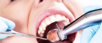
From the first group of diseases we can distinguish caries . If it is not treated and the carious area is not filled, tooth decay is inevitable. Caries is superficial damage to dental tissues, which gradually moves to deeper layers. Over time, if carious cavities are not removed, bacteria reach the “heart” of the tooth – its nerve. This disease is called pulpitis and also leads to tooth loss.
Tooth extraction is also indicated in the presence of jaw tumors - cysts or granulomas . These formations appear, as a rule, as a result of untreated pulpitis and form in the bone tissue near the root of the tooth. If the disease is neglected and treatment is not started at the initial stage, you can easily lose a once healthy, living tooth.
Inflammatory gum diseases are also no less dangerous. These include gingivitis and its more serious stage – periodontitis, which is incredibly dangerous for the condition of teeth. If gingivitis is a slight inflammation of the gums exclusively, then periodontitis is an inflammation of the tissues around the tooth. The disease begins with inflammation of the gums, then continues with the destruction of the bone around the teeth, they become mobile, and then fall out completely. The reasons for the development of periodontitis are: stress, malocclusion pathologies, advanced dental diseases, and most importantly, improper oral care.
The main stages of the development of this pathology
There are three stages of pathology development in which the disease can still be successfully cured. Symptoms of the disease are determined by two factors - the stage of its development and the form of the disease itself.
Table No. 1. Phases of the disease
| Phases | Description |
| First phase | The pathology develops “insidiously”, without clear symptoms. At first you won’t even notice it if you don’t diagnose it by making an appointment with a specialist. |
| Second phase | It is possible that there are symptoms that can easily be confused with a lack of vitamins or simply chronic fatigue. Such as, for example, fragility and hair loss, nail splitting, yellowing of teeth, cramps during sleep, a more frequent pulse than normal. |
| Third phase | When the disease has already entered the incurable phase, pronounced symptoms can be noticed. For example, a painful joint begins to ache almost constantly. Small swelling may also appear there. And physical activity can cause a crunch in it. |
This stage is the last one at which medical assistance can still be provided. If the pathology continues to be ignored, it progresses, becomes neglected, and the patient may even become disabled. The disease can paralyze, deform and enlarge the affected joint, or even the entire rest of the arm or leg.
Dental implants – contraindications
Let's start with the good news: the list of contraindications is shrinking as new protocols are developed literally every year. Therefore, if some disease is now on the list of prohibitions, there is hope that in a couple of years it will not be there. However, it is important to consider contraindications for several reasons:
- Patient's health.
This is a surgical operation in which a foreign object is introduced into the body and provokes a response, so in some pathologies unpleasant consequences can occur.
- Implant rejection.
A long rehabilitation period may end in nothing if the factors that impede successful implantation were not initially taken into account.
Signs of implant failure in the image
It is necessary for the doctor to know the contraindications in order to take them into account during the treatment process. It is also important for the patient to inform the implantologist about his condition. It is dangerous to hide common diseases, because... it can undo all the work.
Features of the symptomatic picture
The symptoms of this disease depend on how widespread it is in the body. The main one is joint pain. They appear on the structure of the bones of the joint. The pathology cannot be noticed with the naked eye for a long time, but diseased bones deteriorate all the time.
And at the same time, with proper vigilance, signs of pathology can be noticed almost from the very beginning. These are the following:
- brittle nails;
- gray and falling hair;
- quickly accumulating fatigue;
- after physical activity, the joints ache and feel discomfort;
- teeth look sick;
- pressure surges;
- paroxysmal increase in heart rate;
- cramps in the leg muscles during sleep;
- hot temper.
Despite the fact that the disease does not manifest itself for a long time, it can be identified by some subtle symptoms
If the pathology is not cured immediately, it makes itself felt in the following ways:
- when moving, joints creak and crunch;
- they have incessant pain;
- the tissues near the joint swell, sometimes the skin turns red;
- stiffness of movement, which is most pronounced after sleep.
During the second degree of pathology, the symptoms of this disease in the affected areas are as follows:
- aching pain;
- crunching sound when moving;
- swelling.
In the third group, the following symptoms of the disease are immediately visible:
- increasing the size of fabrics;
- paralysis of diseased joints;
- changing the shape of the arms and legs.
Due to the gradual decrease in the working capacity of the limbs, up to its complete loss, and, consequently, the loss of the patient’s ability to move and care for himself, this stage is a serious reason for registering a disability for the patient. An advanced disease gradually deforms the joints, causing subluxations to appear in them.
The general form can also be identified by the appearance of the following signs:
- reduction of the patient's height;
- slouch;
- scoliosis.
Severity
There are several degrees of osteoporosis. They characterize the severity of the disease;
- Grade 1. Initial stage, at which there may be no clinical symptoms. Changes in bones can be detected during a random diagnosis for another disease. In some cases, brittle hair and nails and dry skin may occur.
- Degree 2. Destructive changes are isolated in nature. Changes are detected in one part of the skeleton. Most often this is the thoracic spine or lower extremities. Clinically, stage 2 osteoporosis is expressed in pain localized in the affected area. There may be convulsions and rapid heartbeat.
- Grade 3: Major changes are found in the bones. The bones become flattened, and when the spine is affected, a decrease in the height of the vertebrae is observed. In this regard, the patient's height decreases. Patients complain of severe pain and a hump forms on the back.
- Grade 4. The most severe stage of osteoporosis. Radiologically it is expressed in pronounced structural changes in the bones. The patient's height may decrease by up to 10 cm. Fractures occur due to minor injuries.
How can the further development of the disease be prevented?
First of all, you need to consider the following: it is the underlying pathology that should be treated, which is the cause of deformation of the bones near the joints. The main thing is to bring calcium metabolism back to normal, as well as restore bone mineral density.
The process and methods of treatment depend on the following factors:
- How severe is the mineral deficiency?
- how old is the patient from birth;
- what condition is his body in?
- Are there any other pathologies in it at the moment?
When inflammation of the joint reaches an acute phase, accompanied by deformation of the adjacent bones, the patient needs a protective regime, and the suffering limb should never be loaded in any way.
If the joint is fixed in a stationary state, this will make the healing of inflammation faster, and will also speed up the healing of the bones. The immobility of the joint can be ensured by applying plaster and splints.
One of the methods for treating pathology is fixing the affected area with splints, plaster or bandage.
In most cases, improving the condition of bones in such a pathology requires a lot of effort on the part of both parties - both doctors and the patient. The patient should be prepared to carefully carry out every day all the procedures that the doctor prescribes, even taking into account the fact that all of them will require a lot of effort, time and often also money.
The entire treatment complex consists of many stages. They have the following directions:
- relieving the patient of pain, which is achieved by using anesthetic drugs;
- relief from inflammation achieved through the use of non-steroidal antiseptics;
- if necessary, you can also use steroidal anti-inflammatory drugs, but only for a very short time;
- To restore density to the patient’s musculoskeletal system, calciotonin and bisphosphonates are used.
How do atrophic processes occur and what are their negative consequences?
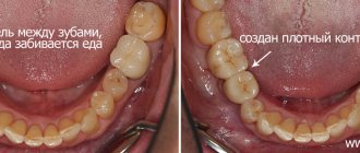
Since there is no pressure on the bone, it shrinks in volume, just like the gums. Moreover, this process occurs regardless of what kind of prosthesis you wore - permanent or removable.
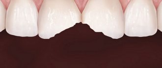
Due to the fact that the bone gradually decreases in size and teeth are completely absent, the overall appearance of a person changes noticeably. Of course, not at all for the better: we can observe sunken cheeks and lips. In addition, facial wrinkles rapidly develop due to the sharp loss of facial muscle tone. Also, the patient cannot avoid a number of problems with the gastrointestinal tract. The patient is forced to adhere to a strictly defined nutritional program, not consuming solid food at all.
A general deterioration in the condition of the body is possible - the development of diseases occurs along a chain, since a limited diet can cause acute vitamin deficiency. What is noteworthy is that bone loss occurs faster when the diet is too poor. Needless to say, the loss of several teeth can lead to the loss of all the others due to their gradual displacement and loosening?
The use of various drugs for periarticular osteoporosis
Conservative treatment of this pathology includes the use of drugs from different pharmacological groups. Treatment of symptoms involves reducing pain, as well as relieving inflammation. This effect is achieved by using the following medications:
- non-steroidal antiseptics, for example, Diclofenac and Movalis;
- analgesics - “Pentalgin” and/or “Baralgin”;
- glucocorticosteroid hormonal drugs, but only in an advanced stage, and for a very short time - “Prednisolone” and its derivatives.
In this case, to carry out blockades inside the diseased joint, the drug is injected directly into its cavity.
Auxiliary techniques
The doctor may prescribe physiotherapeutic procedures. These are electro- and phonophoresis with antiseptic, anesthetic, vascular drugs, the use of therapeutic mud, paraffin baths, magnetic therapy, even quartz treatment. The latter is only dosed.
You should also pay attention to exercise therapy. The entire set of exercises is developed by a doctor who takes into account the patient’s age and the condition of his musculoskeletal system. It also determines how many exercises will be in the complex, as well as how often they should be repeated. Exercises should not tire the patient or give him unpleasant emotions. On the contrary, it will be good if they give him more strength and lift his spirits.
Exercise therapy is an equally effective method of treating periarticular osteoporosis
You can apply lotions of dimethyl sulfoxide (“Dimexide”), medical bile, and gently warm up the sore spot using warming and anti-inflammatory ointments.
When the patient is already reliably on the mend, doctors can recommend the following procedures:
- gentle painless massage with elements of light joint development;
- long-term use of chondroprotectors, for example, “Chondrolon” and/or “Teraflex”;
- courses of calcium supplements, including vitamins and antioxidants.
The complex treatment method also includes a change in diet. It should contain enough of the following products:
- dairy products;
- lean meat;
- fruits and vegetables;
- greenery;
- nutty.
In this case, the patient should eat fractionally, he should eat four to five small portions of food per day. At the same time, the following are strictly contraindicated:
- beer, vodka, wine and other alcoholic products;
- gas. beverages;
- everything is fried and fatty.
You can use it, but only a little:
- coffee;
- cocoa and products containing it (chocolate);
- sweet.
But he should consume as much kelp (seaweed) as possible, since there are a lot of microelements there.
How can teeth be restored after tissue atrophy?
There are 3 proven ways to combat this disease:
- basal type implantation;
- traditional implantation (classical method)
- Implantation All-on-4 or all on six implants with a prosthesis on a titanium bar
In itself, implantation promotes uniform distribution of the load on the bone tissue by securing the implant in it. This process does not cause rejection of the body systems, since implants replace dental roots, exactly copying their role.
Dental implantation with delayed loading
This method makes it possible to restore lost teeth (the entire dentition can be installed). The most important distinguishing feature of the procedure is that in order to secure the implants, it is necessary to first grow the bone by planting donor or artificial material.
Upon completion of the procedure, the bone should take root and adapt to the updated environment. Then the specialist can proceed to install the implant in the traditional way. The implant will take up to six months to fuse with the bone, after which crowns should be installed. In total, implantation using the classical method requires up to one and a half years for complete restoration of teeth.
Momentary load: specifics of the procedure
The most advanced method of implantation today is implantation with immediate loading. The birthplace of this medical achievement is Switzerland. The method makes it possible to fully restore teeth without the need to build up tissue.
In order for the method under consideration to work correctly, special individual designs are used, made for patients with acute atrophy. The process is characterized by a fast and one-step installation - the implants are screwed into punctures in the gums, in places where the teeth have been removed.
Based on the current state of the bone affected by atrophy, a careful selection of implants with a special type of fixation and an improved type of fastening is made. Among other things, in areas of acute tissue atrophy, it is possible to install implants at an angle to achieve increased reliability of fastening.
Already two days after implantation, a metal-plastic prosthesis is installed. This element serves as a finishing structural detail that provides the patient with the ability to fully chew food after 7 days.
This method is good for its specificity and allows patients to quickly restore the original performance of the tissue, and also stimulates regenerative processes in it. Chewability will return gradually.
ethnoscience
To treat this disease, there are also traditional medicine recipes. You can make bones stronger, ease pain, and prevent the development of pathology using the following means:
- mixtures that include eggshells and lemon juice;
- decoctions including parsley and dill;
- wraps with tinctures of comfrey and chamomile made with alcohol.
Some folk remedies will help get rid of discomfort caused by pathology
To normalize calcium levels, you can use the following traditional medicine:
- one piece of egg shell;
- one part propolis;
- two parts honey.
Fry the shells in the oven. Mix all three ingredients. Take the result 1 tsp. three times a day.
And here is another recipe that may be useful in the treatment of periarticular osteoporosis:
- one volume of fresh parsley/dill;
- ten volumes of boiling water.
Finely chop the greens, then pour boiling water over them. All this should sit for at least three hours. Take daily between meals, two to three times a day, one glass at a time.
You can also make lotions from tinctures/decoctions of horsetail, knotweed or comfrey. Use them like this for about 21 days.
What are the obvious advantages of immediate loading implantation?
- no need for bone tissue augmentation. The volume of basal type implants is selected individually. They can be installed at an angle for reliable fixation, regardless of the size of the affected area;
- thanks to an individual approach, the master can select a design according to all parameters. It is also important that with this method the doctor can correctly calculate the interimplant pressure so that they take root as quickly as possible;
- bone tissue actively restores its original appearance - since the chewing load is immediate, this method quickly restores blood circulation, increasing the intensity of regenerative processes;
- guarantee of a positive outcome - due to the instant restoration of bone tissue, implants take root in the shortest possible time. The result will delight the patient with aesthetics, practicality and high functionality just a few months after the procedure;
- high degree of resistance to infections;
- has virtually no contraindications.
Warning
It is easier to prevent any pathology than to treat it, and osteoporosis is no exception. Moreover, preventing the disease is most important for those with a hereditary tendency towards it.
To avoid osteoporosis, it is enough to follow the general rules of a healthy lifestyle:
- include in your menu the maximum number of healthy products listed above;
- regularly give yourself some physical activity;
- take a walk every day;
- exclude from the diet all carbonated, sweet and alcoholic drinks, as well as strong coffee;
- follow the daily routine.
Reasons for women
Osteoporosis is a disease that develops under the influence of many risk factors. There are primary and secondary osteoporosis. Primary osteoporosis develops with age. The reasons are considered to be:
- Complicated family history;
- Mature age;
- Short stature;
- Early menopause and late onset of menstruation;
- Instability of the menstrual cycle;
- Infertility;
- Breastfeeding for longer than one and a half years;
- History of bone fractures;
- Taking hormonal medications;
- Low physical activity;
- Pathology of the endocrine system (thyrotoxicosis, hyperparathyroidism, type 1 diabetes mellitus);
- Excessive drinking and smoking.
Secondary osteoporosis can be caused by the following diseases:
- Itsenko-Cushing's disease and syndrome;
- Thyrotoxicosis;
- Rheumatoid arthritis;
- Diabetes mellitus type 1;
- Conditions after gastric resection,
- Chronic renal failure;
- Chronic liver diseases.
There is a high risk of developing osteoporosis in people taking medications whose side effect is weight loss (immunosuppressants, glucocorticosteroids, anticonvulsants, gonadotropin-releasing hormone antagonists, aluminum-containing antacids).
Smoking, alcohol abuse, caffeine, excess or insufficient physical activity are factors that can cause osteoporosis. The likelihood of developing osteoporosis is higher in people who eat insufficient amounts of calcium, vitamin D, a lot of meat, or cannot tolerate dairy products. Generally recognized factors that increase the risk of developing osteoporosis are the presence of osteoporosis in parents, sisters and brothers, early menopause, and a sedentary lifestyle. Make an appointment
Forecast of treatment results
For a considerable number of people, the diagnosis of “periarticular osteoporosis” is associated with disability. But more than half the success of treatment is determined precisely by the attitude with which the patient takes up the matter and how much effort the patient himself puts in, and not by the doctor and medications. So this pathology should be diagnosed in time so that the joints do not have time to become overgrown with thorns and growths.
It is important to detect the disease in time to avoid dire consequences
As soon as the patient is prescribed medication and rehabilitation, he is obliged to follow each recommendation as clearly and patiently as possible. But if there is even a slight deviation from at least one, only then can a person really become disabled, unable to even get out of bed. The prognosis of treatment is determined by when the pathology is diagnosed and how it is treated.
Complications
According to statistics, osteoporosis is one of the leading causes of disability and mortality. The main complications of the pathology are fractures:
- femoral neck;
- radius bones;
- vertebrae
Bone fractures force patients to remain in bed for a long time.
As a result, the risk of bedsores and pneumonia increases. Severe stages of osteoporosis lead to external physical defects that contribute to impaired self-care. Therefore, it is important to diagnose the pathology in time and carry out correct treatment. If osteoporosis is detected, you must strictly follow your doctor's recommendations. Make an appointment
Answers to questions people ask
What is the specialty of a doctor who specializes in osteoporosis called?
As soon as pain appears in the joints, you need to go to the clinic at your place of residence. There, describe all complaints to the local therapist. He will do an initial examination, after which he will issue directions for tests and x-rays. If osteoporosis, which is a consequence of a joint disease, is suspected, the doctor will prescribe densitometry, as well as consultations with colleagues and specialists in other, narrower areas of medicine. In this case, the patient’s “main” doctors will be rheumatologists. If necessary, he can be examined by an orthopedist or a traumatologist.
First of all, you need to contact a therapist - he will subsequently refer the patient to a doctor with a more narrow specialization
What is the reason that the development of osteoporosis goes hand in hand with rheumatoid arthritis?
There are several reasons for this:
- hormones and immunosuppressants that the doctor prescribes for the treatment of rheumatoid arthritis accelerate the development of osteoporosis;
- hormonal intra-articular blockades, used as painkillers and anti-inflammatory drugs, perform their tasks perfectly, but their side effect is the destruction of bones near the joints, so that they become brittle and brittle;
- Pain and decreased mobility are another reason why joints become weaker.
Is arthritis related to osteoporosis?
Calcium deficiency causes joints to deteriorate faster, making arthritis worse and more difficult to treat.
What is hidden behind the expression “joint replacement ”?
This is the installation of a prosthesis for a deformed joint. It requires a long rehabilitation.
Cheap screening method
In America, it is even recommended that smokers aged 65 years and older undergo a preventive examination using a densitometer (this is an instrument for measuring the optical density of developed photographic materials). However, in countries where this healthcare system is not covered by health insurance, patients are left without the necessary expertise. Many people (especially women) find out about their disease only when the bone tissue is already seriously damaged, for example, with fractures.
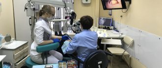
The study, in which researchers wanted to test the benefit of using dental X-rays in diagnosing osteoporosis, included 652 women aged 45-70 years. All women underwent DXA (densitometry) and received a panoramic image of the teeth from the lower jaw. A total of 140 women were diagnosed with osteoporosis by DXA, and more than half of these patients showed early signs of osteoporosis on dental X-rays.
Experts say further research is needed to validate this “screening method” for diagnosing osteoporosis. This is a completely new approach to the patient when the first suspicion of osteoporosis arises when visiting the dentist.