Exostosis of the jaw is a benign formation that manifests itself in the form of an osteochondral growth (osteophyte). The protrusions can be single or multiple and are located on the jaw bone. Maxillary osteophytes most often form on the outer (buccal) surface of the alveolar ridge, and exostosis on the lower jaw - from the inside, on the lingual side. A palatal bone growth (palatal torus) is also found.
Exostoses are painless and do not cause any discomfort at an early stage. But as it increases, the formation begins to cause a lot of discomfort - it complicates eating, affects sound pronunciation, interferes with prosthetics, etc. Treatment of osteophytes is only surgical. Removing exostosis on the gum is a low-traumatic operation that takes about an hour. The operation is performed under anesthesia, so the patient does not experience any discomfort. The method of surgical intervention depends on the location of the osteophyte.
Reasons for appearance
It is not precisely determined why osteophytes appear on the gums. But factors contributing to the disease include:
- Hereditary predisposition;
- frequent inflammation, purulent processes leading to atrophy, deformation of the jaw bone and nearby tissues;
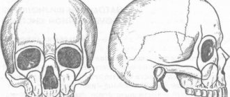
- injuries of the dental system, especially accompanied by fractures of the facial part of the skull with improper reposition of bone fragments;
- complex tooth extraction;
- advanced periodontitis, periodontal disease;
- bite pathologies;
- congenital abnormalities of the jaw.
Often the pathology appears in childhood or adolescence. Also, the appearance of jaw growths may be associated with dysfunction of the endocrine system.
Why do bone growths form on the gums?
Benign formation - exostosis - on the lower jaw
Exostosis in anatomy - a bone located on the outside, a common dental pathology. The neoplasm does not degenerate into a malignant tumor, but requires constant medical supervision. Painless growths in the form of small round or sharp lumps can be located at the base of the tooth, on the palate, or under the tongue. The ICD-10 code is K10.
With exostosis, there is a sensation of a foreign object in the mouth, the mucous membrane changes color. Sometimes the mobility of the lower jaw is impaired, and facial asymmetry is observed.
Reasons for the appearance of the torus of the lower jaw:
- congenital defects of the jaw structure, malocclusion;
- genetic pathologies that are accompanied by impaired bone growth;
- strict diets;
- long-term inflammatory processes, abscesses in the oral cavity;
- injuries and fractures of the jaw followed by improper reposition of broken jaw fragments;
- hormonal imbalance, diseases of the endocrine system;
- periodontal displacement due to improper tooth extraction;
- edentia – partial or complete absence of molars;
- herpes, other viral infections;
- dysplastic growths along the edge of the jaw are a consequence of osteoma of the skull bones.
Lines of small bumps on the upper and lower jaw
Bone spines in the mouth are formed from cartilage tissue, less often affecting the base of the jaw. Externally they look like small white tubercles, similar to foci of inflammation. Most often, several symmetrical growths appear or a line of small neoplasms is formed.
Solitary single osteochondral exostosis is a sedentary neoplasm. Occurs due to injury and inflammation or during puberty. Multiple exostotic chondrodysplasia - numerous spines in different areas. This genetic disorder is most often diagnosed in infants, children, and adolescents.
From a psychosomatic point of view, bone growths in the mouth indicate a fear of losing support or not providing adequate assistance to others. The problem arises among people who underestimate themselves and cannot build their own lives on their own.
Symptoms
Exostosis of the tooth appears in the form of a convex growth that appears for no apparent reason. Main symptoms:
- Sensation of a foreign body in the mouth;
- discomfort when eating, talking (with large osteophytes);
- pain when pressing on the tumor;
- redness, thinning of the mucous membrane in the pathological area.
A small anomaly can only be detected during a dental examination, since visually it does not manifest itself in any way.
Tori of skeletonized maxillae
Of the 600 examined skeletonized maxillae, in 90 (15%) we found tori of various shapes, sizes and locations. The height, length, and width of the palatine ridge were measured. Characteristics of the severity of the bony elevation of the hard palate are given according to a 3-point system.
The torus, which belongs to the first point, is characterized by its small size and height (up to 15 mm). It can be flat, tuberous or pointed ridge and is noted at various parts of the median suture. Such dimensions of the palatal eminence were present in 36 skeletonized upper jaws. Typically, such a bone thickening in the patient’s mouth is detected only by palpation. As a rule, a weakly defined ridge does not cause any particular difficulties during prosthetics. If a slight imbalance of the prosthesis appears, it is easy to correct the base.
With the second score, there is a significant severity of palatal elevation. In this case, the height of the bone protrusion varies between 10-20 mm. Such bony elevations were found on 22 skeletonized jaws. The shape of the torus can be ovoid and is often located in the middle of the hard palate. There are bony protrusions of the main localization on the horizontal plate of the palatine bone with a small transition of the posterior part of the palatine processes along the intersection of the transverse and sagittal sutures of the upper jaw.
At the third point of the torus, a large swelling of the bone was noted. The protrusion covers a large area of the hard palate, reaching a height of more than 20 mm. Large torus sizes were found in 32 skeletonized maxillae.
The results of measuring the length and width of the skeletonized upper jaws and the horizontal plate of the palatine bone made it possible to distinguish them into 3 groups.
Group I included jaws with a short hard palate along the midline: 150 upper jaws, of which 18 were toothless. The main feature of this group of jaws is their short length along the sagittal suture (33-44 mm); there are almost no large bony protrusions. As a rule, the arch of the palate of the first group of jaws is mainly wide-bottomed with a flat surface - a good prerequisite for success in prosthetics.
Group II includes 240 skeletonized jaws of medium size (45-48 mm in length) with different shapes of the roof of the palate, of which 52 are toothless. More often, this group includes jaws with a deep dome-shaped arch of the palate, which is an unfavorable factor for fixing the prosthesis.
Group III consisted of large maxillae (from 45 to 56 mm or more): 210 jaws, of which 30 were skeletonized toothless maxillae. Basically, this group of jaws has unfavorable conditions for the anatomical retention of a removable denture.
In the clinic, bone protrusions are found not only in the middle of the hard palate (torus), but also in the area of the tubercle, the vestibular surface of the sockets of extracted teeth of the upper jaw.
Why should jaw osteophytes be removed?
A bone growth on the gum is not dangerous until it begins to grow. Increasing in volume, the osteophyte puts pressure on the dentition and bone structures. This leads to tooth displacement, malocclusion, and jaw deformation. Large growths impede the movements of the tongue, complicate diction, and interfere with normal chewing of food. Large growths prevent prosthetics and implantation. Osteophyte of the jaw will not disappear on its own. The only effective method of treatment is surgical removal of the pathological formation.
How dangerous are growths on the gums?
A large growth can cause erosion of soft tissues in the mouth.
Due to the absence of pain, elevated temperature, and signs of inflammation, patients consider exostosis to be a harmless disease. But growths can provoke the development of complications.
- As the osteophyte increases, the tongue does not have enough room to maneuver, problems arise with the pronunciation of certain sounds, and the person begins to whistle.
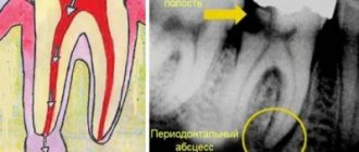
- Due to rubbing of the inner surface of the cheek against the thorn, erosions are formed that do not heal for a long time, phlegmon, abscesses.
- Some osteophytes grow constantly, reaching the size of a chicken egg.
- The functions of the jaw joint are impaired.
- In advanced cases, there is a constant aching pain in the jaw.
A sharp bony protrusion on the upper or lower jaw interferes with prosthetics and leads to permanent destruction of fillings. If the neoplasm passes upward through the cartilaginous plates, chronic rhinitis and sinusitis develop.
There are no specific methods for preventing exostosis. People who have had jaw injuries need to visit the orthodontist 2-3 times a year. It is necessary to regularly examine the mucous membranes of the oral cavity, check the integrity and elasticity of the gums. You should not forget about the rules of dental care, use high-quality toothbrushes and toothpastes.
Indications
- Rapid growth of osteophyte;
- exostosis after wisdom tooth removal;
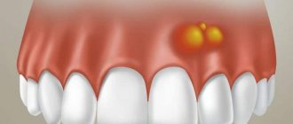
- discomfort, pain;
- the appearance of cosmetic defects on the lower or upper jaw (they look like white balls on the jaw, noticeable when smiling or talking);
- the need for implantation, removable or fixed prosthetics;
- the risk of tumor transformation from benign to malignant.
If you need to install prostheses or implants, exostoses will become an obstacle to the procedure. Dentures will injure the bone growth on the gum, and implants will not be able to take root normally in the bone due to the pressure of osteophytes.
Causes and symptoms of the disease
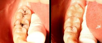
Jaw exostosis often occurs as a result of tooth extraction surgery. If after removal the socket is not smoothed correctly, and the extirpation (complete tooth removal) was traumatic and difficult, bone protrusions-spikes may form as a result. Other causes of the formation of growths include trauma, incorrect alignment of damaged jaw fragments, and old, improperly healed fractures.
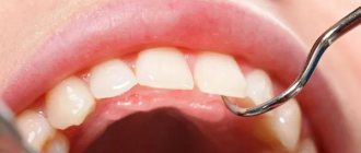
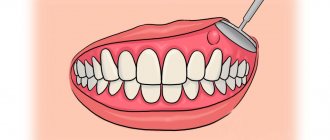
Bone growth is usually detected before prosthetics, when it is necessary to assess how pliable the mucous membrane is and determine the degree of bone tissue atrophy.
A patient with exostosis opens his jaw freely and does not complain of any discomfort. The mucous membrane over the osteophyte tends to have a pale pink color. Usually there are no pathological changes, the mucous membrane remains mobile.
Indications and contraindications
The doctor decides on the need for surgical intervention if the following complaints are present:
- Pain and discomfort.
- Rapid growth of the thorn.
- Bone growth after wisdom tooth extraction.
- Formation of aesthetic defects in the lower or upper jaw.
- The need for implantation and prosthetics.
- Probability of malignancy of formation.
Spikes prevent prosthetics or implantation. Prosthetic structures injure the growth, and implants are not able to take root in the bone tissue due to the pressure of the spikes.
There are two types of contraindications to surgery:
- Relative - manipulation is carried out with increased caution and control. These include: infectious diseases in the acute stage, diseases of the heart and blood vessels, inflammatory processes of the dentition and oral mucosa, pregnancy.
- Absolute – it is prohibited to perform the operation. The list includes: neoplasms, tuberculosis, osteoporosis, pathologies of blood clotting, heart disease, vascular disease and the endocrine system in the acute stage.
General overview, causes and symptoms
Bone tumors are painless and do not cause discomfort at the initial stage. As the growths grow, they begin to cause discomfort to the patient - difficulties arise with chewing food, sound pronunciation deteriorates, it is impossible to install a prosthesis, etc.
The palatal torus is a common pathology in children of the newborn period. As the child grows older, the volume of the growth increases. Treatment of pathology is aimed at eliminating the defect and smoothing the surface.
Removal of exostosis of the jaw is a minimally invasive surgical procedure that takes up to 60 minutes. The manipulation is performed under anesthesia and does not cause any discomfort to the patient. The doctor chooses the surgical procedure based on the location of the osteophyte.
Exostosis is an acquired pathology. Overgrowth of bone tissue occurs due to various injuries:
- After the extraction of the unit. Ignoring the procedure to smooth the edges of the wound leads to the appearance of bone spines.
- Old injuries.
Growths of osteogenic dysplastic origin may appear along the edge of the jaw.