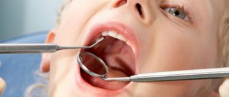
Leptotrichosis is a disease of the oropharynx caused by the bacterium Leptothrix buccalis. This is a conditionally pathogenic microflora, since leptotrichia constantly live in the human oral cavity. In order for the disease to develop, some provoking factor is necessary. Children are very often susceptible to this disease. The bacteria multiply quickly, usually affecting the back of the tongue, but can also develop on other surfaces of the oral cavity.
Causes
The provoking factors causing leptotrichosis are:
- Diseases of the immune system, incl. AIDS.
- Fungal infections, candidiasis.
- Diabetes.
- Diseases of the gastrointestinal tract.
- Diseases of the oral cavity leading to degeneration of the epithelium of the mucous membrane.
- Metabolic disorders, carbohydrate-protein metabolism.
- Lack of vitamins C and B.
- Taking certain medications (corticosteroids, antibiotics).
Insufficient oral hygiene can also provoke the development of the disease. It is especially often the cause of leptotrichosis in children.
How dangerous is leptothrix and where does it come from?
Long-term parasitism of the bacterium on the oral mucosa leads to infection of nearby tissues and the development of leptotrichosis sepsis. Often manifested in the form of oral stomatitis. Some experts consider leptotrichosis to be similar to HIV infection.
The disease was found in 5% of women undergoing gynecological tests - vaginal smears. Recent foreign studies have made it possible to determine the connection between leptotrichia and bacterial vaginosis (vaginal dysbiosis). The established connection suggests that leptothrix is one of the signs of asymptomatic vaginosis in women.
It is quite easy to catch this infectious pathogen. The microorganism is transmitted sexually and domestically: through the water of stagnant reservoirs, insufficiently clean tap water, dirty bed linen, towels, etc. That is, it is possible to infect a sexual partner with leptotrichosis during sex. It is advisable for both to undergo treatment.
There is an opinion that intrauterine devices are a common cause for the development of the disease.
Back to contents
Symptoms
Leptotrichosis has pronounced symptoms:
- White deposits form on the surface of the mucous membrane. As a rule, they are localized on the root of the tongue, tonsils, and dorsum of the tongue.
- Dense plaques develop under the plaque, which are clearly visible if the deposits are removed.
- The patient feels a burning sensation and pain in the oral cavity. Sometimes these symptoms are so severe that it becomes difficult for the patient to speak or eat.
- There are small “tweezers” on the surface of the plaques, which cause a sore throat and a feeling of a foreign body.
Modern aspects of the treatment of inflammatory diseases of the pelvic organs in women
Inflammatory diseases of the pelvic organs are characterized by various manifestations, depending on the level of damage and the strength of the inflammatory reaction. The disease develops when a pathogen (enterococci, bacteroides, chlamydia, mycoplasma, ureaplasma, trichomonas) penetrates into the genital tract and in the presence of favorable conditions for its development and reproduction. These conditions occur in the postpartum or post-abortion period, during menstruation, during various intrauterine manipulations (introduction of intrauterine contraceptives (IUC), hysteroscopy, hysterosalpingography, diagnostic curettage) [1, 5].
Existing natural protective mechanisms, such as anatomical features, local immunity, the acidic environment of the vagina, the absence of endocrine disorders or serious extragenital diseases, can in the vast majority of cases prevent the development of genital infection.
In response to the invasion of a particular microorganism, an inflammatory response occurs, which, based on the latest concepts of the development of the septic process, is usually called a “systemic inflammatory response” [16, 17, 18].
Endometritis
Acute endometritis always requires antibacterial therapy. The basal layer of the endometrium is affected by the inflammatory process due to the invasion of specific or nonspecific pathogens. Endometrial protective mechanisms, congenital or acquired, such as T-lymphocytes and other elements of cellular immunity, are directly related to the action of sex hormones, especially estradiol, act in conjunction with the macrophage population and protect the body from damaging factors. With the onset of menstruation, this barrier on a large surface of the mucous membrane disappears, which makes it possible to become infected. Another source of protection in the uterus is the infiltration of the underlying tissues with polymorphonuclear leukocytes and the rich blood supply of the uterus, which promotes adequate perfusion of the organ with blood and nonspecific humoral protective elements contained in its serum: transferrin, lysozyme, opsonins [16].
The inflammatory process can spread to the muscle layer, and metroendometritis and metrothrombophlebitis occur with a severe clinical course. The inflammatory reaction is characterized by a disorder of microcirculation in the affected tissues, expressed by exudation; with the addition of anaerobic flora, necrotic destruction of the myometrium can occur [12].
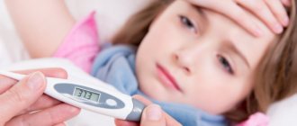
Clinical manifestations of acute endometritis are characterized already on the 3rd–4th day after infection by an increase in body temperature, tachycardia, leukocytosis with a band shift, and an increase in the erythrocyte sedimentation rate (ESR). Moderate enlargement of the uterus is accompanied by pain, especially along its ribs (along the blood and lymphatic vessels). Purulent-bloody discharge appears. The acute stage of endometritis lasts 8–10 days and requires quite serious treatment. With proper treatment, the process ends, less often it turns into a subacute and chronic form, and even less often, with independent and indiscriminate antibiotic therapy, endometritis can take a milder abortive course [5, 12].
Treatment of acute endometritis, regardless of the severity of its manifestations, begins with antibacterial infusion, desensitizing and restorative therapy.
Antibiotics are best prescribed taking into account the sensitivity of the pathogen to them; the dose and duration of antibiotic use are determined by the severity of the disease. Due to the threat of anaerobic infection, additional use of metronidazole is recommended. Given the very rapid course of endometritis, the preferred antibiotics are cephalosporins with aminoglycosides and metronidazole. For example, cefamandole (or cefuroxime, cefotaxime) 1.0–2.0 g 3–4 times a day intramuscularly or intravenously drip + gentamicin 80 mg 3 times a day intramuscularly + metronidazole 100 ml intravenously drip.
Instead of cephalosporins, you can use semi-synthetic penicillins (for abortive cases), for example, ampicillin 1.0 g 6 times a day. The duration of such combined antibacterial therapy depends on the clinic and laboratory response, but not less than 7–10 days.
To prevent dysbacteriosis, from the first days of antibiotic treatment, nystatin 250,000 units 4 times a day or fluconazole 50 mg per day for 1–2 weeks orally or intravenously is used [5].
Detoxification infusion therapy may include the administration of infusion agents, for example: Ringer's solution - 500 ml, polyionic solutions - 400 ml, 5% glucose solution - 500 ml, 10% calcium chloride solution - 10 ml, unithiol with 5% ascorbic acid solution, 5 ml 3 times a day. In the presence of hypoproteinemia, it is advisable to carry out infusions of protein solutions (albumin), blood replacement solutions, plasma, red blood cells, and amino acid preparations [12].
Physiotherapeutic treatment occupies one of the leading places in the treatment of acute endometritis. It not only reduces the inflammatory process in the endometrium, but also stimulates ovarian function. When normalizing the temperature reaction, it is advisable to prescribe low-intensity ultrasound, inductothermy with a high-frequency or ultra-high frequency (UHF) electromagnetic field, magnetic therapy, and laser therapy.
- Nonsteroidal anti-inflammatory drugs (have anti-inflammatory, analgesic effects):
– paracetamol + ibuprofen 1-2 tablets 3 times a day – 10 days;– diclofenac rectally in suppositories or orally 50 mg 2 times a day – 10–15 days;
– indomethacin rectally in suppositories or orally, 50 mg 2 times a day – 10–15 days;
– naproxen 500 mg 2 times a day rectally in suppositories or orally – 10–15 days.
- Preparations of recombinant interferons (have an immunomodulatory, antiviral effect, enhance the effect of antibiotics): interferon a-2b or interferon a 500,000 IU 2 times a day rectally in suppositories - 10 days.
- Interferon inducers (have immunomodulatory, antiviral effects):
– methylglucamine acridone acetate 250 mg intramuscularly every other day – 10 days;– sodium oxodihydroacridinyl acetate 250 mg intramuscularly every other day for 10 days.
is recommended .
- Combined enzyme preparation (has anti-inflammatory, trophic effect): Wobenzym 3-5 tablets 3 times a day.
- Homeopathic remedies (have an anti-inflammatory effect, in combination with other drugs normalizes ovarian function): gynecohel 10 drops 3 times a day.
- Traditional methods of therapy: physiotherapy, herbal medicine, hirudotherapy, acupuncture, physical therapy.
- Methods of gravitational blood surgery: plasmapheresis, endovascular laser irradiation of blood (ELBI), ultraviolet irradiation of blood, intravenous administration of ozonized 0.9% sodium chloride solution.
- Combined oral contraceptives (medium-, low-dose, monophasic) 1 tablet per day - from the 5th to the 25th day of the cycle for 3-6 months:
– ethinyl estradiol 30 mcg + levonorgestrel 150 mcg (rigevidon);– ethinyl estradiol 35 mcg + norgestimate 250 mcg (Sileste);
– ethinyl estradiol 30 mcg + gestodene 75 mcg (femoden);
– ethinyl estradiol 30 mcg + desogestrel 150 mcg (Marvelon).
Additional treatment on menstrual days includes the following.
Tetracyclines (have a wide spectrum of action: gram-positive cocci, spore-forming bacteria, non-spore-forming bacteria, gram-negative cocci and bacilli, chlamydia, mycoplasma): doxycycline 100 mg 2 times a day.
Macrolides (active against gram-positive cocci, gram-negative bacteria, gardnerella, chlamydia, mycoplasmas, ureaplasmas):
– azithromycin 500 mg 2 times a day;
– roxithromycin 150 mg 2 times a day;
– clarithromycin 250 mg 2 times a day.
Fluoroquinolones (active against all gram-positive and gram-negative bacteria): ciprofloxacin 500 mg 2 times a day; ofloxacin - 800 mg once a day for 10-14 days.
Nitroimidazole derivatives (active against anaerobes, protozoa): metronidazole 500 mg 4 times a day.
Antifungal agents (active against fungi of the genus Candida):
– nystatin 250,000 units 4 times a day;
– natamycin 100 mg 4 times a day;
– fluconazole – 150 mg once.
Acute salpingoophoritis
It is one of the most common diseases of inflammatory etiology in women. Every fifth woman who has suffered salpingo-oophoritis is at risk of infertility. Adnexitis can cause a high risk of ectopic pregnancy and pathological course of pregnancy and childbirth. The fallopian tubes are the first to be affected, and the inflammatory process can involve all layers of the mucous membrane of one or both tubes, but more often catarrhal inflammation of the mucous membrane of the tube occurs - endosalpingitis. Inflammatory exudate, accumulating in the tube, often flows through the ampullary opening into the abdominal cavity, adhesions form around the tube, and the abdominal opening of the tube closes. A saccular tumor develops in the form of a hydrosalpinx with transparent serous contents or in the form of a pyosalpinx with purulent contents. Subsequently, the serous exudate of the hydrosalpinx resolves as a result of treatment, and the purulent pyosalpinx can perforate into the abdominal cavity. The purulent process can involve wider areas of the pelvis, spreading to all nearby organs [9, 10, 13].
Inflammation of the ovaries (oophoritis) as a primary disease is rare; infection occurs in the area of the ruptured follicle, since the rest of the ovarian tissue is well protected by the covering germinal epithelium. In the acute stage, swelling and small cell infiltration are observed. Sometimes, in the cavity of the follicle of the corpus luteum or small follicular cysts, ulcers and microabscesses form, which, merging, form an ovarian abscess or pyovarium. In practice, it is impossible to diagnose an isolated inflammatory process in the ovary, and this is not necessary. Currently, only 25–30% of patients with acute adnexitis have a pronounced picture of inflammation; the remaining patients experience a transition to a chronic form, when therapy is stopped after a rapid subsidence of inflammation.
Acute salpingoophoritis is also treated with antibiotics (preferably third generation fluoroquinolones - ciprofloxacin, ofloxacin, pefloxacin), as it is often accompanied by pelvioperitonitis - inflammation of the pelvic peritoneum.
For mild forms, the following is prescribed.
1. Antibacterial therapy orally for 5–7 days.
- A combination of penicillins and b-lactamase inhibitors (have a wide spectrum of action (staphylococci, Escherichia coli, Proteus, Klebsiella, Shigella, gonococcus, bacteroides, salmonella): amoxicillin + clavulanic acid 625 mg 3 times a day.
- Tetracyclines (have a wide spectrum of action: gram-positive cocci, spore-forming bacteria, non-spore-forming bacteria, gram-negative cocci and bacilli, chlamydia, mycoplasma): doxycycline 100 mg 2 times a day.
- Macrolides (active against gram-positive cocci, gram-negative bacteria, gardnerella, chlamydia, mycoplasmas, ureaplasmas):
– azithromycin 500 mg 2 times a day;– roxithromycin 150 mg 2 times a day;
– clarithromycin 250 mg 2 times a day.
- Fluoroquinolones (active against all gram-positive and gram-negative bacteria):
– ciprofloxacin 500 mg 2 times a day;– ofloxacin – 800 mg once a day – 10–14 days.
2. Oral nitroimidazole derivatives (active against anaerobes, protozoa):
– metronidazole 500 mg 3 times a day;
– ornidazole 500 mg 3 times a day.
3. Oral antifungals (active against Candida fungi):
– nystatin 500,000 units 4 times a day;
– natamycin 100 mg 4 times a day;
– fluconazole – 150 mg once.
4. Oral antihistamines (prevent the development of allergic reactions):
– fexofenadine 180 mg 1 time per day;
– chloropyramine 25 mg 2 times a day.
Additional treatments include the following.
- Nonsteroidal anti-inflammatory drugs (have anti-inflammatory, analgesic effects):
– paracetamol + ibuprofen 1-2 tablets 3 times a day;– diclofenac or indomethacin rectally in suppositories or orally, 50 mg 2 times a day – 10–15 days;
– naproxen 500 mg 2 times a day rectally in suppositories or orally – 10–15 days.
- Preparations of recombinant interferons (have an immunomodulatory, antiviral effect): interferon α-2β or interferon α 500,000 IU 2 times a day in suppositories for 10 days.
- Multivitamin preparations with antioxidant effects: Vitrum, Centrum, Duovit, Supradin, 1 tablet for 1 month.
In severe cases, the following groups of drugs are prescribed.
1. Antibacterial therapy orally for 7–10 days. During antibacterial therapy, the clinical effectiveness of the drug combination is assessed after 3 days, and if necessary, drugs are changed after 5–7 days.
- Cephalosporins of the III, IV generations (active against gram-negative bacteria, staphylococci): cefotaxime, ceftriaxone, cefepime 0.5–1 g 2 times a day intravenously.
- A combination of penicillins and β-lactamase inhibitors (has a wide spectrum of action: staphylococci, Escherichia coli, Proteus, Klebsiella, Shigella, gonococcus, bacteroides, salmonella): amoxicillin + clavulanic acid 1.2 g 3 times a day intravenously.
- Fluoroquinolones (active against all gram-positive and gram-negative bacteria):
– ciprofloxacin 1000 mg once a day;– pefloxacin, ofloxacin 200 mg 2 times a day intravenously.
- Aminoglycosides (have a wide spectrum of action: gram-positive cocci, gram-negative aerobes):
– gentamicin 240 mg 1 time per day intravenously;– amikacin 500 mg 2 times a day intravenously.
- Carbapenems (active against gram-positive and gram-negative aerobes and anaerobes): imipenem/cilastatin or meropenem 500–1000 mg 2–3 times daily intravenously.
- Lincosamides (active against gram-positive aerobes and gram-negative anaerobes): lincomycin 600 mg 3 times a day intravenously.
2. Antifungal agents (active against fungi of the genus Candida): fluconazole 150 mg once orally.
3. Nitroimidazole derivatives (active against anaerobes, protozoa): metronidazole 500 mg 2 times a day intravenously.
4. Colloidal, crystalloid solutions (intravenous drip):
– rheopolyglucin 400 ml;
– reogluman 400 ml;
– glucose 5% solution 400 ml.
5. Vitamins and vitamin-like substances (have an antioxidant effect). Intravenous stream or drip in 0.9% sodium chloride solution:
– ascorbic acid 5% solution 5 ml;
– cocarboxylase 100 mg.
Additional treatments include the following.
- Human immunoglobulins - normal human immunoglobulin (contains immunoglobulin G, complements antibacterial therapy for severe infections), intravenously at a dose of 0.2–0.8 g/kg body weight.
- Preparations of recombinant interferons (have an antiviral, immunomodulatory effect, enhance the effect of antibiotics): interferon α-2β 500,000 IU 2 times a day rectally in suppositories - 10 days.
- Interferon inducers (have antiviral, immunomodulatory effects):
– methylglucamine acridone acetate 250 mg intramuscularly every other day – 10 days;– sodium oxodihydroacridinyl acetate 250 mg intramuscularly every other day for 10 days.
- Methods of gravitational blood surgery (have detoxification, immunostimulating, antimicrobial, antiviral effects): plasmapheresis, intravenous administration of ozonated 0.9% sodium chloride solution.
- Laparoscopy, inspection and sanitation of the pelvic cavity, rinsing the pelvic cavity with ozonated 0.9% sodium chloride solution.
Treatment for chronic salpingoophoritis includes the following.
- Nonsteroidal anti-inflammatory drugs (have anti-inflammatory, analgesic effects):
– paracetamol + ibuprofen 1-2 tablets 3 times a day after meals – 10 days;- diclofenac or indomethacin rectally in suppositories or orally 50 mg 2 times a day - 10-15 days;
– naproxen 500 mg 2 times a day rectally in suppositories or orally – 10–15 days.
- Preparations of recombinant interferons (have an immunomodulatory, antiviral effect, enhance the effect of antibiotics): interferon α-2β or interferon α 500,000 IU 2 times a day rectally in suppositories (10 days).
- Interferon inducers (have an immunomodulatory, antiviral effect): methylglucamine acridone acetate or sodium oxodihydroacridinyl acetate 250 mg intramuscularly every other day - 10 days.
Additional treatment is recommended.
- Combined enzyme preparation (has anti-inflammatory, trophic effect): Wobenzym 3-5 tablets 3 times a day.
- Traditional methods of therapy: physiotherapy, herbal medicine, hirudotherapy, acupuncture, physical therapy.
- Methods of gravitational blood surgery: plasmapheresis, ELBI, ultraviolet irradiation of blood, intravenous administration of ozonized 0.9% sodium chloride solution.
- Combined oral contraceptives (medium-, low-dose, monophasic) 1 tablet per day - from the 5th to the 25th day of the cycle for 3-6 months:
– ethinyl estradiol 30 mcg + levonorgestrel 150 mcg (rigevidon)– ethinyl estradiol 35 mcg + norgestimate 250 mcg (Sileste).
– ethinyl estradiol 30 mcg + gestodene 75 mcg (femoden)
– ethinyl estradiol 30 mcg + desogestrel 150 mcg (Marvelon).
Low-dose oral contraceptive drugs normalize the function of the hypothalamic-pituitary-ovarian system. With long-term use, monitoring of hemostasis and liver function is necessary.
- Homeopathic remedies (have an anti-inflammatory effect, in combination with other drugs normalize ovarian function): gynecohel 10 drops 3 times a day.
Pelvioperitonitis
Inflammation of the pelvic peritoneum most often occurs secondary to the penetration of infection into the abdominal cavity from an infected uterus (with endometritis, infected abortion, ascending gonorrhea), from the fallopian tubes, ovaries, from the intestines, with appendicitis, especially with its pelvic location. In this case, an inflammatory reaction of the peritoneum is observed with the formation of serous, serous-purulent or purulent effusion. The condition of patients with moderate pelvioperitonitis, the temperature rises, the pulse quickens, but the function of the cardiovascular system is slightly impaired. With pelvioperitonitis, the intestine remains unbloated, palpation of the upper half of the abdominal organs is painless, and symptoms of peritoneal irritation are determined only above the pubis and in the iliac regions. However, patients note severe pain in the lower abdomen, there may be retention of stool and gas, and sometimes vomiting. The level of leukocytes is increased, the leukocyte formula shifts to the left, the ESR is accelerated. Gradually increasing intoxication worsens the condition of patients [14, 15].
Treatment of salpingoophoritis with or without pelvioperitonitis begins with a mandatory examination of the patient for flora and sensitivity to antibiotics. The most important thing is to determine the etiology of inflammation. Today, benzylpenicillin is widely used for the treatment of specific gonorrheal process, although drugs such as ceftriaxone, perazone, ceftazidime are preferable.
The “gold standard” in the treatment of salpingoophoritis from antibiotic therapy is the administration of cefotaxime at a dose of 1.0–2.0 g 2–4 times a day intramuscularly or 1 dose - 2.0 g intravenously in combination with gentamicin 80 mg 3 times a day (Gentamicin can be administered once at a dose of 160 mg intramuscularly). It is imperative to combine these drugs with intravenous administration of metronidazole 100 ml 1-3 times a day. The course of antibiotic treatment should be carried out for at least 5–7 days and you can vary mainly the basic drug by prescribing cephalosporins of the second and third generation (cefamandole, cefuroxime, ceftriaxone, perazone, ceftazidime and others at a dose of 2–4 g per day) [14].
If standard antibiotic therapy is ineffective, ciprofloxacin is used at a dosage of 500 mg 2 times a day for 7–10 days.
In case of acute inflammation of the uterine appendages, complicated by pelvioperitonitis, oral administration of antibiotics is possible only after the main course, and only if the need arises. As a rule, there is no such need, and the persistence of previous clinical symptoms may indicate the progression of inflammation and a possible suppurative process.
Detoxification therapy is mainly carried out with crystalloid and detoxification solutions in an amount of 2–2.5 liters with the inclusion of solutions of rheopolyglucin, Ringer, polyionic solutions - acessol, etc. Antioxidant therapy is carried out with a solution of unithiol 5.0 ml with a 5% solution of ascorbic acid 3 times a day intravenously [14].
In order to normalize the rheological and coagulation properties of blood and improve microcirculation, acetylsalicylic acid 0.25 g/day is used for 7–10 days, as well as intravenous administration of rheopolyglucin 200 ml (2–3 times per course). Subsequently, a whole complex of resorption therapy and physiotherapeutic treatment is used (calcium gluconate, autohemotherapy, sodium thiosulfate, humisol, plasmol, aloe, fiBS) [3, 15]. Physiotherapeutic procedures for acute processes include ultrasound, which provides analgesic, desensitizing, fibrolytic effects, increased metabolic processes and tissue trophism, inductothermy, UHF therapy, magnetotherapy, laser therapy, and subsequently, sanatorium-resort treatment.
Purulent tubo-ovarian formations
Among 20–25% of inpatients with inflammatory diseases of the uterine appendages, 5–9% develop purulent complications requiring surgical interventions [9, 13].
The following features regarding the formation of purulent tubo-ovarian abscesses can be highlighted:
- chronic salpingitis in patients with tubo-ovarian abscesses is observed in 100% of cases and precedes them;
- the spread of infection occurs predominantly through the intracanalicular route from endometritis (with IUD, abortion, intrauterine interventions) to purulent salpingitis and oophoritis;
- there is a frequent combination of cystic transformations in the ovaries with chronic salpingitis;
- there is a mandatory combination of ovarian abscesses with exacerbation of purulent salpingitis;
- Ovarian abscesses (pyovarium) are formed mainly from cystic formations, often microabscesses merge with each other.
The following morphological forms of purulent tubo-ovarian formations are found:
- pyosalpinx - predominant lesion of the fallopian tube;
- pyovarium - predominant damage to the ovary;
- tubo-ovarian tumor.
All other combinations are complications of these processes and can occur:
- without perforation;
- with perforation of ulcers;
- with pelvioperitonitis;
- with peritonitis (limited, diffuse, serous, purulent);
- with pelvic abscess;
- with parametritis (posterior, anterior, lateral);
- with secondary lesions of adjacent organs (sigmoiditis, secondary appendicitis, omentitis, interintestinal abscesses with the formation of fistulas).
Clinically differentiating each of these localizations is practically impossible and impractical, since the treatment is fundamentally the same - antibacterial therapy occupies a leading place both in the use of the most active antibiotics and in the duration of their use. In purulent processes, the consequences of the inflammatory reaction in tissues are often irreversible. Irreversibility is due to morphological changes, their depth and severity. Severe renal dysfunction is common [3, 9].
Conservative treatment of irreversible changes in the uterine appendages is unpromising, since if it is carried out, it creates the preconditions for the occurrence of new relapses and aggravation of impaired metabolic processes in patients, increases the risk of upcoming surgery in terms of damage to adjacent organs and the inability to perform the required volume of surgery [9].
Purulent tubo-ovarian formations are a difficult diagnostic and clinical process. Nevertheless, characteristic syndromes can be identified.
- Clinically, intoxication syndrome manifests itself in the phenomena of intoxication encephalopathy, headaches, heaviness in the head and severity of the general condition. Dyspeptic disorders (dry mouth, nausea, vomiting), tachycardia, and sometimes hypertension (or hypotension during the onset of septic shock, which is one of its early symptoms, along with cyanosis and facial hyperemia against the background of severe pallor) are noted [4].
- Pain syndrome is present in almost all patients and is of increasing nature, accompanied by a deterioration in general condition and well-being, there is pain during a special examination and symptoms of irritation of the peritoneum around the palpable formation. Pulsating increasing pain, persistent fever with a body temperature above 38°C, tenesmus, loose stools, lack of clear contours of the tumor, ineffectiveness of treatment - all this indicates the threat of perforation or its presence, which is an absolute indication for urgent surgical treatment.
- The infectious syndrome is present in all patients, manifested in most of them by high body temperature (38°C and above), tachycardia corresponds to fever, as well as an increase in leukocytosis, ESR and leukocyte index of intoxication increase, the number of lymphocytes decreases, and the shift of the leukocyte formula to the left increases , the number of molecules of average mass increases, reflecting increasing intoxication.
- Kidney function often suffers due to impaired urine passage.
- Metabolic disorders manifest themselves in dysproteinemia, acidosis, electrolyte disturbances, and changes in the antioxidant system.
The treatment strategy for this group of patients is based on organ-preserving operations, but with radical removal of the main source of infection. Therefore, for each specific patient, both the time of the operation and the choice of its volume should be optimal. Clarifying the diagnosis sometimes takes several days, especially when differentiating it from an oncological process. Antibacterial therapy is required at each stage of treatment [1, 2].
Preoperative therapy and preparation for surgery include:
- antibiotics (use cefoperazone 2.0 g/day, ceftazidime 2.0–4.0 g/day, cefazolin 2.0 g/day, amoxicillin + clavulanic acid 1.2 g intravenous drip once a day, clindamycin 2.0 –4.0 g/day, etc.). They must be combined with gentamicin 80 mg intramuscularly 3 times a day and metronidazole infusion 100 ml intravenously 3 times;
- detoxification therapy with infusion correction of volemic and metabolic disorders;
- mandatory assessment of the effectiveness of treatment based on the dynamics of body temperature, peritoneal symptoms, general condition and blood counts.
The surgical stage also includes ongoing antibacterial therapy. It is especially advisable to administer one daily dose of antibiotics on the operating table, immediately after the end of the operation. This concentration is necessary and creates a barrier to further spread of infection, since penetration into the area of inflammation is no longer prevented by dense purulent capsules of tubo-ovarian abscesses. β-lactam antibiotics (cefoperazone, ceftriaxone, ceftazidime, cefotaxime, imipinem/cilastatin, amoxicillin + clavulanic acid) pass these barriers well.
Postoperative therapy includes continuation of antibacterial therapy with the same antibiotics in combination with antiprotozoal, antimycotic drugs and uroseptics. The course of treatment is prescribed in accordance with the clinical picture and laboratory data; it should not be stopped earlier than 7–10 days. Infusion therapy should be aimed at combating hypovolemia, intoxication and metabolic disorders. Normalization of gastrointestinal motility (intestinal stimulation, hyperbaric oxygenation, hemosorption or plasmapheresis, enzymes, epidural blockade, gastric lavage, etc.) is very important. Hepatotropic, restorative, antianemic therapy is combined with immunostimulating therapy (ultraviolet irradiation, laser irradiation of blood, immunocorrectors) [2, 9, 11].
All patients who have undergone surgery for purulent tubo-ovarian abscesses require post-hospital rehabilitation in order to restore organ function and prevention.
Literature
- Abramchenko V.V., Kostyuchek D.F., Perfileva G.N. Purulent-septic infection in obstetric and gynecological practice. St. Petersburg, 1994. 137 p.
- Bashmakova M. A., Korkhov V. V. Antibiotics in obstetrics and perinatology. M., 1996. P. 6.
- Bondarev N. E. Optimization of diagnosis and treatment of mixed sexually transmitted diseases in gynecological practice: abstract. dis. ...cand. honey. Sci. St. Petersburg, 1997. 20 p.
- Ventsela R.P. Nosocomial infections. M., 1990. 656 p.
- Gurtovoy B. L., Serov V. N., Makatsaria A. D. Purulent-septic diseases in obstetrics. M., 1981. 256 p.
- Keith L. G., Berger G. S., Edelman D. A. Reproductive health. T. 2: Rare infections. M., 1988. 416 p.
- Krasnopolsky V.I., Kulakov V.I. Surgical treatment of inflammatory diseases of the uterine appendages. M., 1984. 234 p.
- Korkhov V.V., Safronova M.M. Modern approaches to the treatment of inflammatory diseases of the vulva and vagina. M., 1995. P. 7–8.
- Kumerle X. P., Brendel K. Clinical pharmacology during pregnancy / ed. X. P. Kumerle, K. Brendel: trans. from English: in 2 volumes. M., 1987. T. 2. 352 p.
- Serov V.N., Strizhakov A.N., Markin S.A. Practical obstetrics: a guide for doctors. M., 1989. 512 p.
- Serov V.N., Zharov E.V., Makatsariya A.D. Obstetric peritonitis: Diagnosis, clinic, treatment. M., 1997. 250 p.
- Strizhakov A. N., Podzolkova N. M. Purulent inflammatory diseases of the uterine appendages. M., 1996. 245 p.
- Khadzhieva E. D. Peritonitis after cesarean section: textbook. allowance. St. Petersburg, 1997. 28 p.
- Sahm DE The role of automation and molecular technology in antimicrobial susceptibility testing // Clin. Microb. And Inf.1997. 3; 2: 37–56.
- Snuth CB, Noble V, Bensch R et al. Bacterial flora of the vagina during the mensternal cycle // Ann. Intern.Med. 1982: 948–951.
- Tenover FC Norel and emerging mechanisms of antimicrobial resistance in nosocomial pathogens // Am. J. Med. 1991; 91: 76–81.
V. N. Kuzmin , Doctor of Medical Sciences, Professor MGMSU, Moscow
Diagnosis and treatment
Despite the pronounced symptoms, visual examination alone is not enough to make an accurate diagnosis. The doctor will definitely prescribe an additional examination of a smear taken from the oral mucosa. The diagnosis is made when leptothrix bacteria are detected in the smear.
In rare cases, leptotrichosis resolves spontaneously. Most often, treatment is required, and the effectiveness of medications varies greatly between patients. As a rule, leptotrichosis is persistent, and complete recovery requires a long time, the patient’s patience and strict adherence to the doctor’s prescriptions. Leptotrichosis can be treated using different methods and their combinations:
- Drug treatment. As a rule, the doctor prescribes a course of anibiotics and, in addition to it, washing the affected areas with antiseptic solutions. Drug treatment is long-term, taking several months.
- Cryotherapy. One of the modern methods, in which the affected areas are frozen. The procedure is performed under local anesthesia. Cryotherapy is most often used as an additional treatment method.
- Physiotherapy. The oral cavity is treated with laser and ultraviolet radiation. As an independent method, physiotherapy is effective only if all plaques are in the irradiation zone. Plaques not irradiated during the session are preserved. Physiotherapy is successfully combined with drug treatment and vitamin therapy.
Combined treatment programs achieve the greatest success. At the first stage, treatment with antibiotics and rinsing the throat and mouth with antiseptic solutions are prescribed. At the same time, treatment of diseases that provoked the development of leptotrichosis is carried out. The course of treatment is long, usually 3-4 months, but in most cases the combined effect gives a good effect.
Features of the pathogen
Leptothrix are gram-negative, filamentous bacteria that do not form spores and prefer an anaerobic environment with very little oxygen (such bacteria are called microaerophilic).
Their name comes from Latin words meaning “fine hair”. In a smear, leptothrix looks like long thin or thick rods, which are arranged in chains resembling hairs. The external resemblance to actinomycete fungi initially led to a classification error: for a long time leptothrix was classified as a fungus.
They are usually grouped under the general name of the genus Leptothrix species (spp.).
Some researchers attribute Leptothrix to a special variety of lactobacilli, modified under the influence of environmental conditions.
They noticed that some women who go to gynecologists with complaints of heavy discharge have bacteria in their smears that form long chains.
In women who do not have symptoms of infection, lactobacilli are grouped in chains ranging from 5 to 15 microns in length. In women with complaints of heavy discharge, the length of one chain can reach 40-75 microns.
Until now, in the medical literature, the names of bacteria leptothrix and leptotrichia are considered synonymous. This is due to the confusion and difficulty of cultivating them.
Attention! In the new classifications, these are two different genera - Leptothrix and Leptotrichia! Be careful, bacteria cause completely different diseases!
Main purposes for the drug Terzhinan
According to the instructions, vaginal tablets are indicated for use:
- for trichomonas and vaginitis caused by pyogenic pathogens
- acute and chronic colpitis of nonspecific or mixed origin, including recurrent ones;
- vaginal candidiasis;
- vaginal dysbiosis;
- ureaplasmosis.
To prevent the development of infectious inflammation in the uterine cavity and adjacent tissues, Terzhinan is prescribed:
- before childbirth;
- insertion of intrauterine devices;
- surgical interventions;
- carrying out abortions, diagnostic curettages;
- before checking the patency of the fallopian tubes.
Medical experience shows that the drug is much more effective than its analogues in treating complicated cases of diseases and rarely causes disturbances in the internal microflora. Terzhinan is better than other intravaginal medications in relieving the symptoms of recurrent thrush and exacerbations of ureaplasmosis.
How is leptotrichosis diagnosed and treated?
The diagnosis is based on a characteristic microscopic picture: the detection of chains of bacteria in the form of a dot-dash. To confirm the diagnosis of a disease associated with Leptotrichia amnionii, culture is used (unlike other leptotrichia, it is not cultured on blood agar) and PCR (16S rDNA). Standard treatment for leptotrichosis has not currently been developed. In typical cases, antibiotic therapy is used. Sensitivity of leptotrichia to beta-lactam antibiotics, clindamycin, metranidazole, tetracycline and chloramphenicol, and resistance to macrolides, aminoglycosides and fluoroquinolones have been noted.
Folk remedies
Leptothrix in a smear (treatment is carried out under the supervision of a gynecologist, therapist, urologist or venereologist) requires a full comprehensive examination and properly selected therapy. In the absence of serious contraindications, auxiliary means can be used.
Recipes from healers and healers are used in complex therapy after consultation with the attending physician. It is not recommended to use folk remedies on your own to avoid complications or an allergic reaction. There is always the possibility of individual sensitivity.
| Name | Recipe | Application |
| Garlic tincture | Chop the garlic, pour 100 g of the product with vodka (1 l). | It is recommended to use the finished tincture 2 times a day, 1 tsp in the morning and evening. for a month. |
| Iceland moss | Pour 1 tbsp. moss with boiling water (1 tbsp.), let stand for 30 minutes and strain. | The resulting product must be taken on an empty stomach, 2 tbsp. 3 times a day. The decoction can also be used for douching or rinsing the mouth. |
| Celandine | Pour dry herb (1 tbsp) with hot water (300 ml), leave for 1 hour and strain. | It is recommended to take 2 tbsp of the finished product. 3 times a day. The warm solution can also be used for douching. |
| Mumiyo | Stir 2 g of product in water (4 tbsp). | It is recommended to take the finished medicine before meals 2 times a day. |
| Herbal collection | Mix 1 tbsp. crushed calamus root, buckthorn, stinging nettle leaves, St. John's wort and thyme. Mix all ingredients well and pour 2 tbsp. herbal mixture with hot water (500 ml). Leave overnight and strain. | The finished product has anti-inflammatory, immunostimulating and antibacterial effects. Adults are recommended to consume 1 tbsp. morning and evening before meals. |
Essential oils have an antibacterial effect (fir, juniper, lemon, lavender). It is enough to dissolve 10 drops in a glass of water and use the resulting mixture for douching.
Forms of leptotrichosis
Considering the area of damage, in medicine the following forms of the disease are distinguished:
| Name | Description |
| Vaginal leptotrichosis | A disease that develops against the background of vaginal lesions with leptotrichia. The pathology does not apply to sexually transmitted infections; infection occurs while swimming in dirty water. Pathogenic microorganisms enter the vaginal mucosa, multiply and provoke inflammation. The anus, labia and perineum swell, and genital itching occurs. |
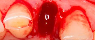
| Leptotrichosis in the mouth | The disease develops in the mouth and is caused by Leptothrix buccalis. The tonsils, tongue, and gums become inflamed, and the crowns of the teeth are destroyed. Oral mucosal lesions in the mouth are often detected in newborns. |
| Choriamnionitis | The pathology is characterized by infection of the walls of the fetal bladder and amniotic fluid. |
Each form of leptotrichosis is accompanied by characteristic symptoms and requires properly selected therapy. In the absence of timely treatment, pathological processes progress and cause serious complications.
How to use Terzhinan
The medication is indicated for use in patients over 16 years of age. Terzhinan is not recommended for adolescents due to the imperfection of the internal vaginal microflora.
- Before use, it is recommended to moisten the tablet in warm water.
- Lying on your back, carefully insert the drug deep into the vagina.
- After administering Terzhinan, do not stand up for 10–15 minutes so that the candle does not accidentally fall out. The best time for the procedure is in the evening, before bed.
General treatment regimen: 1 tablet once every day for 6–20 days. For a preventive course, a week of use is enough. For acute conditions and recurrent pathologies, it is advisable to use the product for at least two weeks in a row. In case of menstrual flow, it is not recommended to interrupt treatment. The exception is an increase in tissue sensitivity and the appearance of irritation of the mucous membrane when exposed to the drug on these days.
During the period of using vaginal tablets, it is important to take good care of hygiene. Due to the specific secretions of the dissolved drug, more frequent changes of underwear are required. It is recommended to use gaskets with a sufficient degree of protection.
Sexual rest during therapy is not necessary, but may be recommended by a gynecologist. It is important to remember that nystatin in Terzhinan may reduce the effectiveness of barrier contraception.
After 5–7 days after the end of the course of treatment, a repeat laboratory test should be done to determine the presence of infection.
Side effects and contraindications
Terzhinan does not cause exacerbations of existing chronic pathologies. Due to the lack of systemic action, it does not interact with other medications. In rare cases, its use may cause itching and irritation of the mucous membranes. The only contraindication is individual intolerance to one or more components. Its signs:
- severe burning sensation;
- redness of the integument;
- swelling of the mucous membrane or severe pain at the injection site.
In isolated cases, Prednisolone in tablets can provoke erosive lesions of the vaginal walls. In addition, this hormone requires careful use of the drug for hypothyroidism, diabetes, and cardiovascular pathologies.