Sometimes situations arise when it is necessary to refill a dental canal. The reasons for such cases may be different. For example, poor-quality root canal filling, incorrect identification of the source of infection during initial treatment. In addition, the structure of the patient’s tooth or a violation of the tightness of the installed filling also affects the result of tooth treatment. We will tell you about the reasons for the need to refill a tooth and the rules for its implementation to obtain a reliable result.
Features of the procedure for re-treatment of a dental canal
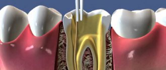
Refilling the canal of a previously treated tooth is a special case in the practice of dentists. Before doing this, it is necessary to remove the old filling, clearing the entire dental canal of the filling material. During such work, the flexible rod may break. This will somewhat complicate the dental treatment process.
Typically, this problem occurs with a very dense filling that is closely adhered to the walls of the canal of the tooth being treated.
Difficulty also arises if it is necessary to refill curved dental canals. When cleaning such channels, you can damage their walls or even break the instrument. Such situations require increased attention from the specialist to ensure high-quality completion of the canal filling. Specialists are required to inform the patient about possible difficulties during treatment before the procedure.
What materials are used to fill canals?
The main requirements for such filling materials are dense, hermetically sealed filling of the canals, chemical inertness (the material should not dissolve under the influence of body fluids), radiopacity (it should be clearly visible in the picture). Today, the following types of materials are used for filling dental canals:
- Solid fillers (fillers).
These include gutta-percha (a latex processing product), silver and titanium pins. Silver pins have recently been rarely used, since despite their good antibacterial properties they have a significant drawback - they do not provide complete tightness. - Polymer and natural pastes (sealers)
. A more preferable option is polymer sealers, which adhere better to the walls, do not stain dental tissue and do not dissolve when interacting with tissue fluids. - Glass ionomer cements.
Good adhesion and radiopacity, high biocompatibility and minimal shrinkage are the main positive qualities of such materials. They also have significant drawbacks: low strength, which is why such fillings are short-lived and not designed for serious functional loads. - Calcium hydroxide cements
. Non-toxic, biocompatible, radiopaque materials that exhibit minimal shrinkage, are easily removed if necessary and have bactericidal properties. However, they are considered not too strong and can break under heavy loads on the dental crown. - Polydimethylsiloxanes
. Modern reliable sealants with good therapeutic and operational parameters. Perhaps their only drawback is that this is a new product on the dental market and experience in using such materials has not yet been accumulated. There is no reliable information yet about the experience of patients after such treatment.
Why undergo a dental canal refilling procedure?
There are several reasons why you need to re-treat a dental canal:
- persistence or resumption of pain after filling;
- X-ray shows the presence of inflammation;
- incomplete closure of dental canals.
In addition, complications may result from errors or inaccuracies that arose during treatment. For example, when preparing the canal for treatment, an infection was introduced, access to the base of the canal is impossible, or there are pathological holes in the walls of the tooth or its bottom. In addition, no one is immune from the mistakes of the doctor performing root canal treatment.
When treating a dental canal before filling, the following problems most often occur:
- Dentim gets into the sawmill,
- the middle part of the canal is greatly expanded,
- tool failure,
- the integrity of the root walls is compromised.
When filling a canal, the main problems arise if it is not completely filled, the filling material extends beyond the hole, or there is a longitudinal fracture at the root of the tooth. Medical errors are also possible at this stage - the doctor may incorrectly estimate the length of the canal or not completely clean it. This may contribute to the development of inflammation.
Preparation stages:
- Treatment of carious cavities is a necessary procedure in order to avoid infection getting to the root. This also allows access to estuaries.
- Pulp removal - this procedure can be carried out using several methods, most often it requires two visits. At the first stage, the pulp is necrosis; at the second, it is directly removed.
- Determining the length is a particularly important stage, since each tooth in an individual patient has its own individual characteristics, both length and relief.
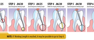
- Processing with mechanical instruments - at this stage, the channels are passed through to expand them. This is the main step towards quality filling. A canal that is not completed to its full length can never be adequately healed.
- The final stage is filling with gutta-percha.
It is important to take into account that the last stage directly depends on the previous ones, and if all standards are not followed, it cannot be carried out.
Channel length measurement
Root canals must be filled exclusively to the apex of the root. What happens if you ignore this point?
The canal will not be completely sealed; unfavorable microflora will begin to multiply in it. This will further lead to inflammation, complex periodontitis and then the tooth will need to be removed.
It will be over-sealed - in this case, there may be various complications. If it is a tooth on the upper jaw, gutta-percha can enter the nasal sinus - this will be odontogenic sinusitis. Another complication may arise due to damage to the nerves - neuralgia, severe pain, and subsequently inflammation.
Therefore, through trial and error, the dentist is obliged to take measurements from beginning to end.
Mechanical restoration
The next stage is mechanical processing of the cavity. The main task is to make the tooth suitable for further filling. To do this, it is necessary to remove all narrowings and expand the channel as much as possible. This is done in two ways.
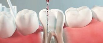
In interaction with the endodontic handpiece - the so-called Pro-files. These devices rotate in the cavity of the root canal, thereby removing all unwanted tissue and expanding it.
With the help of a hand instrument, the dentist independently rotates such instruments in the tooth.
Gutta-percha filling
The final stage is filling with gutta-percha.
There are several methods for this:
- Thermofil system - filling is carried out with heated material, and already in the canal it begins to harden. This is a highly effective method, but it also has disadvantages. This is a difficult procedure to carry out, expensive and requires high professionalism of the doctor.
- Single paste method - this method involves filling the canal with a plastic material. To date, a more dangerous filling method is not yet known, since complications occur in almost every case.
- Lateral condensation method - this technique is the safest and allows you to get good results. In this case, the canals are filled very tightly with gutta-percha, thereby guaranteeing that the entire canal is filled.
In case of pulpitis and periodontitis, the quality of treatment is assessed not only by the patient’s complaints. In addition to pain, a feeling of fullness and discomfort, a control image can tell a lot.
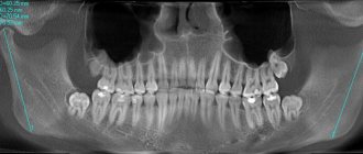
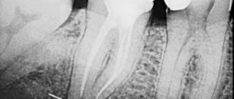
A mandatory conclusion of dental filling is radiographic control. It is carried out both before the procedure, during the treatment process, and after filling. In the picture you can see the quality of the work performed, and if necessary, re-sealing occurs.
How does the process of refilling dental canals take place?
The current level of development of medicine in the field of dentistry is quite high. But dentists make mistakes, which is why filling dental canals again is quite common. At this point in time, doctors use several methods for performing this procedure.
Mechanical methods involve the use of a special instrument - an endodontic motor and an apex locator. In this case, antiseptic agents are added to the filling material. To prevent the development of inflammation, the doctor must very carefully and completely fill the dental canal with filling material.
Medicinal methods involve the use of products with organic solvents. They are able to soften and destroy the filling in a few minutes.
The cement filling can be removed from the canal using ultrasound and an endo-nozzle. This canal cleaning is usually carried out in one procedure.
How can you make sure that the canals are sealed correctly?
There are only two ways.
1. Focus on your own feelings.
You should be wary:
- pain that does not go away after several days;
- excessive tooth sensitivity and discomfort while eating;
- any changes in the color and density of the oral mucosa.
However, pain may be an individual reaction and a variant of the norm, so be sure to consult a doctor. And best of all→
2. Take a control x-ray.
From the image, you can even independently determine how well the dentist did his work, whether there are voids or excess material in the canals. It is clearly visible in the photographs: the sealed canals have a bright white color.
And to finally calm down, come for a free consultation at our clinic with pictures. It’s possible without them - after all, we also have our own state-of-the-art X-ray machine, with which we can check the quality of the previous treatment in a few seconds!
Or immediately in order to fill teeth using 100% reliable methods, using modern equipment and an experienced doctor.
Modern technologies in the field of tooth refilling
Dental treatment under a microscope is one of the newest services that has appeared in dentistry. High-tech equipment allows specialists to fill the dental canal with better quality, more accuracy and efficiency. Thanks to the use of such a device, the likelihood of causing harm to healthy tissue is close to zero. In addition, the treatment is carried out very delicately. Microscope glasses allow you to view the tooth at multiple zooms. This will allow you to see all the affected areas that may simply not be visible during normal examination.
A microscope allows dentists to perform root canal treatment faster and safer for the patient.
Why does a tooth hurt after filling and what to do at home
The situation when a tooth hurts under a filling is common. It is not always possible to see a dentist right away. You have to dull the pain with painkillers or traditional medicine. To be able to help yourself, you need to understand the causes of pain.
Dentists have some criteria by which they determine whether pain after tooth filling is normal or not. The nature of the sensations, intensity, duration, frequency, etc. are taken into account. Patients can also understand the causes of pain by the nature of the sensations and accompanying symptoms. And some causes of the disease may mean that the dentist needs to be changed.
Removing the seal under the anchor pin
Obturation of the tooth root canal is done in parallel using anchor pins. If the filling in the lower part of the root is placed correctly and hermetically, then it is left to install the pin structure. This procedure is carried out over two trips to the dental office.
On the first day, the doctor treats the dental canal mechanically, softens the filling with medications and cleans the passage with an endo-instrument. The doctor then soaks a cotton swab in the medicine and inserts it into the canal. The patient takes the medicine for a couple of days and then makes another visit to the doctor, during which the cotton swab is removed and the canal is refilled.
The nerve was removed, but the canal was not sealed
Such situation. On the X-ray of my teeth, they saw that in my six, in which the nerves were ripped out, only one canal was sealed. My employee’s nerve was removed and the canal was also not sealed - the filling was only placed on top. The fillings are not temporary, but permanent. It turned out to be completely random - during a check when another tooth (mine) was hurting. And the employee was sick after that, the hollow one.
Is there any reasonable explanation for the fact that the nerve is removed, but the canal is not filled and the filling is left only on top, or is this real negligence?
There are several options. Possibly negligence. But if one channel is sealed, then most likely there is another option. I haven't completed channels 5 and 6 yet. This turned out to be the case in the USA. The previous dentist in the USA turned a blind eye to this. We moved and took the new one to a specialist to do a refilling. Well. I did it in a cool place, with a cool specialist, a professor. I paid more than 3 tons of dollars for two teeth (5 and 6 on the lower left). And still, the channels have not been completed completely. He told me that you have very narrow and winding canals, plus 4 roots. The Russians used a different technology to fill the filling, but it surprisingly didn’t work too badly. Don't touch your teeth anymore. If inflammation occurs, you will have one way out - open the gum, bite off the tip of the root and fill it at the other end.
