Treatment of the upper chewing teeth is associated with the risk of damage to the maxillary sinuses due to their close anatomical proximity. When treating root canals, filling material may end up in the sinus cavity, which entails the development of serious purulent processes. Our Center has been specializing for many years in providing assistance to patients who have become victims of unsuccessful therapy. Such situations are corrected by experienced maxillofacial surgeons with ENT training using modern gentle surgical techniques
Causes and consequences of filling material in the maxillary sinus
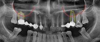
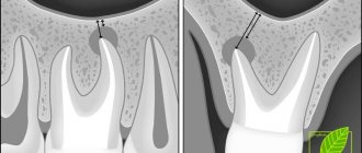
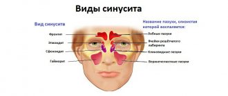
In the maxillary region there are the maxillary sinuses, which occupy most of the internal space. Sometimes they are separated from the upper chewing teeth only by a thin bone septum (bottom of the sinus). During endodontic treatment, especially if the roots of the tooth have grown into the sinus cavity, the dentist may not calculate the effort, damage the apex of the root and the bottom of the sinus, as a result, the filling material falls into the sinus, “taking” purulent masses with it.
It is good if the problem is identified immediately and the patient is referred to a maxillofacial surgeon for immediate removal of the material. Otherwise, inflammation occurs in the sinus and the person comes for help himself, but with consequences.
Long-term presence of filling material in the maxillary sinus is dangerous due to the development of complications:
- chronic odontogenic sinusitis and sinusitis;
- mycetoma (fungal infection of the nasal sinuses);
- inflammatory processes on the roots of adjacent teeth;
- encephalitis and meningitis;
- osteomyelitis of the jaw.
Treatment procedure
Regulations for actions during treatment of a pathological condition:
- The foreign body must be removed; this is the main task of treatment.
- With such a lesion, inflammatory processes regularly develop. They are generated not only by filling material that has found its way into an unusual place, but also by oral microflora that has penetrated into the resulting hole or by swelling from inflammatory processes in the gums. They need to be cleaned.
- The patient needs to undergo treatment of both the maxillary cavities and the oral cavity, eliminating the causes of the disease.
Removal of a foreign body always occurs only through surgery. In this case, the cavity is penetrated through a hole and the object is directly removed. There are two such methods:
- laparoscopic;
- endoscopic.
The first method is more traditional. In this case, an incision is made between the upper lip and gum and the material is removed through the resulting hole. In the second case, the paranasal sinus is punctured in the place where the bone is thinnest and a foreign particle is captured through the resulting hole with a special tool and then brought out.
The operation is performed under local anesthesia and lasts no more than thirty minutes. The second method is most widespread. Laparoscopy is usually used in situations where, for some reason, endoscopy is not recommended.
After this, drug treatment of the inflammatory process is carried out. For this, immunostimulants or antibiotics, as well as other medications, can be used. Additionally, antibacterial solutions or an antiseptic are used to wash the damaged paranasal sinus.
Vitamin complexes are also used in treatment.
Symptoms of complications
A person feels the presence of a foreign body in the sinus 2-3 days after treatment due to the characteristic symptoms of sinusitis. In some cases, it may not manifest itself for months or even years with a strong immune system, but makes itself felt when the body’s defenses are weakened. Also, the rate of development of the pathological process depends on the composition of the filling material that ends up in the sinus, the presence of components in it that can provoke inflammation, and the growth of fungal flora.
Characteristic symptoms:
- chronic nasal congestion;
- aching headaches;
- pain in the upper jaw, worsening with chewing;
- pain when lightly tapping the facial bone under the eye closer to the nose;
- thick purulent nasal discharge with an unpleasant odor;
- increase in body temperature in advanced conditions.
The difference between sinus inflammation due to the presence of filling material in it and sinusitis-sinusitis that accompanies influenza and colds is a one-sided manifestation of symptoms. Signs of odontogenic sinusitis are usually only on the side on which the tooth was treated.
Consequences
Finding a foreign object inside the maxillary cavity is very dangerous due to its possible consequences. Doctors include acute symptoms:
- Sinusitis in bacterial or fungal form.
- Encapsulation of material in the form of a cyst or benign disease; often in such situations, an exacerbation is diagnosed; it turns into a swollen form.
- Sometimes granuloma formation occurs.
Minor symptoms:
- Meningitis disease.
- Cold-related inflammatory disorders of various types.
- Hypertrophy of the mucous membrane is possible; the pathology is accompanied by impaired nasal breathing.
- The source of inflammation can transmit coccal infection to other parts of the body. One result may be myocarditis of the heart muscle.
- Other complications.
Taking care of your dental health is the best prevention. In addition, you need to avoid facial injuries, not subject your body to severe stress, organize a healthy diet and lead an active lifestyle.
Regular dental checkups will allow you to fully monitor the health of your teeth.
How to warn
Most often, underexamined patients encounter perforation of the maxillary sinus during dental treatment, when the doctor, without a clear understanding of the location of the roots, carries out treatment according to the standard protocol.
Rest assured that in our Center you will not encounter such risks.
The following allows us to prevent the penetration of filling material into the maxillary sinus: CT diagnostics before the start of tooth treatment.
Based on high-precision data, the doctor’s actions are planned and a safe endodontic protocol is selected.
Unfortunately, x-rays, which most clinics resort to due to the lack of expensive equipment, do not provide a complete picture. In our Center, the examination is carried out using a modern CT scanner Sirona Gallileos (Germany) with advanced settings in ENT mode. Such 3D images allow you to study in detail the location of the maxillary sinuses and dental roots, calculate the thickness of the bone septum, and determine the presence of dental cysts. Qualified endodontists
Treatment of teeth bordering the maxillary sinuses should be extremely careful, careful and meticulous. A doctor without experience, sufficient theoretical knowledge and practical skills to work in emergency situations is likely to make mistakes. In our Center, endodontic dental treatment is carried out by specialists with at least 10 years of experience; we do not hire students “off the street” or students immediately after graduation. Minimally invasive techniques for canal treatment under a microscope and 3D filling eliminate the risk of damage to the root canals and sinuses.
CT monitoring after treatment is also required . Before sending the patient home, the doctor must make sure that there is no perforation of the maxillary sinus, no dental material has entered it, and also evaluate the quality of canal filling. If pathological aspects are detected, the patient will have the opportunity to receive immediate help and avoid complications.
In case of unforeseen situations, when the bottom of the maxillary sinus is damaged and filling material gets into it, the patient should be immediately referred to a maxillofacial surgeon to remove the foreign body. Failure to provide timely assistance is fraught with the development of an acute inflammatory process with all the ensuing consequences .
Reasons for appearance
Many patients do not understand how filling material intended for use in dental treatment gets from the gums into the paranasal cavities?
Medicine knows cases of penetration of foreign substances under the following conditions:
- prosthetics;
- installation of seals.
Sometimes the walls between the gums and sinuses are quite thin, so pieces of filling material can easily get into the tissue through their damage. Injuries often occur during the dental treatment process.
The bone at the treatment site can be very thin, and in this case the roots will be located close to the paranasal cavities.
If there is not enough bone tissue at the treatment site, then in some cases the dentist artificially builds up the material. This is necessary for reliable dental treatment. In this situation, damage to the walls and penetration of a piece of dental substance into the sinuses is also often observed.
Filling material, once in the gum layer, often causes pain; a person feels severe pain while chewing food, discomfort after touching the damaged area.
The case when a foreign substance does not cause any reaction in the body is quite rare, but sooner or later they end in edema.
Why is it better to contact the ENT department of dentistry?
If you still find yourself in a difficult situation after unsuccessful dental treatment, you can only eliminate the consequences with a guarantee from an oral and maxillofacial surgeon with ENT training.
Otolaryngologists and ordinary dentists have a fundamentally different approach to the issue of diagnosis and treatment of diseases of the paranasal sinuses of odontogenic (“dental” origin). Each of them solves the problem only in its own part, while it requires an integrated approach.
The complexity of interaction between these two specialists in city clinics, the reluctance of each to delve into the problems of related specialties, and the lack of joint consultations lead to errors in diagnosis and choice of treatment tactics. As a result, the patient goes in a vicious circle from one doctor to another to no avail.
ENT dentistry has a number of undeniable advantages in this regard.:
- an integrated approach to solving the problem;
- accurate diagnosis using CT in ENT mode;
- treatment is carried out by a specialist equally knowledgeable in dentistry and otolaryngology;
- Carrying out gentle dental operations without mandatory hospitalization.
In our Center, operations to eliminate combined dental and ENT pathologies are performed by experienced surgeons with ENT training, candidates of medical sciences.
KolchinSergey Alexandrovich
Maxillofacial surgeon, 7 years of experience
Surgeon with ENT training. He specializes in ENT dentistry, including performing endoscopic operations and carefully removing tumors in the sinuses.
More about the doctor
Introduction
Endoscopic surgery of the maxillary sinus (MSS) has already become an integral part of the professional life of an otolaryngologist. Gradually, this approach is replacing traditional methods of surgical treatment of intracranial sphincter in maxillofacial hospitals.
An undeniable advantage of endonasal treatment of maxillary sinusitis is its greater physiology compared to intraoral approaches. In addition, maintaining the continuity of the oral mucosa and the integrity of the bone of the anterior wall of the upper jaw is an important factor for the successful implementation of dental implantation in the upper jaw in the future.
One of the reasons for the development of maxillary sinusitis is foreign bodies [1]. They can have different origins (traumatic, dental, fungal), be represented by various materials (filling material, impression mass, tooth fragments, dental implants, glass, iron, wood, bullets, etc.) and enter the sinus cavity in various ways (as a result of trauma, dental treatment, gunshot injury, etc.). In addition, as a result of the activity of different types of fungi (for example, Aspergillus spp.), fungal masses or bodies can form in the sinus cavity [2], which are also regarded as sinus foreign bodies. In some cases, such bodies contain areas of calcification and fragments of filling material.
In addition, foreign bodies in the maxillary sinuses are often an incidental finding during preparation for sinus lift surgery. All patients undergoing treatment by an implant dentist are required to undergo cone beam (CBCT) or multislice (MSCT) computed tomography (CT).
The actual diagnosis of foreign bodies in the maxillary sinuses involves a careful collection of anamnesis, especially dental history, and radiological diagnostics. The diagnostic method of choice for this nosology is CT (CBCT and MSCT) [3]. It is this type of instrumental examination that allows you to see the foreign contents of the sinuses in three-dimensional space.
There are several approaches to the upper nasal cavity using the endoscopic endonasal approach: prelacrimal, through the lower nasal meatus, through the middle nasal meatus, in the area of the natural anastomosis. Currently, there is no clear guidance on the choice of approach and access to HCP. As a rule, the surgeon is based on his own experience and the equipment of the clinic. The method of access to the sinus should depend not only on the availability of the necessary equipment, but also on the localization of the pathological process in the sinus and its nature. The presence of a foreign body in the projection of the natural anastomosis implies access to the sinus in the area of the natural anastomosis. The location of the pathological contents in the area of the bottom of the upper nasal sinus makes it possible to perform a maxillary sinusotomy using access through the lower nasal meatus.
The Caldwell-Luc intraoral approach will be a good option in cases of large foreign bodies in the upper jaw and the impossibility of fragmenting them [4, 5]. However, this technique is now used less and less due to improvements in endoscopic technologies and instruments.
An additional modern method of intraoperative control is navigation equipment. In world practice, in cases of foreign bodies of the maxillary sinuses, navigation equipment is rarely used. This is due to the fact that the technique is relatively new and was developed primarily for the work of the neurosurgical operating room. The modern navigation station was developed in the 1990s. The widespread introduction of equipment into clinics occurred in the early 2000s. Currently, there are two types of navigation stations: electromagnetic and optical. Both types of stations have their own list of advantages and disadvantages [6].
For intraoperative navigation, preoperative MSCT or CBCT is necessary. Both types of tomography are suitable for use in navigators. This allows you to control the movements of the instrument in the surgical field.
Indications for the use of navigation control are foreign bodies located under the mucous membrane, fixed in polyps, cysts, etc., the location of foreign bodies in bays, in the area of the sinus septa, during repeated surgical interventions on the sinuses [7].
In the postoperative period after endonasal maxillary sinusotomy, as a rule, there is no need to prescribe systemic antibacterial therapy. To prevent postoperative infectious and inflammatory complications, patients are prescribed local antimicrobial therapy with Isofra for 5-7 days, 1 injection into each nostril 4 times a day. Isofra is an antibacterial nasal spray with the antibiotic framycetin with a broad spectrum of action against the main pathogens of upper respiratory tract infections, including strains of staphylococcus resistant to penicillins and other antibiotics. Currently, there is no registered decrease in the effectiveness of Isofra, and no resistance has been identified. It is important that the drug does not have systemic absorption, which ensures a safe topical effect at the site of drug administration and eliminates overdose.
In cases of more pronounced symptoms of difficulty in nasal breathing, patients are prescribed the combined drug Polydex with phenylephrine, 1 injection into each nostril 3 times a day for 5-10 days. Polydexa with phenylephrine is the only nasal spray that has a unique combination of two antibiotics, anti-inflammatory and vasoconstrictor components. Two antibacterial components in the drug, neomycin and polymyxin B, complement each other and provide a powerful bactericidal effect against pathogens of acute and chronic diseases of the upper respiratory tract, including Pseudomonas aeruginosa. The dehydrated form of dexamethasone - dexamethasone sodium metasulfobenzoate - eliminates inflammation and swelling of the nasal mucosa without the risk of systemic glucocorticosteroid action. Phenylephrine is a vasoconstrictor that improves breathing and delivery of active components to the site of inflammation.
Both drugs have an individual bottle shape that has no analogues in Russia. A distinctive feature of the bottle is a one-piece body with a spray nozzle, entirely consisting of high quality polyethylene, without metal elements in the design inherent in other sprays. High dispersion rates of the spray cloud and small fraction sizes of the medicinal substance throughout the entire time of spraying of the sprays help to increase the efficiency of delivery and action of drugs in patients in the postoperative period.
The possibility of local use of antibacterial sprays Isofra and Polydex with phenylephrine makes it possible to reduce the time of rehabilitation of patients in the postoperative period and, in most cases, to refrain from using systemic antibacterial drugs [8].
Diagnosis of complications in the maxillary sinus
Diagnostic measures are aimed at determining the size and localization of dental material, the degree and prevalence of inflammation of the mucous and bone tissues of the sinus.
- Visual inspection Performed using a nasal speculum, it allows you to determine the presence of a foreign body in the nasal sinus.
- Computed tomography in a special ENT mode is the most accurate type of diagnosis of combined dental and ENT pathologies today. The advanced settings of our tomograph allow us to evaluate the maxillary sinuses in full. This allows the maxillofacial surgeon not only to determine the presence/absence of a foreign body, but also to assess the condition of the mucous membrane, determine the amount of purulent masses, and the extent of inflammation.
A 3D tomogram gives a complete picture of the condition of the maxillary sinuses, the location of the filling material, and the degree of inflammation
Diagnostics and its features
The most effective way to make a correct diagnosis is computed tomography, but this does not exclude the use of other, more traditional methods:
- conducting a patient interview;
- use of rhinotherapy to study the maxillary sinus;
- X-ray examination;
- MRI;
- puncture of the maxillary sinus.
An experienced doctor can reliably assume the presence of such a disease, but it can be confirmed using computed tomography.
Stages of removing material from the sinuses
Treatment at the Center is comprehensive; we try to combine all procedures in one visit
- Preparation It is important to create sterile conditions to prevent infection of the surgical wound during the intervention. Hygienic cleaning of the oral cavity and treatment of compromised teeth are carried out.
- The operation is performed according to the selected protocol in the operating room of the ENT department. The patient is in a state of drug-induced sleep. After the operation, a CT scan is repeated to assess the quality of the work performed.
- Prosthetics If it is not possible to preserve the causative tooth, digital impressions are immediately taken and a temporary immediate prosthesis is made to hide the aesthetic defect until permanent prosthetics are installed.
To put you to sleep, we use safe ultra-thin sedatives - Propofol or Diprivan. We do not use general anesthesia, since it has a severe effect on the body and is associated with certain risks. Our full-time anesthesiologist monitors the patient’s well-being during the operation. Upon completion of treatment, a person wakes up rested, full of strength and energy, and within half an hour after drinking tea he can return home.
Symptoms of perforation of the maxillary sinuses
Damage to the maxillary sinuses during tooth extraction can be diagnosed if the patient has:
- blood with air bubbles released from the tooth socket;
- the appearance of blood from the nose on the side of removal;
- the appearance of “nasality” in the voice.
If this damage occurred during endodontic treatment or implantation, the doctor can diagnose it based on the following signs:
- failure of an instrument or implant into a tooth socket;
- the appearance of air bubbles in the blood;
- changing the position of the instrument when working in a wound.
Recovery period
Recovery after surgery is the most unpleasant moment for patients. We made sure that this period was as short and painless as possible
No hospitalization
The use of gentle surgical techniques using an ultrasound protocol and microscopic equipment allows us to carry out treatment with the utmost delicacy and minimal trauma. Coming out of sedation is not accompanied by the consequences of general anesthesia; you will not be haunted by nausea, dizziness and clouding of consciousness. Therefore, hospitalization and hospital stay for 3-4 days will not be required ; 30 minutes after the operation you will go home.
For patients with cardiovascular pathologies who require postoperative observation, the Center operates a day hospital department.
A set of procedures for accelerated rehabilitation
For quick recovery after surgery, our Center offers its own method of accelerated rehabilitation. The procedures are aimed at preventing swelling, hematomas, and eliminating pain. The program includes:
Home care
You will receive at home all the medications you need for recovery and instructions for taking them free of charge. In this way, we prevented the purchase of counterfeit products, and you will not need to run to pharmacies on your own after the operation.
The medication kit, selected by a doctor, includes all the necessary medications to improve your well-being and prevent complications.
Please do not violate the frequency of use and follow the recommendations from the leaflet that you will receive along with the medications.
If you have any questions, please contact the phone number listed in the brochure. The postoperative patient support service operates around the clock 24/7.
Treatment of damage to the maxillary sinuses
Treatment is determined by the degree of damage, the presence of foreign bodies in the cavity, as well as the speed of diagnosis and initiation of treatment. The only non-surgical treatment is one that was diagnosed at the time of tooth extraction and does not have signs of infection or the presence of foreign bodies in the sinus. In this case, the doctor does everything to keep the blood clot that closes it in the tooth socket and prevent it from becoming infected. For this purpose, a gauze swab soaked in iodine solution or a special plastic plate is placed in the hole; in rare cases, sutures are required.
These dental procedures are performed in combination with a course of antibiotics, drops with a vasoconstrictor effect and anti-inflammatory drugs. If a foreign object gets inside the maxillary cavity, treatment is carried out surgically through opening and removal of the foreign object and non-viable tissue. The specialists of the Implantmaster clinic not only effectively treat such injuries, but also do everything possible so that their patients know about them only by hearsay.