Full text of the article:
Working with diseases of the internal organs is especially difficult because they cannot be seen. Previously, doctors had to treat patients literally “blindly,” since it was possible to examine a person’s internal organs only during surgery. Nowadays, doctors don’t have to pick up a scalpel; various types of scans help make a diagnosis. However, patients are wary of this type of examination. This is due to the high cost of some procedures and the fear of radiation. Let's try to figure out what the features of certain scans are and when to resort to them.
X-ray
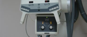
The oldest and most common method of visualizing the human body. X-rays are used everywhere, from surgery to dentistry. The method is simple and clear: a person is irradiated with special rays that easily pass through soft tissues and linger in hard ones. Thanks to this principle, an image is transmitted to a photographic film or sensor located on the side opposite the ray source, and radiography or fluoroscopy is available to the doctor.
Main advantages
such an examination: speed and cost. Almost all hospitals are equipped with X-ray machines; the procedure is quick and inexpensive.
Main disadvantages:
exposure and image quality. When performing radiography, the patient is irradiated, and the picture is two-dimensional. The doctor can hardly see the internal organs individually, since their shadows overlap each other. It is also impossible to see the cartilage tissue and brain in detail. The cartilage practically does not block the rays, the brain is securely closed by the skull. X-rays are not suitable for their examination.
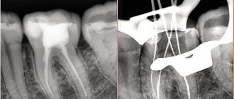
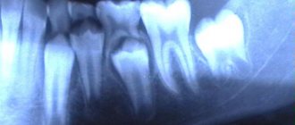
It will be most effective to carry out radiography for damage to bones, joints and teeth.
What is the difference between X-ray and digital X-ray?
The principle of digital diagnostics in medicine is completely similar to the method that is used for research in industry, construction and even, for example, at customs, in veterinary medicine. Modern X-rays in any advanced clinic or hospital are a special method of radiation diagnostics, during which all images are processed digitally.
Radiography of this type has many advantages, thanks to which it is successfully displacing analogue techniques from absolutely all areas of medicine. Even hospitals are striving to acquire such equipment, not to mention advanced paid medical centers, which have long forgotten about the old analog equipment.
The essence of the digital radiography method in medicine
The main and, perhaps, the most fundamental change affected the very method of capturing the image. If previously a special material with a special photosensitive coating was needed, now such a need has disappeared completely. The image is immediately generated in electronic form and is ready for viewing and processing.
During the shooting process, a special system of converters and detectors is used, which allows you to display the image immediately on a computer monitor. The sensitivity of flat panel detectors is several times higher than that of film, so a specialist can reduce exposure time and the radiation dose itself, and still get an image of exceptional quality.
Fluorography
Another type of examination that all residents of our country regularly undergo. Fluorography was “invented” almost a hundred years ago. This is a kind of accelerated radiography. Scientists proposed photographing a screen with an image obtained from radiography. This made the procedure faster and more widespread. Screening tests began to be administered to everyone in order to detect latent pulmonary tuberculosis.
Main plus
The procedure is fast,
the main disadvantage
is the image quality. The patient also receives a dose of radiation, and the doctor has a rather blurry picture, so fluorography is recommended to be supplemented with questionnaires and laboratory tests for the presence of tuberculosis.
Expert opinions
Ilya Gipp, Ph.D., Head of MRI-guided therapy:
— Many of these devices can be used for treatment. For example, a special installation is attached to an MRI machine. It focuses ultrasound waves inside the body, increasing the temperature in a targeted manner, and burns out tumors - for example, uterine fibroids.
Kirill Shalyaev, director of the largest Dutch manufacturer of medical equipment:
- What seemed impossible yesterday is reality today. Previously, during CT scans, a drug was administered to slow down the heart. The newest CT scanners make 4 rotations per second, so there is no need to slow down the heart.
| What radiation doses do we receive* | ||
| Action | Dose in mSv** | Over what period of time will we receive this radiation in nature? |
| X-ray of a hand | 0,001 | Less than 1 day |
| X-ray of a hand using the very first machine in 1896. | 1,5 | 5 months |
| Fluorography | 0,06 | 30 days |
| Mammography | 0,6 | 2 months |
| Mammography with MicroDose characteristic | 0,03 | 3 days |
| Whole body CT scan | 10 | 3 years |
| Live in a brick or concrete house for a year | 0,08 | 40 days |
| Annual norm from all natural radiation sources | 2,4 | 1 year |
| Dose received by liquidators of the Chernobyl accident | 200 | 60 years |
| Acute radiation sickness | 1000 | 300 years |
| Epicenter of a nuclear explosion, death on the spot | 50 000 | 15 thousand years |
| *According to Philips **Microsievert (mSv) is a unit of measurement of ionizing radiation. One sievert is the amount of energy absorbed by a kilogram of biological tissue. | ||
Mammography
A separate type of radiography designed to diagnose breast diseases, which is why women undergo mammography. There is no consensus on the recommended age for the procedure. Mammography helps ensure the absence of a malignant tumor with an accuracy of 89%. It is believed that women should be screened regularly starting at age 39, although some cancer societies recommend screening at a younger age.
Mammography is prescribed to diagnose breast cancer, the procedure is quick
, this is a plus, but the patient
is irradiated
, and the risk of an incorrect diagnosis remains, this is a minus. Mammography can be digital or film; digital mammography provides a clearer image.
How many times can an x-ray be taken?
If we are talking about analog devices, then experts recommend a break between irradiations of 3 weeks and take one photo per visit.
. However, it happens that it is necessary to increase the number of studies, then they are carried out every couple of days, reducing the negative impact as much as possible. Several x-rays on an analog device in one day can have a bad effect on your health.
The invention of digital equipment has made it possible to greatly reduce risks and allow for more frequent x-ray examinations. There is no longer any need to make compromises between harm and health benefits; doctors prescribe as many procedures as necessary to effectively monitor the progress of treatment.
Computed tomography (CT)
Computed tomography is also carried out on the principle of radiography, but as a result the doctor receives not a flat two-dimensional picture, but a three-dimensional image. This is achieved by simultaneously taking a large number of images, which are assembled into a single image. CT scanner sensors are highly sensitive and distinguish a huge number of shades, so the doctor can examine in detail all the bones and organs of the patient. The image quality can be further improved by injecting the patient with a special substance, the so-called “contrast”. The contrast helps to distinguish healthy tissues from altered ones and detect abnormal structures in the body, and also makes it possible to study the condition of blood vessels in detail. CT with contrast is not prescribed in every case; often a simple computed tomography is sufficient.
CT scan is done quickly
, with its help to screen for lung cancer. You can also use computed tomography directly during surgery.
Disadvantages of CT
can be considered a high radiation dose to the patient. Therefore, CT is not prescribed to pregnant women, children and overweight patients (more than 200 kilograms).
Ultrasound examination (ultrasound)
X-ray is not the only way to “look inside” the human body; another technology is ultrasound. Some animals, such as bats, use sound waves to orient themselves in space. People have also learned to use waves to solve some problems, including in medicine. A picture of the internal organs can be obtained by sending a sound wave into the human body and monitoring its return. The computer helps process the results and present them in the form of a three-dimensional picture.
The main advantage of this research method is safety.
. Ultrasound can be performed even on pregnant women; in addition, ultrasound devices are mobile and can easily be placed in the patient’s room to monitor the condition of organs and blood flow in real time.
However, ultrasound cannot provide a high-definition image, so the use of this method of research is limited, for example, gastrointestinal diseases cannot be diagnosed using ultrasound.
Digital X-ray: capabilities and practical applications in medicine
Digital technologies in radiography open up quite broad horizons for physicians. The technique is absolutely not inferior to the analog method of research and is excellent for:
- Assessing the condition of soft tissue and bone tissue after injury or any negative impact.
- Checks for various types of neoplasms. Can be used to work with both benign and malignant tumors.
- Search for foci of active inflammatory processes.
- Assessment of pathological deviations from the norm, abnormal scenarios of development of the congenital type.
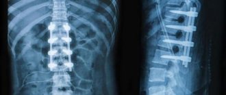
- Diagnosis of the condition of the spinal column.
- Finding the location of foreign bodies if they are swallowed. This is especially true when working with young children, for whom such situations are not uncommon.
- In preparation for surgical treatment.
For example, by checking the head area, a specialist can detect metastases, lesions, strokes, or even a hematoma. When checking the lungs using digital radiography, bronchitis, changes due to oncology or tuberculosis, and fibrosis can be detected. When assessing the condition of the spine, the doctor will see a hernia, disc displacement, cancerous lesions and many other abnormalities.
Digital X-rays can be used not only to quickly make a diagnosis, but also to track progress in the treatment process. Timely assessment of dynamics allows you to quickly adjust prescriptions, avoid side effects and achieve the best possible result.
The use of digital x-rays today not only simplifies the work of specialists and reduces diagnostic costs. With its help, it is possible to detect those pathologies for the search for which previously it was necessary to use more serious invasive methods.
Magnetic resonance imaging (MRI)
The principle of MRI is based on the property of atomic nuclei to respond to a strong magnetic field. The calculation is based on the reaction of hydrogen nuclei, of which there are many in the composition of water molecules, and the human body, as is known, consists of 60% water. When entering a magnetic field, the nuclei of atoms are oriented along it; they can be excited and the energy that they will give off when the influence weakens can be recorded, i.e. “relaxation”. Computer analysis allows you to convert the information received and determine the location, density and structure of tissues in the body.
MRI allows you to “see” cartilage, soft tissue and the human brain without causing harmful effects
, so the procedure can be performed by anyone and as many times as desired.
However, the examination takes a long time
, and closed-type tomographs can cause attacks of claustrophobia. True, there are open-type devices. MRI procedures should not be performed on people who have electrical devices (such as pacemakers) or metal implants implanted in their bodies.
MRI will be effective in studying tumors, brain and vascular abnormalities.
Scintigraphy, SPECT, PET
Perhaps these are one of the rarest procedures on our list. These examination methods are based on radiation diagnostics, but it is used in reverse. The patient is not irradiated from the outside, but a special radioactive drug is injected into him to make him “glow from the inside.” First, scientists invented and tested scintigraphy. With its help, it was possible to obtain two-dimensional images. Then research went further and single photon emission computed tomography ( SPECT)
), followed by positron emission tomography (
PET
). The difference between these methods is rather technical; they use different radiopharmaceuticals and different types of detectors that record radiation from the patient’s body.
The question arises: “Why such difficulties?” The fact is that thanks to these procedures, you can see formations in the pictures that are not visible in pictures obtained by external irradiation. Metastases and tumors can appear inside bones or organs and remain silent for a long time. The radiopharmaceutical is injected into the body and accumulates in the tissues, which allows it to “illuminate” certain areas.
Main disadvantage
of this examination method is the cost. The radiopharmaceutical is developed individually for each patient; in addition, the patient receives radiation exposure, and the procedure itself is more complex than those we described earlier. However, in some cases it cannot be avoided, for example, in cases of oncological and neurological diseases, diagnosis of heart and thyroid diseases.
X-ray is an accessible diagnostic method
An X-ray image has long become synonymous with a generally recognized diagnostic method - accessible, informative, accurate, and inexpensive. Many modern research methods have been created based on X-ray radiation - computed tomography, angiography, densitometry, mammography, digital radiography. And even a new branch of medicine - x-ray surgical methods of diagnosis and treatment. What is the basis of all of the above?
A little history:
An article by Wilhelm Conrad Röntgen (as his last name is correctly spelled), in which he described the discovery of rays that would later be named after him, was published in 1896. X-rays can penetrate many opaque materials; however, it is not reflected or refracted. The transparency of substances in relation to the studied rays depended not only on the thickness of the layer, but also on the composition of the substance. Although the eye does not react to radiation, it illuminates photographic plates; he took the first photographs using X-rays. The discovery of the German scientist greatly influenced the development of science. After a short period of time, X-ray tubes found application in medicine and various fields of technology. For this discovery he was awarded the Nobel Prize in Physics in 1901.
X-ray diagnostics are widely used in medicine. Let's recall some of the terms that you may have heard in medical institutions.
Radiography is an image of the internal structure of the object under study, created by x-rays, on film or paper.
Fluoroscopy is an image of the internal structure of the object under study, created by X-rays, displayed on a luminous screen.
Digital radiography is the recording of studies obtained using X-rays on a digital medium, which makes remote X-ray diagnostics possible.
Computed tomography is a modern diagnostic method that allows you to obtain 3D images of organs and tissues.
General X-ray - for example, an overview X-ray of the chest organs. Allows you to assess the condition of a significant part of the body as a whole.
A targeted photograph is a photograph of a specific organ or its area. As a rule, special styling is performed for a targeted shot to achieve maximum visualization.
An image with an X-ray contrast agent - for a more accurate diagnosis, a drug with X-ray contrast properties is injected into organs or vessels. You must tell your doctor if you have previously had an allergic reaction to iodine or barium.
What can a doctor see in the image? Using X-rays, injuries are successfully diagnosed, the lungs are examined, various formations (stones, tumors), and areas of obstruction can be identified. With a contrast study of blood vessels, the doctor sees aneurysms, areas of blood vessels affected by atherosclerosis, etc. Different tissues transmit X-rays differently: bone tissue absorbs them almost completely, soft tissue partially retains them, and air allows them to pass through completely. Depending on this, shadows of different intensities are obtained on the film: in place of bones there are white areas, in place of soft tissues they are gray, layers of air appear black on an x-ray. X-rays are essentially negatives, so the lighter areas on them are called “shades.” For example, healthy lungs filled with air appear black on x-rays. The area of pneumonia (pneumonia) is a lighter spot, which doctors will call a shadow (see photo).
Using X-rays, you can make an accurate diagnosis for various injuries. For example, a fracture is visible as a darker “break” in a lighter “field” of the bone. Inflammation is usually visualized as a lighter area. Intestinal obstruction can be judged by the presence of gases in the organ or by changes in the shape of intestinal loops. Stones in organs look like light formations with clear boundaries and contours. If an X-ray is taken with a contrast agent and the image shows uneven filling of the organ, the doctor may assume the presence of a benign or malignant tumor. When examining vessels with a contrast agent, dilations are clearly visible - ruptures (aneurysms) are possible in this place.
How is an X-ray taken? Before the procedure, the patient must take off his jewelry, belt, remove all metal objects, phone, etc. from his pockets. In some cases, such as when examining the chest or spine, the doctor may ask you to undress to the waist. X-rays of the extremities can be performed with clothes on. Those parts of the body that are not examined are covered with special protective lead aprons. The doctor also puts on a protective suit and goes into the next room. Pictures are taken in different positions - mostly lying down or standing. Depending on the projection in which the image is needed, the doctor may ask you to change the position. Studies with contrast agents usually require some preparation. Your doctor will provide instructions for preparing for the study.
Modern X-ray machines (including those used in our clinics) provide minimal radiation exposure. For example, a general X-ray of the chest organs is no more than 0.15-0.3 mSv. We employ experienced x-ray technicians who minimize the impact of ionizing radiation on the patient’s body. X-ray examinations are performed in the left bank emergency room of the TERVE clinic on Partizana Zheleznyaka, 21A and the right bank emergency room of the TERVE clinic on Krasnoyarsky Rabochiy Ave., 150 p. 48 .
Prices for tests are listed on the pages of emergency rooms.
Hybrid Imaging Techniques
Probably, science would not be science if it did not constantly move forward and try to create something new from the old. So, doctors began to combine different scanning methods to obtain even more detailed and high-quality images. PET and SPECT are combined with CT, MRI complements PET. Such experiments are not cheap, but can sometimes help make decisions about further treatment for the patient.