X-ray examination is carried out in any clinic. This is one of the reliable ways to identify the presence of any pathologies and establish a diagnosis. Of course, the method also has a number of disadvantages, but, nevertheless, radiography is one of the first prescribed if necessary . It is especially often used to diagnose the condition of a person’s bones, and can also refer the patient for an X-ray of the spine. What does this study show?
X-ray of the spine - what does it show?
Radiography – what kind of method?
X-ray examination is a fairly common diagnostic method based on the passage of X-rays emitted by special equipment through the body. A study using radiation makes it possible to obtain a black-and-white image in which the elements of the examined area will be clearly visible. Depending on the type of tissue and its density, darker or lighter elements will be visible in the X-ray image. Bone tissue appears white in the image, while softer muscle and ligament tissue appears dark, grayish or black.
Nature of X-rays
On a note! X-rays were discovered by physicist V.K. X-ray and were named after him.
By studying the image, the doctor can see certain tissue changes, if any. Sometimes even minor disorders can be detected, which makes it possible to diagnose any disease at an early stage of development. The doctor will also see where the changes are localized and, in accordance with this, prescribe the appropriate treatment tactics.
X-ray of the spine
Advantages of X-ray examination:
- simplicity and low cost of implementation - radiographic equipment is available in any clinic. In a municipal medical institution, you can take a photo free of charge according to your policy;
- high speed of obtaining results - the image can be collected in a few minutes;
- modern equipment makes it possible to obtain an image in digital format instead of a photograph on film;
- Most studies do not require any prior preparation.
X-ray shows osteochondrosis of the cervical spine
Disadvantages of this diagnostic method:
- the negative impact of radiation on the body, so X-rays are often not allowed;
- the presence of a number of contraindications;
- inability to assess the condition of soft tissues;
- insufficient information content compared to a number of other methods.
Digital X-ray
Only the doctor interprets the images. He is the one who is able to notice even minor manifestations of the disease. But in some cases, an ordinary person can see the problem in the picture and understand what is wrong with his body. In the case of an X-ray of the spine, even the patient himself is able to discern scoliosis and fractures.
If you want to learn in more detail how they do it and what an x-ray of the lumbosacral spine shows, as well as learn about the contraindications and consequences, you can read an article about it on our portal.
Prices for lumbosacral corset
X-ray of the lungs: preparation, technique, meaning, how to read, problems in the images
Chest X-ray (X-ray) is a simple, effective test that has remained relevant for over 100 years in helping to see any abnormalities in the chest cavity.
The value of the method is that it can show almost all serious pathologies at an early stage, suitable for emergency diagnosis and treatment monitoring. Therefore, it is a frequently performed method and can be performed on an outpatient basis or in a department using a stationary or portable X-ray machine.
Snapshot of healthy lungs
In a healthy person, the R-image clearly shows the fields of the lungs located on the sides of the spine. They do not block X-rays, so they look uniform and have no spots.
There is a heart in the center; on the right side its shadow should protrude no more than one centimeter. The collarbone is visible at the top. The dome-shaped diaphragm is clearly visible in the lower part. Normally, one half of it is raised relative to the other. Horizontal shadows are edges.
The bronchi are usually not visible. Their appearance indicates “smoker’s lungs” or chronic diseases (bronchiectasis or cavitary formations in them).
Also, the vascular pattern should not be visible; its strengthening indicates a violation of blood circulation, an increase in pressure in them, which is typical for cardiac pathology. Normally, the vessels are visible only near the roots in the central part. The roots should be clearly visible, have a standard size, and not be expanded. The trachea should be centrally located.
Important
If the radiologist sees a similar picture (without disturbances in blood supply, cavitary and cystic formations, signs of stagnation), then in the description protocol he will write a conclusion: the pulmonary pattern is not changed, has a clear shape, no pathological formations have been identified.
When required
Radiography, unlike fluorography, is a more informative method. Allows you to resolve doubts that arose after FLG. Therefore, doctors often refer for x-rays if strange “findings” are detected on fluorography. In these cases, you cannot do without an overview photo.
It is also preferable if a person works in hazardous work or has contact with a patient with tuberculosis.
X-rays are required in the following conditions:
- difficulty breathing, shortness of breath;
- prolonged coughing;
- chest injury and pain;
- elevated temperature of unknown origin;
- severe sweating;
- monitoring the effectiveness of treatment of various pulmonary diseases, such as pneumonia, cancer;
- the presence of blood or pus in the sputum;
- chest pain for no apparent reason;
- after an accident with multiple bruises.
What diseases does it detect?
Pathologies
- Chronic obstructive diseases (COPD) - emphysema and chronic bronchitis.
- Air outside the lungs (in the pleural cavity). The pathology is called pneumothorax.
- Infection (pneumonia).
- An abscess is a ring-shaped shadow that indicates a cavity.
- Fluid in an organ, which is a sign of heart failure
- Fluid around (pleural effusion).
- Blackouts (cancer, tuberculosis infection).
- Edema.
Chest bone problems:
- Chest contusion.
- Rib fractures.
- Clavicle fracture.
Heart problems:
- Dilated and enlarged (cardiomegaly) indicates heart failure, problems with the valves.
- Abnormal position of the heart (dextrocardia), when it is located in the right half of the chest
- To monitor the operation of pacemakers and the position of catheters
- Fluid around the heart (pericardial effusion) is pericarditis.
- Salt deposits - calcifications of the heart or blood vessels.
- Dilation or narrowing of the arteries, which suggests an increase in blood pressure in the pulmonary arteries, indicates pathology of the right side of the heart.
Aperture problems:
- Lowering or raising its dome.
- Protrusion (hernia).
- Wall sealing.
- The presence of gas or liquid under the diaphragm dome.
Normally, it has the shape of a slightly concave dome. Its right half should be higher than the left because of the liver. The difference is about 3 cm. Its outline should be smooth. A flattened (drooped) diaphragm is often seen in patients with chronic obstructive pulmonary disease (COPD) or pneumothorax.
Important
Chest X-ray allows you to practically make the correct diagnosis, start treatment in a timely manner and save a person’s life. It reveals the pathology of all organs of the chest, starting with the lungs and ending with the diaphragm. On an x-ray, shadows of 2 mm in size can be distinguished, and with fluorographic examination - at least 5 mm.
What kind of preparation
The study does not require special preparation. Smoking does not affect the diagnosis. It is advisable to reduce food intake, since with a full stomach the diaphragm moves slightly upward, therefore, the result may be distorted.
Pregnant women and children are of concern. Usually the attending physician decides on the need for x-rays in this category of patients.
Before the procedure, you should inform about the presence of a pacemaker or other implants. Leave jewelry at home, remove glasses, remove dentures.
Technique
X-rays are similar to photography. It is performed by a radiologist in a special room. The entire stage of implementation includes several mandatory steps:
- The patient is registered in the journal.
- Frees the upper half of the body from jewelry and clothing.
- If necessary, covers areas of the body that are not subject to examination with a lead apron.
- Placed between the film screen and the X-ray tube. Presses your chest or back tightly against the device.
- Takes a deep breath and holds it for a few seconds while the photo is taken.
At this time, the specialist is in another room and turns on the emitting device.
The procedure takes no more than five minutes and does not cause discomfort.
The examination can be performed in a standing, lying or sitting position. The radiologist begins the description immediately and gives the result to the patient. The image can then be shown to your doctor or any other specialists if necessary.
General radiography is usually done, but the direction may indicate the need for targeted R-graphy. It provides more information on a specific area, allowing a better study of the pathological area.
Why do you need x-rays in two projections?
There are two types of R-images - frontal and lateral projections. In a straight position, it is not always possible to see infiltrates of the upper lobe of the lung. A side shot allows you to study them in more detail. It also shows the paths to the roots better, indicating, for example, tuberculosis.
The direct position does not always “see” pneumonia, since the lung consists of segments, and in the direct projection they are layered on top of each other. Diagnostics from both sides allows you to better determine the size of the heart. To more accurately identify central and peripheral cancer.
Also, on lateral photographs, small foci of infiltration, abscesses, and cysts are more clearly visible; in this case, they are not covered by the sternum.
Are X-rays dangerous?
X-rays are a form of radiation such as light or radio waves. They pass well through most objects, including the body. Some patients are concerned that they are contributing to cancer.
The amount of radiation a person receives from a chest x-ray is much less than the natural radiation from the Earth that we are exposed to every day.
Radiation is dangerous if its total dose exceeds a threshold level (more than 1 millisievert in one session).
During FLG, the patient receives a dose of up to 0.8 mSv, and for one x-ray – 0.3 mSv. A 16-hour flight is equivalent to one x-ray.
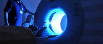
The highest dose is received from a CT scan (computed tomography) - about 4 mSv, which is 30-40 times higher than the dose from an X-ray. So, CT scans must be done according to strict indications.
For life-threatening diseases, harm from radiation will be the least evil for the patient. The diagnosis has no contraindications, but there are age restrictions. It is not advisable to perform the procedure on children under 14 years of age and pregnant women.
How to “read” an R-image
To correctly describe an image, you need knowledge of various forms of diseases and extensive practical experience. It is difficult for the average person to “read” an x-ray. Even doctors sometimes disagree on the images they receive.
Still, some concepts are worth getting acquainted with. Various types of compactions, white spots (accumulation of air or liquid), shadows in the area of the lung fields are considered dangerous. They talk about pathological processes.
Different parts of the body transmit rays differently. Dense bone (ribs and spine) absorbs most of the radiation and appears white in the image, while soft tissue such as muscle, fat, and organs allows x-rays to pass through and absorbs less, so they appear darker.
As a result, on an x-ray we see bones as white, soft tissue as gray, and air as black.
Do not panic if a shadow is detected in the lung. It can indicate not only a dangerous disease, but also a defective film. The occurrence of shadows can be caused by many reasons and are not always life-threatening.
Radiologists have a whole classification of shadows according to size, degree of compaction, shape, and location. For example, a spot that is ominous for us may not cause alarm to a radiologist, and may be interpreted as residual effects of a killed infection. Distinguishing signs that seem insignificant at first glance will tell him about this.
The table below presents a list of diagnoses with the corresponding x-ray pattern for each of them:
| Disease | Characteristic changes |
| Tuberculosis | A large number of small blackouts. A large, round shadow may indicate advanced tuberculosis. |
| Edema | Unevenly scattered shadows resembling flakes. |
| Exudative pleurisy | A thin dark line, usually along the lower edge of the costal arch. The trachea is displaced or stretched |
| Venous stagnation | The basal zones resemble the wings of a moth, which indicates stagnation of blood in the pulmonary circulation (or basal pneumonia). |
| Cancer | Round formations, different in size, having a strictly defined line along the contour. |
| Emphysema | Consolidation of the diaphragm, high airiness of the lung fields. |
| Peritonitis | Due to the concentration of gases in the peritoneal area, there is no clearing under the dome of the diaphragm. |
| Abscess | Ring shadow. |
| Atelectasis (collapsed lung) | Intense (black) uniform darkening of the entire lung, lobe or segment. On the affected side, the dome of the diaphragm is raised high. |
| Heart failure | The heart's shadow takes on a rounded shape on the right or left. With an enlarged right ventricle, darkening is visualized on the left |
| Silicosis (dust) | Finely scattered diffuse nodular opacities with relative sparing of the lower lung zones |
What diseases look like on R-images
| Problems: | |
| 1. Chronic obstructive pulmonary diseases (COPD) - emphysema and chronic bronchitis. The image shows flattening of the diaphragm, excessive clearing of the lung fields, and expansion of the roots. An x-ray will not show acute bronchitis. | |
| 2. Air behind the lungs (in the pleural cavity). As a result of air entering the pleural cavity, we see partial or complete collapse. The pathology is called pneumothorax. | |
| 3. Infection (pneumonia). An area of clearing is visible. | |
| 4. Abscess. A ring-shaped shadow indicates a cavity. | |
| 5. Fluid in the organ (heart failure). Bilateral pleural effusions are present in 70% of patients with congestive heart failure. | |
| 6. A lesion in the lungs (cancer, tuberculosis infection). We see a spot. | |
Chest bone problems: | |
| 1. Chest contusion. There are small inflammatory infiltrates in both lungs. | |
| 2. Rib fractures. | |
| 3. Fracture of the collarbone. | |
Heart problems: | |
| 1. Expansion. Enlarged (cardiomegaly). Occupies 50% of the size of the chest, an increase suggests its expansion (cardiomegaly) or pericardial effusion. | |
| 2. Calcium deposits. Calcium deposits in the organ are most often associated with an old, cured infection. This nodule appears firm and white and has the same density as bone. | |
Aperture problems: | |
| 1. Flattening, indicating dilated lungs, characteristic of COPD and emphysema. | |
| 2. Right-sided pneumothorax without mediastinal displacement and diaphragmatic hernia (indicated by an arrow). | |
| 3. Free air under the diaphragm (indicated in yellow) | |
Types of R-visualization, their advantages
Most often, conventional film X-rays are used in medical institutions. For this, it is necessary to purchase film on which X-rays passing through the human body form an image of the lung.
Such a film requires development using harmful chemicals and exists in a single copy. The main disadvantage of analog X-rays is the higher radiation dose and the contact of medical personnel with harmful substances during the development process. Artifacts of the film itself due to pulsation of large vessels and movements during the act of breathing cannot be excluded.
Digital X-ray is a completely different matter. Instead of film, a digital receiver is used, which converts the rays into an image on a computer. It can be copied, processed, sent electronically, and recorded on other digital media. The radiation dose on a digital device is 8-10 times less.
Recording an image on a digital medium provides a unique opportunity to view images over time (important for chronic diseases), make the required number of copies, highlight individual areas, enlarge and print the resulting image.
The method will be of great help to specialists in the therapeutic department, accurately detecting pneumonia, bronchitis, pleurisy, and tuberculosis. Cardiologists will show heart defects.
A gastroenterologist will help diagnose a diaphragmatic hernia, and a pediatrician will help diagnose spinal curvature. Endocrinologists using a digital device will be able to identify thyroid diseases at an early stage.
Important
Digital radiography increases the efficiency of research and has a low radiation dose. Due to the high resolution, the images are clearer and more contrasty, which allows you to better assess the nature of the compaction and identify the shadow. Diagnose a tumor in the initial stage of development.
In severe cases, when the patient is not transportable, it is possible to take an x-ray at home or in the ward. The procedure is performed on a portable mobile or portable X-ray device. The device runs on a battery. It is suitable for all radiographic procedures in emergency and intensive care wards, ideal for pediatric wards.
Another radiological method is fluoroscopy. It allows visualization of the organ in real time. You can examine the pathology in the breathing process and evaluate the mobility of the diaphragm. R-copy is used in case of loss of elasticity of the lungs and suspected bronchial obstruction.
In controversial and unclear situations, this additional examination method is used. The decision to conduct it remains with the attending physician.
What are the benefits and risks of R-graphy
Chest radiography greatly aids in diagnosis by providing general guidance as an initial diagnostic test and is particularly useful in diagnosing pneumonia, cancer, and chronic obstructive disease.
Advantages of the method:
- accessible and relatively safe;
- inexpensive, highly informative method;
- acts as a document important in resolving issues of disability;
- especially useful in emergency diagnosis;
- free of side effects in the typical diagnostic range;
- The equipment used is inexpensive and therefore available for emergency departments, doctors' offices, ambulatory care centers, and nursing homes.
At the same time, the risks are minimal, and the benefits significantly outweigh the harm. Don't be afraid of R-examination. It should be done in case of sudden complaints of shortness of breath, persistent cough with hemoptysis, chest pain associated with breathing, despite the fact that FLG or radiography was recently done.
Be attentive to yourself and your health, use this informative and safe diagnostic method in a timely manner.
X-ray danger
Previously, X-rays were taken without question by all patients as prescribed by the doctor. Now, having read about the dangers of this research method, many are beginning to refuse to conduct it. But in vain, since in fact, a rarely performed x-ray will have a negative impact no greater than the consumption of unhealthy foods purchased daily in stores. The theory traveling on the Internet that X-rays can cause tissue cells to mutate to the point of becoming cancerous has no scientific basis as such.
Is X-ray dangerous?
Important! The dose that can cause cell mutation is about 150 roentgens received by the body during the year.
With an X-ray of the spine, a person will receive only 1.5 mSv (millisievert). Each organism receives approximately the same amount from the natural background radiation of the Earth during 6 months of life. Thus, by taking a single image of the spine, a person does not risk anything, and he certainly should not be afraid of developing cancerous tumors if he does not visit the X-ray room every day.
Harm of X-ray exposure
Attention! If the purpose of the study is to assess the condition of the bones, then radiography is necessary, except in cases where there are absolute contraindications to its implementation.
Learning to describe x-rays of the sinuses
Anyone can describe X-ray images of the sinuses with sinusitis (inflammatory accumulation of fluid) independently if they study the X-ray syndromes of the disease. It is enough to remember the normal intensity of the clearings formed by the frontal and maxillary sinuses in order to learn how to identify sinusitis or cysts (cavity formations filled with fluid) on an x-ray.
X-ray of the paranasal sinuses. The arrows indicate the maxillary (maxillary) and frontal sinuses
It will be easy for the reader to read the radiograph if he remembers that the sinuses on it are black. With pathological accumulations of fluid, white shadows appear (see picture).
A fragment of a radiograph shows how to detect bilateral sinusitis
If you compare Figure 2 with the previous one, you can detect intense darkening (white) in the projection of both maxillary sinuses against the background of bilateral sinusitis. They are formed by the accumulation of fluid.
To summarize: you can easily read X-ray images of the lungs, nose and even teeth only after studying the x-ray anatomy of the normal areas of study. This is what representatives of the Leningrad School of Radiologists advise, and we agree with them. Practical experience is required to identify fractures on photographs.
What does an x-ray show?
X-ray examination allows us to detect a large number of diseases quite quickly. These include:
- injuries (including fractures, bone cracks, etc.);
- osteoporosis;
Osteoporosis - osteochondrosis;
- inflammatory processes of bones;
- tumors;
- tuberculosis of the spinal column;
- curvatures, including scoliosis;
- any genetic abnormalities.
Chest x-ray
X-rays are performed only if there are certain complaints and indications. Nobody sends anyone for such research just like that.
Contraindications and indications
X-rays will be required in certain cases. Mainly:
- presence of pain in the back area;
- curvature of posture;
- injuries of various origins;
- pain in the buttocks or thighs;
- numbness of the limbs;
- suspicion of a number of diseases - intervertebral hernia, arthritis, osteochondrosis, etc.
Radicular syndrome in cervical osteochondrosis
Attention! Since radiography is not able to accurately show the condition of the soft tissues of the body, from the point of view of the reliability and accuracy of the results, when examining the spine for the presence of hernias and similar diseases, it is recommended to conduct an MRI. The method is more expensive, but in this case more effective.
X-ray diagnosis of the patient
In some cases, x-rays cannot be used as a research method. For example, contraindications, although not absolute in most cases, are:
- the first months of pregnancy, and the entire pregnancy in general . X-rays can have a negative impact on the developing fetus and can cause negative and irreversible changes in the body of the unborn child;
- small age . It is not recommended for very young children to do such a study. Either radiography in this case is important to carry out with a number of precautions and only if there are serious indications;
- the patient's inability to remain motionless for a short time . In this case, the picture will be blurry and uninformative;
- inadequate condition of the subject.
Because a child's spine is flexible, serious injuries in children are rare.
On a note! Significant excess weight will not give reliable results. X-rays will be ineffective.
How is the procedure performed?
It’s worth learning how an X-ray of the cervical or thoracic spine
before undergoing such examination. To begin research, you need to remove all metal accessories and jewelry, as well as clothing that contains metal elements. This will allow you to get a clear image.
How the procedure will be performed depends on the projection in which the image is taken. There may be such projections:
- straight - the patient lies or stands straight, arms along the body, the x-ray beam is directed at an angle of 15-20 degrees onto the thyroid cartilage and passes through the neck;
- lateral – the patient needs to stand sideways and slightly raise the chin, the procedure can also be performed lying down;
- X-rays of the cervical spine can be done through the mouth - this is a type of direct projection that allows you to see the upper cervical vertebrae;
- oblique - rarely prescribed, intended to study the intervertebral foramina.
X-ray of the cervical spine in two projections
Most often, the procedure is performed in the usual frontal and lateral projections. As a rule, such a study is enough to assess the condition of the vertebrae and determine the cause of the patient’s complaints. Upon receipt of the images, additional studies may be prescribed that will allow the condition of the soft tissues of the neck to be assessed, or treatment will be prescribed if there is enough information.
X-ray of the cervical spine with functional tests
X-ray of the cervical spine with functional tests
involves taking pictures while the patient is in certain positions. The doctor may ask you to tilt your head left or right, forward or backward. During such a study, it will be possible to determine the mobility of the vertebrae in the cervical region and assess their condition. Often this procedure is prescribed for suspected osteochondrosis.
How is an x-ray done?
X-rays will be performed by a doctor depending on the X-ray machine used. The patient will need to either lie down in the position that the doctor tells him to take, or stand near the device. The specialist may ask you to bend over, turn sideways, or take some other position. These measures are needed to take the most informative picture of a particular part of the spine, so all the doctor’s instructions must be followed.
X-ray machine
The part of the body that does not need X-ray examination is covered with special shielding materials that will protect the patient from the negative effects of the X-ray machine. Next, the doctor will leave the office, go behind a special protective stand, and turn on the device. The irradiation lasts a few seconds, after which you can get up (with the doctor’s permission), get dressed and wait for your shot.
Taking a photo takes several minutes. It then needs to be taken to the doctor who gave the referral for examination. Tissue evaluation should be carried out by a specialist who understands radiography and can read images correctly.
How to do an x-ray
An X-ray of the spinal column can be taken at almost any medical institution. This is also a municipal clinic, where X-rays are done free of charge, but there are usually long queues there. This is also a paid medical center where you can get an x-ray without a queue, but you will need to pay money.
How to quickly understand an x-ray
Every reader who will not spare us precious time will be able to quickly understand X-ray images.
An x-ray is a summary image of the anatomical structures through which x-rays pass. The degree of absorption by tissues varies, so the X-ray image consists of black and white shades of varying intensity (see figure).
Brightness on radiographs of various anatomical structures (according to Matthias Hofer)
Organs and tissues are represented by a cluster of shadows and clearings of varying intensity, to which the eye of a radiologist (radiologist) must “get used to”.
Preparation
No serious preparation is required on the part of the patient for radiography. You just need to come to the x-ray room at the appointed time and follow the doctor’s instructions. Only with radiography of the lumbosacral region may a cleansing enema be required. Also, before examining these parts, it is necessary to reduce the level of gas formation in the intestines through a special diet - 2 days before the examination, it is important to exclude baked goods, fatty foods, milk, cheeses, legumes, raw fruits and vegetables from the diet. You can additionally drink activated carbon.
Before taking an x-ray, you need to prepare
The evening before the X-ray examination, you can eat no later than 7-8 hours. Next, the intestines are cleansed through enemas or taking laxatives. Cleansing is carried out several hours before the examination.
How to prepare for and attend an x-ray?
Step 1. You need to visit a doctor and take a referral for an X-ray examination of the spine.
Take a direction first
Step 2. If it is necessary to examine the lumbosacral region, it is important to adhere to the above-described diet for 2 days before the x-ray.
Follow a special diet
Step 3. You need to do a colon cleanse. If other parts of the spine are to be examined, then steps 2 and 3 can be neglected.
Cleanse your colon
Step 4: It is recommended to wear comfortable clothing that can be easily taken off and put on.
Wear comfortable clothes
Prices for orthopedic shoes
Step 5. At the appointed time, you need to come to the clinic, not forgetting the directions, and wait for the doctor to be invited to the office.
Visit a doctor
Step 6. In the office, it is important to do everything that the specialist says - undress, take off your jewelry, take the desired position.