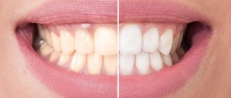
Stomatitis
This is a group of diseases characterized by inflammation of the oral mucosa with hyperemia, swelling, and an increase in the amount of mucus in the oral cavity.
Depending on the severity and depth of the lesion, even ulcers or foci of necrosis may form in the oral cavity, sharply disrupting the general condition - fever, weakness, anxiety, refusal to eat. There are many causes of the disease: mechanical, chemical, thermal, bacterial factors. Often the cause of the disease in infancy is contaminated nipples, toys and other objects that fall into the child’s mouth. Stomatitis often develops as a result of infectious diseases (measles, scarlet fever, influenza, whooping cough, etc.). The mucous membrane of the oral cavity acquires a bright red color, becomes swollen, and tooth marks are visible on the mucous membrane of the cheeks and tongue. Saliva becomes viscous and viscous. The mucous membrane is covered with a whitish coating. The tongue is dry, swollen, often with a brown tint, chewing is painful. The duration of the disease is from 1 to 3 weeks, the prognosis is favorable.
A general preventive rule for children and adults is to maintain good oral hygiene.
Medical Internet conferences
Relevance: According to WHO, the number of acute and chronic poisonings in children in economically developed countries is increasing from year to year, the cause of death of 20% of children under the age of 15 years is exogenous intoxication. In our country, over the past 5 years, the number of poisonings in children has doubled (an annual increase of 3-15%). Among nosological forms, more than 80% are poisonings with medicinal substances, food and household chemicals, with the maximum number of poisonings (from 77.2% to 85%) occurring between the ages of 1 and 3 years. Accidental use of various chemical substances by children is often accompanied by the development of burns of the upper digestive tract (UGT) of varying severity. In recent years, there has been a trend toward an increase in the number of chemical burns of the esophagus in children, which is explained by an increase in the number and types of aggressive substances, as well as their easy availability to the consumer.
Purpose: to study the outcomes of damage to the upper parts of the VOPT due to the accidental use of various chemical substances by children.
Materials and methods: According to the archives of the KB named after. S.R. Mirotvortsev SSMU conducted a retrospective analysis of 612 case histories of children aged 9 months to 15 years hospitalized with suspected oral poisoning by various chemicals and drugs (2009 – 2013). Signs of poisoning were diagnosed in the emergency room in 544 children and sent for treatment to the children's intensive care unit (DORIT). Due to a suspected chemical burn of the VAPT, 68 children were urgently hospitalized at the children's surgical clinic (DSC). In the conditions of the DHC, all children (100%) underwent a general clinical laboratory examination (CBC, OAM, ECG); instrumental examination was carried out in 43 (63.2%) patients: FGDS - 38 (55.8%) patients, fluoroscopy of the upper digestive tract (UGDT) with Susp. BaSO4 – 5 (7.3%) patients. The children were consulted by an ENT doctor and a pediatrician. The depth of tissue damage in a burn of the esophagus (endoscopic picture) was assessed according to the classification (S.D. Ternovsky, E.N. Vantsyan, 1971). Both conservative and surgical treatment methods were used (bougienage of the esophagus with COOK bougies, gastrostomy placement).
Results. All patients hospitalized at DHC ingested the chemical accidentally due to adult carelessness. Gender distribution: 45 boys (66.2%), 23 girls (33.8%); by age: up to 1 year – 1 (1.5%) child; from 1 year to 3 years - 54 (79.4%) people; from 3 to 6 years – 10 (14.7%) people; 6 to 9 years old – 3 (4.4%) people (diagram 1); average age of patients – 2.17±0.12 years.
A burn to the esophageal mucosa was detected when using acetic essence (acetic acid) in 36 (52.9%) children, alkali - in 10 (14.7%) patients, potassium permanganate crystals - in 17 (25%) patients, and other chemicals ( glue, ammonia-anise drops, ammonia, tincture of iodine, etc.) – in 5 (7.4%) children.
Chemical burns of the mucous membrane of the oral cavity with acid and alkali were clinically manifested by swelling and hyperemia of the lips, hyperemia and bleeding of the oral mucosa, hypersalivation; when consuming potassium permanganate crystals, the mucous membranes of the mouth and tongue turn black. In severe cases, fibrin deposits on the oral mucosa, dysphagia, and dysphonia were noted in 8 (11.7%) people. Complications in the form of acute renal and liver failure were not registered in these children.
Primary FGDS was performed in 47 (69.1%) patients, 4 patients underwent fluoroscopic examination of the VOPT with Susp. BaSO4 within 1 to 7 days. Parents of 12 children refused endoscopic examination (FGDS). The endoscopic picture of a 1st degree burn was diagnosed in 12 children (25.5%), a 2nd degree burn in 8 patients (17%), a 3rd degree burn in 11 patients (23.4%), a burn in 16 cases (34.1%). the esophagus was not endoscopically confirmed (diagram 2). As a result of FGDS, the localization of the burn surface was verified: a burn of the mucous membrane of the upper third of the esophagus was diagnosed in 12 patients, a burn of the mucous membrane of the middle third of the esophagus - in 4 children, a burn at the border of the upper and middle third of the esophagus - in 3 patients, a burn at the border of the middle and lower third of the esophagus - in 1 child, burn of the mucous membrane of the lower third of the esophagus – in 2 patients.
Primary FGDS was not performed in 21 patients. In 5 children there were no clinical signs of a chemical burn of the VA.
Esophageal stenosis as a complication of a chemical burn due to accidental use of chemical substances was diagnosed in 14 (45.2%) patients. All patients with esophageal stenosis underwent therapeutic bougienage using a conductor string, Cook bougies, and in severe cases (III degree burn) - in combination with the application of a gastrostomy.
Chemical burns of the esophageal mucosa were accompanied by inflammatory changes in the hemogram (ESR acceleration - 17.2±2.05 mm/h, leukocytosis - 13.3±1.7*109/l). All children received conservative treatment: gastric lavage, early antibacterial therapy to prevent secondary infection, in order to accelerate epithelization of the mucous membrane, oral sea buckthorn oil, short courses of glucocorticosteroids (5-7 days), enveloping agents (Almagel), infusion therapy as a component of anti-shock therapy ( according to indications). To remove unabsorbed crystals of potassium permanganate and reduce the cauterizing effect of the substance on tissue, the oral mucosa was treated with a swab with 1% ascorbic acid solution.
Conclusions:
1. FGDS is the defining diagnostic method that verifies the diagnosis of “Chemical burn of the esophagus.”
2. Every third child examined endoscopically (22 people (32.3%)) was diagnosed with a burn of the esophageal mucosa due to accidental use of various chemicals. 3. Symptoms of damage to the mucous membrane, revealed during examination of the oral cavity and pharynx, are not reliable signs of the severity of damage to the mucous membrane of the esophagus and stomach.
4. Accidental use of aggressive chemicals (acids, alkalis) by children can lead to the formation of cicatricial stenosis of the esophagus, which negatively affects the child’s quality of life.
Gingivitis
An inflammatory process that causes swelling and tenderness of the soft tissues. If not treated in a timely manner, the problem worsens and becomes chronic.
The main causes of gingivitis:
- insufficient oral hygiene;
- thermal or chemical burns;
- use of certain medications;
- unbalanced diet (insufficient amount of vitamins in food)
- smoking;
- some infectious diseases;
- gastritis;
- ulcerative processes in the digestive system;
- caries.
Forms and types of gingivitis
Depending on the clinical situation and the nature of the development of the disease, acute and chronic gingivitis are distinguished. Acute gingivitis
manifests itself in the form of classic signs of the disease: redness, swelling and bleeding of the gums.
Chronic gingivitis
develops more quietly, without pronounced signs, but gradually leads to the growth of gum tissue (hyperplasia), which entails partial and complete coverage of the surface of the tooth crown by the gum.
Prevention measures
By following simple rules you can reduce the likelihood of serious oral diseases:
- Brushing your teeth at least 2 times a day after eating;
- Using dental floss and mouthwash;
- Balanced diet;
- Rejection of bad habits;
- Visit the dentist at least once every six months.
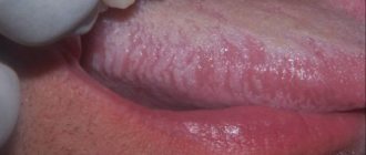
Symptoms
The first signs of the disease often go unnoticed because they do not cause any pain or discomfort in the patient. Nevertheless, a specialist will be able to determine the onset of leukoplakia by the appearance of the mucous membrane, lips and the area where the teeth meet.
The first sign of the disease is the appearance of a keratinized gray area, which can appear on the palate (in smokers), in the corners of the mouth, on the inside of the cheek, etc. An easily removable white plaque forms in this area, but after a few days the formation makes itself felt again . The patient may feel tightness in the mouth, but, as practice shows, most people simply do not pay attention to this.
Plaques with a diameter of no more than 4 centimeters are formed. They may appear:
- on the inner surface of the cheeks;
- on the tongue (on the back or sides);
- in the sky;
- on the gums;
- in the corners of the mouth.
The process of plaque formation takes up to one month. At the first stage, the area of the future formation seems slightly swollen; when you feel it with your fingers, the compaction is not felt. However, over time, another symptom of oral leukoplakia appears - the mucous membrane at the site of the swelling loses its original shine and becomes rough, which is noticeable when touched.
There is no pain in this case: only sometimes a feeling of dryness at the site of the outbreak is possible.
Gradually, the color of the spots changes from gray to bright white. The spots in most cases have clear boundaries. Their increase is possible when the disease enters its second stage, called verrucous.
The disease often causes candidiasis and malignant cancers. In an advanced state, leukoplakia is very difficult to treat: the affected areas become even more keratinized, ulcers can form, and the infection gradually spreads to other areas of the mouth.
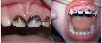
Periodontitis
Periodontitis is an inflammation of periodontal tissues, which includes the teeth, ligaments, cementum and gums. Periodontitis as a disease is a consequence of gingivitis - a minor inflammation of the gums, the main cause of which is neglect of oral hygiene. If with gingivitis the inflammation spreads exclusively to the soft mucous membranes, then with periodontitis the ligaments that hold the teeth in the sockets are affected. This is why in 90% of cases when this disease is diagnosed, tooth mobility is observed, which eventually leads to their loss.
The most common causes of the disease are the following:
1. Improper or irregular oral care
. Dental plaque, which is present on the surface of the teeth and in the spaces between teeth, is not as safe a substance as it might seem at first glance. Soft and easily removed at the beginning, it goes through certain cycles of “development”. The result is the mineralization of plaque and its transformation into hard tartar. This process in most cases is observed in those who do not pay due attention to daily oral care or use an incorrectly selected toothbrush, toothpaste and mouthwash.
2. Poor blood supply to the gums
. Periodontitis is one of the most common problems among smokers. Substances contained in tobacco smoke lead to a narrowing of the blood vessels in the oral mucosa and their fragility, which impairs the blood supply to the gum tissue and supporting apparatus of the teeth. A slowdown in blood circulation and, as a consequence, the development of periodontitis is also facilitated by a lack of chewing load caused by eating habits (for example, the predominance of soft foods in the diet).
3. Nutrient Deficiency
. The lack of fresh vegetables, fruits, herbs, a sufficient amount of fish, meat and dairy products in the diet quickly leads to a lack of essential substances in the gum tissue. If poor nutrition is a permanent habit, then over time the metabolic processes in the gums are disrupted, which creates the ground for inflammation and periodontitis. A deficiency of vitamins A, C and group B can lead to negative consequences.
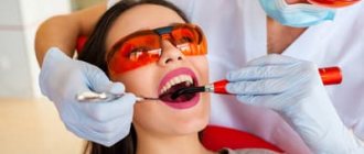
Treatment of periodontitis
Professional teeth cleaning is an integral step in the treatment of periodontitis. This procedure removes physical obstacles (plaque and tartar) that prevent the gums from returning to their previous position and tightly covering the teeth.
Drug treatment - the use of antiseptics for topical use. This need is due to the high risk of spread of inflammation and infection to other tissues.
Surgery
At an advanced stage of periodontitis, when the inflammation has spread deep into the bone tissue, surgical intervention becomes necessary. Such manipulations include partial excision of the gums (gingivectomy), washing of periodontal pockets with medicinal solutions, removal of stones, and flap operations. In some cases, surgical treatment of periodontitis involves the implantation of bone tissue substitutes or the application of collagen or artificial membranes to restore the supporting apparatus of the tooth.
Compliance with oral care rules
Without regularly removing plaque and protecting the oral cavity from bacteria, it is impossible to achieve sustainable results in the treatment of periodontitis. Hygiene procedures twice a day with properly selected products, the use of dental floss and rinses will help make recovery faster.
Periodontal disease
Dental periodontal disease is a serious disease in which the last stage of gum inflammation occurs. This is often the cause of the development of infectious diseases, gastritis, stomach ulcers or cirrhosis of the liver. Even more often, the patient’s teeth simply fall out, and he cannot lead his usual lifestyle or eat his favorite food.
How to recognize periodontal disease
The signs of this dental disease are unclear and blurred. The patient is most often concerned about:
- exposure of the necks of the teeth;
- presence of tartar;
- burning gums;
- discomfort when eating.
There are 3 stages of periodontal disease:
- Easy. The patient has no complaints; very rarely there is a reaction to cold or hot food. The presence of periodontal disease can be determined during a dental examination. The mild stage of the disease is best treated.
- Average. The roots of the teeth are exposed by an average of 4-6 mm. The patient begins to experience a burning sensation in the mouth, and there is an acute reaction to eating hot, cold or sour foods.
- Heavy. The roots of the teeth are exposed by 8-10 mm. Chewing food causes severe pain.
Treatment methods
Diagnostics
Before starting treatment for periodontal disease, the dentist conducts an initial examination, during which he determines the extent of damage to the teeth and gums: which teeth can be restored and which will have to be removed. This is necessary in order to draw up an algorithm for further actions. The patient is then sent to the diagnostic room to take targeted and panoramic X-rays. Using them, the periodontist determines the depth of the pockets and the condition of the bone tissue.
Removing plaque and tartar
Inflammation of the gums, which is always observed with periodontal disease, mainly occurs due to soft plaque, subgingival and supragingival calculus. The main reason for their appearance is poor oral hygiene. Therefore, the specialist’s task is not only to treat the disease, but also to teach the patient proper hygiene.
General and local therapy
To increase immunity, the patient is prescribed a complex of vitamins and anti-inflammatory drugs. If the inflammation is minor, the dentist will prescribe a course of local therapy, which can be done independently at home.
Splinting teeth
An increase in tooth mobility indicates that the jawbone and soft tissue around them have begun to rapidly deteriorate. To prevent the teeth from changing position and falling out (for example, they may fan out), they are held together with fiberglass tape and filling material. This is also necessary before surgical treatment.
Surgical operations
If periodontal pockets reach 5-10 mm, it is impossible to prevent the progression of the disease without surgical intervention. First, the pockets are cleaned of granulations and food deposits. This procedure is called curettage. It comes in two types - open and closed.
Closed is carried out with special instruments, curettes. It is carried out only for periodontal disease at the initial stage (pockets reach 3 mm), when there is slight inflammation of the gums.
Open curettage is necessary in advanced stages of periodontal disease. With its help, all granulations and food deposits are completely removed. This operation is more difficult to perform. To completely clean out the pockets, incisions are made in the gum. Flaps of the mucous membrane are peeled away from the bone and the root surface is cleaned with curettes and an ultrasonic scaler. To restore bone tissue, the periodontist implants synthetic bone.
Next, the patient undergoes flap surgery to prevent receding gums. The doctor removes a 1.5 mm marginal strip of gum, since after prolonged inflammation the gum changes in such a way that it can no longer adhere normally to the tooth. After this, the mucous membrane flaps are pulled to the neck of the tooth.
Timely diagnosis and choosing the right treatment will help stop periodontal disease and maintain healthy teeth!