Anatomical structure of the tooth
Anatomy distinguishes three elements of tooth structure:
Tooth root
- This is an invisible “part” that is hidden in the jaw. A tooth can have from one to three roots, depending on its functions. However, there are known cases when one tooth had up to 5 roots. The root is attached to the alveolus (tooth socket), tightly surrounded by connective tissue.
Tooth neck
- This is the transitional part of the tooth from the root to the crown. It is also covered by the mucous membrane of the gums and connected to the bone substance of the alveoli.
Crown of the tooth
– this is the visible part, in fact, what we call a tooth.
The shape of teeth depends on the functions they perform. Nature has provided for all stages of chewing here.
A man takes a bite of food. The front teeth come into play. They feature a thin edge and cut off pieces of food. Such teeth are called incisors. Then the pieces are sent to the pointed outer teeth. The fangs tear them into smaller pieces. Premolars and molars - large lateral teeth - complete the process - chewing food, grinding it, so that the food ground into porridge is sent into the esophagus.
Upper first molar
Average age of teething: 6-7 years
Average age of root formation: 9-10 years
Average length: 20.8 mm
The largest in size, with a complex anatomy of the root and root canal system, the so-called “6-year molar” is the most frequently treated, while presenting the greatest difficulties in treatment among the posterior teeth. During its treatment, the largest number of endodontic errors and complications arise, and it is undoubtedly one of the functionally important teeth.
The three separate roots of the maxillary first molar form a trifurcation: the palatal root is the longest, and the distal buccal and mesiobuccal roots are approximately the same length.
The palatal root in the apical third often curves in a buccal direction. Of the three channels, it has the largest diameter and is the easiest to access. Its mouth is shifted to the palatal wall of the crown. The root deviates sharply from the median axis of the tooth. In cross section, the root is flattened and has a ribbon-like shape, which requires special attention when cleaning and instrumenting it. Fortunately, it rarely has more than one apical foramen.
The distal buccal root is conical and usually straight. It always has one channel.
The mesiobuccal root of the first molar has generated more research, clinical inquiry, and frustration than any other root in the oral cavity. Green showed that 14% of the mesiobuccal roots of the maxillary first molars studied had two apical foramina, and 36% of the roots had two orifices. Pineda reported that 42% of these roots had two canals and two apical foramina. Slowey confirmed Pineda's data within a few percent difference. The fact that nearly half of these roots have two canals, whether they end in a single opening or not, is reason enough to always assume two canals until careful examination proves otherwise.
The additional orifice lies centrally, between the orifices of the mesiobuccal and palatine canals. The search is facilitated by using fiber optics and by identifying the anastomosis between the orifices of the mesiobuccal and palatal canals. The second canal in the mesiobuccal root will always be narrower than the other canals, so it is more difficult to clean and shape. Access to the main mesiobuccal root canal is easier when a straight entry is properly created.
All carious tissue, leaking fillings and denticles must be removed before endodontic treatment begins.
After treatment, complete closure of the approach is necessary to prevent vertical coronal or crown-root fractures. If indicated, internal reinforcement with intraradicular pins is recommended.
Diagnosis of apical periodontitis
It should be remembered that the symptoms of apical periodontitis are similar to the symptoms of some other oral diseases: pulpitis, sinusitis, hilar cyst and others. To make an accurate diagnosis, the following studies are necessary:
- electroodontometry
- helps to assess the degree of pulp necrosis; - radiography
- allows you to see tissue changes in the apical region; for chronic apical periodontitis, x-ray is the best diagnostic method, but it may not detect acute inflammation at an early stage; - A blood test
is an additional diagnostic method. With apical periodontitis, an increase in the level of leukocytes and ESR is observed.
Treatment methods for apical periodontitis of the tooth
How to treat chronic apical periodontitis and how does the treatment method for acute apical periodontitis differ? In both cases, the patient will need several visits to the doctor, since the therapy is carried out in several stages.
- The first stage:
opening the tooth, thoroughly cleaning the dental canals from the remains of necrotic pulp and areas affected by caries. Expansion of channels. - Second stage:
relief of inflammation. It is produced by filling the channels with antiseptic and anti-inflammatory materials. Additionally, the doctor may prescribe medicinal rinses, and in advanced cases, a course of antibiotics. - Third stage:
filling the canals and monitoring the results of treatment using radiography.
In particularly difficult cases, surgical intervention may be required to gain access to the affected root through an opening in the alveolar bone. If none of the treatment methods brings results, the tooth is removed.
The neck of the tooth is exposed, how to treat it
After the doctor has diagnosed the cause of the exposed cervix, he recommends the patient one of the following treatment methods:
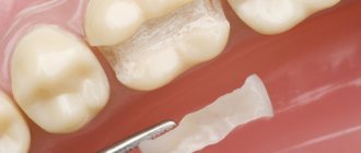
- Filling the exposed area. This procedure is indicated at the initial stage of the pathological process. When a wedge-shaped defect has formed, there is nothing to attach the filling material to, so filling is irrelevant.
- Remineralization of enamel. A product containing calcium is used. The patient will not feel pain during the procedure. The process lasts about 10 minutes. A drug is applied to the affected tooth to help strengthen the enamel, protect the roots from exposure, and prevent tooth loss. In addition, the advantage of remineralization is the return of the enamel to its former shine. How many sessions a person needs is determined by the doctor.
- The enamel is coated with fluoride (fluoridation). Special varnishes or gels that contain fluoride are used. Fluoridation can be simple or deep. With the help of such methods, the dental neck is restored and tooth sensitivity is reduced. With deep fluoridation, the drug strengthens the enamel as it penetrates inside it.
- Installation of veneers. This is a thin ceramic plate. It is applied to a previously ground tooth. This technique is indicated for advanced cases. With the help of veneers, it is possible to completely eliminate the disease and minimize the risk of its recurrence.
- Crowns. Before installing crowns, the tooth is ground down. It is a radical way to protect the tooth root and prevent further progression of the pathology.
- Gum plastic surgery.
Drug and surgical treatment
If the cause of exposure of the neck of the tooth is a lesion or pathology of the mucous membrane, the patient is consulted by an implant surgeon. During the operation, tissue taken from the palate of the mouth is implanted under the tooth root. This helps to increase the volume of gum tissue and cover exposed areas of the tooth. At the end of the surgical intervention, sutures are placed on the gums. After this, the patient is scheduled to regularly visit the attending physician, who will monitor the healing process.
Use an antiseptic solution to rinse your mouth. You can eliminate swelling and redness of the gums with the help of medicinal gels. If pus is present, an antibacterial drug is prescribed. The choice of medication is made only by the attending physician; self-medication is prohibited.
Apical periodontitis of the tooth - what is it?
The diagnosis of periodontitis is made when the patient's periodontal tissues near the apex of the tooth root become inflamed, which is why it is called apical. It affects deep gingival and bone structures, so it is not easy to treat - unlike, for example, caries. Nevertheless, this disease requires immediate intervention, because if the patient is not in a hurry to treat apical periodontitis, then he risks encountering unpleasant consequences of the disease: the appearance of granulomas and cysts, destruction of the alveolar bone, and in the most severe cases, even sepsis.
Symptoms of apical foramen enlargement
Aching pain in the mild stage is the first and main symptom of enlargement of the apical foramen. Already at this stage you should contact your dentist.
Deviations in the development of the apical foramina
In some cases, the size of the holes is initially not large enough, which makes it difficult for the tooth root to receive the required amount of nutrients. This condition requires artificial expansion of the hole to its normal state.
Problems in the apical foramina cause inflammation of the pulp.
Symptoms of pulpitis
The initial stage is painful sensations in the teeth due to temperature changes and other irritants.
The progressive stage is acute paroxysmal pain, often spreading to the entire jaw.
External symptoms are bleeding and tissue growth in the open cavity of the tooth affected by caries, tooth enamel has a gray tint.
Causes of pulpitis
Injuries during periodontal treatment, caries, temperature changes and other opportunities for the development of infection are the causes of pulpitis.
Lack of timely treatment of pulpitis leads to apical periodontitis, a more serious disease. Inflammation during periodontitis moves to the apex of the tooth root.
Complications after treatment of apical periodontitis
Treatment of acute apical periodontitis (as well as chronic) is a rather complex process that requires accuracy and concentration from the doctor. Like any intervention, it can lead to complications under unfavorable circumstances. What symptoms should you pay attention to after treatment of apical periodontitis?
- A sharp pain that occurs immediately after the end of the anesthesia may indicate that when filling the roots, the filling mass has gone beyond the boundaries of the tooth and is pressing on the gums. There is also a possibility of root perforation and breakage of the thin end of the endoscopic instrument in the root canal.
- After putting the medicine into the canals, the tooth hurts when pressed, and the gums are slightly swollen. These symptoms indicate that the doctor has used a strong medication that irritates periodontal tissue. It could also be a sign of an allergic reaction.
However, in most cases, timely treatment of apical periodontitis ends successfully and does not cause complications.
Classification of pulpitis
In dentistry, pulpitis is divided into varieties, each with its own characteristics. Each one can be identified with the help of serious diagnostics. Types of pulpitis:
- Spicy:
- focal;
- diffuse.
- Chronic:
- fibrous;
- gangrenous;
- hypertrophic or proliferative;
- chronic with exacerbation.
Exacerbations of chronic pulpitis are difficult to diagnose. They are often mistakenly interpreted as one of the forms of acute pulpitis. In such cases, the history of the disease (history) plays an important role.