Tumor of the lower jaw involves complete or partial resection of the lower jaw. If reconstructive surgery is not performed, the person becomes disabled and drops out of society.
Reconstructive surgery is the restoration of the form and function of a body part using a person’s own tissue.
Reconstruction of the lower jaw allows you to completely restore the patient’s appearance, chewing and speech function, without resorting to removable dentures.
Cut options
There are three options for accessing the tumor:
- The classic approach involves an incision along the chin and along the neck fold;
- In most cases, an incision along the chin can be avoided and the operation can only be performed through an “invisible” incision in the neck fold;
- In the case of marginal resection of the lower jaw, the operation can be performed transorally - access through the mouth, without external incisions.
Incision options for resection of the lower jaw
The classical approach is becoming a thing of the past, but in Russia it is still widely used, because Most surgeons do not have the necessary skills to perform an operation only through an incision in the cervical fold.
In my practice, I use all access options depending on the indications.
Reasons for appearance
It is not precisely determined why osteophytes appear on the gums. But factors contributing to the disease include:
- Hereditary predisposition;
- frequent inflammation, purulent processes leading to atrophy, deformation of the jaw bone and nearby tissues;
- injuries of the dental system, especially accompanied by fractures of the facial part of the skull with improper reposition of bone fragments;
- complex tooth extraction;
- advanced periodontitis, periodontal disease;
- bite pathologies;
- congenital abnormalities of the jaw.
Often the pathology appears in childhood or adolescence. Also, the appearance of jaw growths may be associated with dysfunction of the endocrine system.
Operation
When reconstructing the lower jaw, a flap from the leg is usually used - the fibular flap. The operation is planned on a computer: the surgeon determines how to correctly select the flap, how to remove the tumor, and how to implant the flap.
Computer-aided planning of mandibular reconstruction
If the patient plans to install dental implants, then an implant surgeon is involved in planning and performing the operation. Tumor removal, reconstruction and implantation are performed in one operation.
Without nutrition, the transplanted flap dies. To avoid this, the feeding vessels of the flap must be sewn into the bloodstream. Since the feeding vessels are very small and fragile (less than a millimeter in diameter), to work with them the surgeon must have microsurgical equipment - be able to suture microscopic vessels under a microscope so that they do not bleed or become clogged.
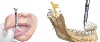
What are the consequences after tooth root apex resection surgery?
Apexectomy can result in various complications, for example, soft tissue swelling (this is a temporary reaction to injury) or even recurrence of the cyst. All negative consequences are usually divided into two types: conditionally normal and pathological.
Fever
This is the body's natural reaction to injury during surgery. An increase in temperature occurs on the first day after apexectomy. Typically varies between 37.5°C and 38°C. In the evening the temperature rises and drops in the morning. It normalizes on its own in 1-2 days. It is not recommended to knock it down if the mark on the thermometer is below 38°C. But if you feel very unwell (general weakness, dizziness, body aches), you are allowed to take an antipyretic drug. Sometimes fever and chills signal the development of serious complications. In this case, the high indicator lasts 2-3 days after the operation, increases every day and is not knocked down by pills. If there are complications, it is accompanied by additional symptoms:
- pain;
- severe swelling of the soft tissues of the face;
- putrid odor from the mouth;
- the presence of purulent plaque on the wound;
- pressure instability.
With such a set of symptoms, you should immediately contact the dentist.
The tooth is loose
result of tooth root resection
Resection involves removing the tip of the root, which often leads to a decrease in the stability of the tooth. If the root is destroyed during surgery, the tooth will become very loose and may fall out. Also, tooth instability is caused by the patient himself if he loads the operated area with solid food.
The dentist returns the stability of the tooth if you contact him immediately after the problem occurs.
To avoid such a complication, it is necessary to follow a gentle diet in the postoperative period. The tissue healing process lasts 4-6 months, during which time the cavity formed during the operation will completely heal, and then the stability of the tooth will become the same. Until this moment, you should not chew nuts, dry foods, or hard candies on the operated side.
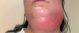
White gums
White plaque can be a sign of a natural healing process or complications: gum inflammation, purulent infection:
- A day after surgery, a white fibrin coating forms on the gum.
- After two to four days, the plaque becomes most noticeable.
- By day 7, swelling subsides, pain disappears, but the gums remain white.
- By day 14, the plaque disappears, new mucous tissue forms underneath it, and the gums acquire a normal color.
The following symptoms indicate the development of a purulent infection:
- severe increasing pain;
- bleeding or profuse ichor (the wound is bleeding);
- rotten smell;
- swelling and redness of the gums for more than a week;
- change in color of plaque to yellowish, gray, brown;
- poor general condition.
This consequence will have to be treated with anti-inflammatory and antibiotics, and can be prevented by rinsing with antiseptic solutions (Miramistin, Chlorhexidine).
Polypous purulent sinusitis
Apexectomy of the upper dental units is accompanied by the risk of breaking through the wall of the maxillary sinus. If this happens, a fistula forms between this cavity and the vestibule of the mouth, which subsequently provokes purulent polypous sinusitis. Its symptoms:
- pain in the lower part of the eye sockets;
- nasal congestion;
- speaking through the nose (nasality);
- it hurts to chew, press on the disturbing area and move your mouth;
- constant headaches;
- weakening or loss of sense of smell;
- purulent fluid from the nose;
- sensation of the presence of a foreign object in the nasopharynx.
This complication is treated with conservative and surgical methods. Conservative treatment consists of taking antibiotics and intranasal hormonal medications (such medications include Mometasone furoate). Surgical treatment involves closing the fistula.
Bleeding from the nose
Nosebleeds during surgery or immediately after indicate perforation (break through) of the bone of the nasal sinus. It can open during resection of the upper teeth, since the roots in the jaw are located close to these sinuses, which is why the sinuses are easily injured at the slightest careless movement of the doctor. If a sinus perforation occurs, the doctor must stop the bleeding, make sure that nothing gets into the hole (perhaps a piece of the root has fallen off and pressed in there), disinfect and close the resulting wound. Then antiseptics for rinsing, antibiotics and anti-inflammatory drugs are prescribed. If fragments and foreign objects get into the sinus, a more serious operation will have to be performed.
Painful sensations
The resection itself is completely painless thanks to anesthesia, but when it wears off (2-3 hours after the procedure), the patient may experience soreness, which is a natural reaction to injury. In the first few hours after surgery, the gums ache very much. The drugs Diclofenac, Dicloberl, Ketanov and the like, which will be recommended by the dentist, will help relieve pain. With each subsequent day, the severity of pain decreases. After 3-5 days it goes away completely. If the pain throbs, the sensation does not go away after 3-5 days, you need to contact the dentist again.
Damage to the maxillary, nasal sinuses, and blood vessels of the jaw
problems associated with resection of the roots of teeth
Damage to the nasal maxillary sinuses during resection of the roots of the upper tooth is manifested by severe bleeding with air bubbles in the blood, a nasal voice of the patient and bleeding from the nose. This postoperative complication is eliminated on the spot. Damage to large blood vessels is manifested by prolonged bleeding from the wound. Stops with sterile swabs with vasoconstrictor medications. If it opens at home, you need to apply a gauze swab to the wound, press it and change it periodically. If the bleeding does not stop within 2-3 hours, you should seek help. To prevent bleeding, you should stop taking blood thinning medications before surgery and not take them after the procedure.
Numbness of the face
This symptom is possible when the trigeminal nerve and its branches are damaged or irritated. Main symptoms:
- nagging pain;
- burning sensation in soft tissues;
- numbness of the soft tissues of the cheek (the cheek hangs and cannot be felt);
- weak sensation when touching the cheek.
Damage to the trigeminal facial nerve is extremely rarely caused by an instrument. More often, irritation occurs due to the pressure that the filling material exerts on the branches. If there is numbness in the face, a course of neurological treatment should be carried out in combination with addressing the cause of the nerve damage.
Suppuration, fistula, lump, abscess
A fistula is a channel connecting foci of infection to the surface of the gum, through which pus comes out. Its appearance indicates poor quality cleaning of the dental canal and wound during the operation. This can be avoided by sealing the perforated area with a special filling, which the dentist must do. Also, the complication is often provoked by the patient by improper oral care in the postoperative period. To prevent gum decay (formation of a purulent lump), it is necessary to regularly disinfect the oral cavity after surgery: rinse with antiseptics (for example, Chlorhexidine) or soda solution. Treatment of complications in the form of a purulent fistula is carried out in two ways:
- Medication with antibiotics (Levomekol, Lincomycin, Metranidazole).
- Surgically (the seal is opened and cleaned so that all the pus comes out).
The success of resection depends not only on the professionalism and experience of the dentist, but also on the patient’s compliance with all prescriptions during the rehabilitation period.
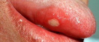
Symptoms
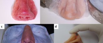
Exostosis of the tooth appears in the form of a convex growth that appears for no apparent reason. Main symptoms:
- Sensation of a foreign body in the mouth;
- discomfort when eating, talking (with large osteophytes);
- pain when pressing on the tumor;
- redness, thinning of the mucous membrane in the pathological area.
A small anomaly can only be detected during a dental examination, since visually it does not manifest itself in any way.
Removal of upper teeth
The bone of the upper jaw is somewhat softer than the lower jaw.
The units are positioned straighter and are easier for the surgeon to grasp. Therefore, removal of the upper molar in many cases is faster and less painful. Patients are often frustrated by the need to have their front teeth removed. The smile zone is always visible, so the absence of one brings a negative attitude and leads to a decrease in self-esteem. But today such an operation is not scary. There are every opportunity to quickly restore the integrity of the dentition.
Removing the front tooth on the upper jaw is simple - it has only one cone-shaped root and is located conveniently for manipulation. The operation will take about 20 minutes, including waiting for the anesthesia to take effect. If there are no contraindications, then simultaneous implantation is recommended - the destroyed unit is removed and an artificial root is immediately installed in the same hole.
Our clinics employ implantologists who are proficient in advanced techniques and have completed internships in foreign clinics. Only premium implants are used. Therefore, you will quickly gain a new beautiful smile. It is possible to install a temporary plastic crown on the pin on the day of removal. The front teeth will be in place, and no one will notice the changes.
Treatment from world-class doctors cannot be cheap, but we have found an opportunity to make help accessible - we treat immediately, and offer to pay for services in installments in convenient amounts, without charging interest.
Removing the upper root of a tooth is a more complex process. The root branch is drilled into several parts, which are removed step by step. This procedure for intervention is typical for molars and wisdom teeth, when the walls have completely crumbled and the root is also not viable.