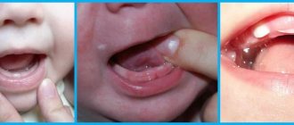
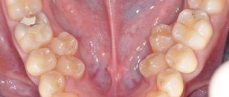
It is believed that at the age of 6-7 years the first milk teeth (lower central incisors) should fall out, and at the same time the first permanent molars grow. But what if the pattern breaks down? Can the upper incisors fall out first or, for example, first the lateral and then the central incisors? What could this mean, and should parents pay special attention to this?
What if my teeth fell out too early? Or did the molars start growing before the incisors fell out? Is it normal for molars to start growing at 4-5 years of age? With what it can be connected? When should a situation alert parents?
Physiological features of the development of baby teeth
The front, or frontal, teeth are located very tightly to each other and only when the child reaches the age of four, diastemas become visible between them - physiological gaps that increase after a year or two against the background of jaw growth. If there are no diastemas at the age of six, this indicates insufficient jaw growth, leads to crowding and requires correction of the bite in children. Physiological abrasion of surfaces, which contributes to the timely development of the masticatory apparatus, begins at three years of age.
Anatomy of different types of temporary teeth
- Incisors.
- Fangs.
- Molars.
Eight temporary incisors have the same structure - a fairly flat crown and 1 root. The incisors of the upper dentition, like the rest of the teeth, are larger than the lower ones. In the center, the incisors have one canal, and in 93% of cases, the lateral ones have two.
The four temporary canines are distinguished by a slightly sharp crown on all sides and the longest root. The crown of the canine preceding the permanent one is shorter and convex. The root has a rounded shape in cross-section and a slightly curved apex towards the buccal direction.
Each of the eight teeth of this type has a multi-cusped chewing surface and ends in several root canals. These are the largest teeth in childhood. The second molars are always larger than the first, which cannot be said about similar molars.
PsyAndNeuro.ru
Early trauma is adverse events (violence, parental deprivation) that occur in childhood and affect later life. Early trauma is thought to increase the risk of developing mental disorders. However, not all people exposed to adverse childhood experiences develop a mental disorder.
There are sensitive periods in psychomotor development during which the brain is especially plastic. If the time of exposure to unfavorable factors coincides with these periods, then the risk of developing a mental disorder increases. On the other hand, the acquisition of new experiences (including unfavorable ones) during such periods has a protective effect. There is no consensus on this issue; scientific research can support both points of view.
To date, there are no reliable methods for assessing the strength of early trauma. Objective research methods, such as identifying DNA methylation disorders and changes in the functioning of the amygdala, are invasive, expensive and time-consuming.
In March 2022, an article by Kathryn A. Davis and co-authors was published in the journal Biological Psychiatry, dedicated to the search for new methods of assessing early trauma. The researchers suggested that teeth could be used for this purpose, which are a kind of “time capsule” that remains unchanged over the years and is imprinted by the environment. The authors of the article summarized the achievements of dentistry, anthropology and archeology, and studied the results of archaeological studies of the human population and primate populations. The collected data were compared with the latest information about the etiology of mental disorders and the role of early trauma in their development.
There are at least five reasons why teeth were chosen as a marker of early trauma.
Firstly, dental development continues during age-related crises. Mineralization of “baby” teeth begins at the 4th month of intrauterine development and ends by 2–3 years. The formation of permanent second molars lasts from 3 to 14–16 years, and permanent third molars, or “wisdom teeth,” complete their formation by 18–25 years. These periods coincide with sensitive periods of formation of the nervous system.
Secondly, teeth retain “traces” of environmental influences (like rings on trees). Odontoblasts and ameloblasts produce proteins that promote the mineralization of dentin and enamel. Traces of this mineralization are visible during the complete formation of the crown in the form of striated exhaustion, Retzius stripes. It is possible that adverse events occurring during the formation of teeth can affect this process and will be “visible” on the enamel.
Thirdly, during the period of tooth formation, exposure to stress factors such as lack of nutrition, illness, poisoning with toxic substances leads to disturbances in the structure of dentin and enamel hypoplasia. According to some studies, similar changes are found both in living people and in archaeological excavations.
Fourthly, presumably, teeth can “retain” the memory of the effects of stress factors, and the main role in this process is played by the so-called neonatal line - a dark stripe in the enamel of baby teeth. There are several studies that have found a correlation between an increase in its width and stress exposure during the perinatal period. Similar studies were conducted on primates, where separation from the mother, death of a sibling, recovery period after surgery, and change of habitat were used as stress factors. A neonatal stress line was also found in the enamel of primates. The idea that teeth store a biological memory of early trauma is supported by studies demonstrating the effect of stress on the condition of hair and nails, which, like tooth enamel, are formed from ectoderm.
Fifthly, the phases of the “life” of teeth coincide with periods of development of major depressive disorder. Thus, the first teeth begin to fall out at the age of 6 years, in the period preceding puberty, in which, as is known, the risk of developing depression increases. Starting at age 13—the age of possible onset of depressive disorders—teeth can be removed for orthodontic reasons. In late adolescence, third molar extraction may occur. All these teeth can be sent for examination to special laboratories to identify “traces” of early trauma. A similar phase pattern can be observed in relation to other mental disorders.
Based on previous research in the fields of psychiatry, dentistry, archeology and anthropology, the authors of the new paper formed the TEETH (Teeth Encoding Experiences and Transforming Health) model. This model is based on the fact that various stress factors acting in childhood affect the development processes of all organs and systems, including the process of dental development (see diagram).
As a result of the study, three postulates were formulated.
First, impaired brain development and dental growth may be associated with early trauma. The fact is that stress factors acting during the development of the nervous system can leave their “mark” in the structure of the brain. Since the brain and tooth enamel are formed from the ectoderm, it is possible that adversity in childhood leaves an imprint on their structure. For example, enamel defects are observed in Down syndrome and cerebral palsy. Moreover, the authors of the article found a study demonstrating that children with low socioeconomic status and decreased salivary cortisol levels have thinner enamel than their advantaged counterparts.
Secondly, exposure to adverse environmental factors at the time of teeth formation can leave a long-term imprint on them, which can later be seen and objectively measured. Pre- and postnatal damage disrupts both the structure (decreased cortical thickness) and function of the brain (decreased activity of the amygdala). The response to stress changes, which is likely due to changes in cortisol levels. Researchers suggest that stress-induced defects in tooth formation are detected in macro signs of damage, such as changes in tooth size, and micro signs, such as changes in the structure and chemical composition of teeth, manifested in the formation of stress lines in the enamel. Since we know the stages of dental development, these signs can be used to trace the time of exposure to a stress factor: macro signs can indicate the presence of early trauma with an accuracy of three to five years, and micro signs with an accuracy of one week.
Thirdly, disturbances in the development of the nervous system can lead to mental disorders. Previously, scientists in their research relied on studying the brain and the level of stress neurotransmitters as markers of the risk of developing psychopathological symptoms. The authors of the new study found at least eight articles describing the effects of various environmental factors on teeth.
To date, the TEETH model has not been tested on a large number of respondents. Despite this, if we continue research in this direction, then perhaps in the future we will have a simple, non-invasive way to diagnose the risk of developing mental disorders.
Author of the translation: Wirth K.O.
Source: Kathryn A. Davis, Rebecca V. Mountain, Olivia R. Pickett, Pamela K. Den Besten, Felicitas B. Bidlack, and Erin C. Dunn. Teeth as Potential New Tools to Measure Early-Life Adversity and Subsequent Mental Health Risk: An Interdisciplinary Review and Conceptual Model. Biological Psychiatry.
What are the anatomical features of “children’s” molars?
- The first upper molars are smaller in size than the second ones. The first has two widely spaced roots, which sometimes merge to the apex. The rudiment of the first “adult” premolar is formed between the roots. Most often, the first upper molars have four root canals, although there are also three or two (in 19 and 5% of cases, respectively).
- The first lower molars differ from others by having an elongated prismatic crown. On their chewing surface there are four tubercles: lingual, higher, and buccal. The first “children’s” molar has 2 roots, with the medial one being longer and wider than the distal one. Often the first lower molars have three canals.
- The structure of the crown of the second upper molars of the dentition resembles the first upper permanent tooth. Four tubercles are clearly visible on the surface, and sometimes a fifth one is noticeable. The buccal surface is almost square with slightly convex sides. An enamel ridge is often visible on the palatal side. Such teeth have three roots, in 85% of cases there are 4 root canals, much less often - 3.
- The second lower molars already have five cusps with less deep grooves than those of “adult” teeth. Both roots are flattened and curved at the top. In 85% of cases, teeth have three canals, although there are exceptions.
With the eruption of permanent, “adult” molars, the change of teeth begins, which lasts from 5-7 to 12-14 years. It is the first molars, which do not have primary analogues, that hold the bite, ensuring the correct placement of the remaining permanent teeth in the arch.
Permanent teeth appeared too early or late
It should be noted that due to the manifestation of acceleration processes, the timing of the eruption of permanent teeth has changed somewhat. Permanent teeth began to emerge earlier, at least 6-8 months. Often, already at the age of five, a child has a mixed dentition, in which the first permanent molars and central incisors are already emerging.
The timing of teething is influenced by both local and general factors.
Local:
- injury,
- premature removal of a temporary tooth,
- edentia,
- tooth retention,
- congenital clefts.
If there is no germ of a permanent replacement tooth or if its position is incorrect, there is no pressure on the bone septum separating it from the roots of the temporary tooth. This leads to a lack of activity of osteoclasts, the cells responsible for root resorption.
Are common:
- heredity,
- genetic factors
- endocrine pathology
- general health.
In particular, with diseases of the thyroid gland, as a result of an imbalance in the mineral balance in the body, there is a delay in the eruption of teeth, both temporary and permanent occlusion. Thus, with hypothyroidism, there is a delay in the eruption of milk teeth by 1-2 years, permanent teeth by 2-3 years.
In general, more and more pediatric dentists and pediatricians are noticing significant differences in the timing and sequence of teeth eruption of temporary and permanent dentition in children. Teething is a multifactorial process. The timing of teething is influenced by race and the general health of the body. None of the currently existing theories fully provides an answer to the mechanisms of teething.
Indisputable, according to many domestic and foreign pediatric dentists, is the fact that the eruption of both temporary and permanent teeth in today’s generation of children occurs earlier than average by at least 6-8 months.
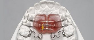
Regular dental checkups will help you avoid serious dental problems.
The pediatric dentist and orthodontist closely monitor the development of the child’s dental system, providing timely assistance if necessary. March 19, 2022


