Content:
- Why does a fragment remain after tooth extraction?
- Symptoms
- What will the doctor do?
- Recovery after surgery
- Do not confuse a jaw bone with a root fragment
- If not deleted
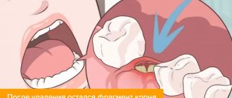
Extraction is a dental operation that is performed quite often. Her technique has been worked out to the smallest detail, so negative consequences are recorded in isolated cases. Sometimes it happens that after a tooth is removed, a fragment remains in the socket. Its presence cannot be ignored. You need to visit the dental surgeon again so that he can remove the forgotten fragment.
Why does a fragment remain after tooth extraction?
Reasons why this problem may occur include:
- A very difficult case. Many people do not go to see a doctor until the last minute. They endure pain and take analgesics. As a result, by the time they visit the dental clinic, an advanced inflammatory process is discovered in their mouth. Visualization of the unit in this case is significantly difficult. The situation is aggravated if the tooth is very tiny. Then, at the slightest pressure, it splits into several fragments. The bleeding that occurs at this moment makes it difficult for the doctor to understand whether all the particles have been removed. To prevent mistakes, the dentist should place all the elements of the tooth on a napkin. This will ensure that the gum tissue is thoroughly cleaned. If he has doubts about the quality of the work performed, radiography comes to the rescue. The image shows whether there are bone formations in the area of dental surgery.
- The root split into a large number of small particles. This is possible with a fracture, progressive inflammation, or malocclusion. Even a very small fragment remaining inside the gum will not allow regenerative processes to proceed normally. The patient will soon experience pain in the operated area and the appearance of bad breath. It is possible that other complications will join the inflammation progressing in the gums.
- Removing the "eight" This tooth is considered the most capricious. It is located in a hard-to-reach place, has long winding roots and a large crown part. It is always difficult to remove it efficiently, so it is important to choose a doctor with extensive experience. It happens that individual elements of the roots grow together with the jaw bone. Then it is very difficult to remove them. After extraction of the figure eight, it is recommended to take x-rays. This simple examination will help avoid complications.
- Anatomical features of the human oral cavity. It happens that the “back” units are very far away. They are difficult to reach.
- During the operation, the adjacent tooth crumbled. If there are two damaged units in a row and you need to remove one of them, the roots of the second may split during extraction. This is important to note. Otherwise, the problem cannot be avoided.
Is it possible to avoid
The success of the operation is largely determined by the experience and professionalism of the doctor. No dentist will remove a tooth without studying in detail the clinical picture of the condition of the oral cavity and the unit itself.
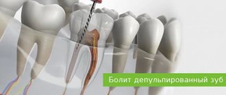
To exclude the possibility of complications developing during the operation and upon its completion, the specialist needs to know the characteristics of the root system, location conditions, find out the quality of the tissue and the degree of destruction of the supragingival part.
If a visual examination does not fully assess the complexity of the case, the patient is recommended to undergo one of the examinations - radiography, CT (computed tomography), orthopantomography or visiography.
If a complex removal is required, the entire range of these examinations is performed. Based on the results obtained, the dentist can clearly determine the number and structure of the root system, find out the direction of their growth and the degree of curvature, calculate the volume and duration of the upcoming manipulation, and prepare the appropriate tools.
An antibiotic sensitivity test is also performed which is very important if the amputation occurs against the background of alveolitis or gumboil.
If the dentist has removed a complex tooth and has some doubts about the cleanliness of the hole, he will definitely refer the patient for a repeat x-ray examination upon completion of the operation.
This is done in order to prevent the development of postoperative consequences associated with leaving a piece of root in the alveolus.
Important! A complete comprehensive examination of the patient before and after the extraction guarantees a safe and quick procedure.
Symptoms
You can understand that a tooth fragment remains after extraction by the following symptoms:
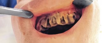
- the gums remain swollen, red, and painful for a long time;
- elevated body temperature persists for 3-4 days;
- worried about joint pain, weakness;
- there is a foul odor coming from the mouth;
- purulent exudate began to separate;
- regenerative processes are slowed down.
Inflammation after extraction normally lasts up to five days. Then the gums should actively recover. If this does not happen, you need to see a doctor. Most likely, serious complications arose after the manipulations.
What will the doctor do?
If you suspect the presence of a fragment, the doctor:
- Conducts a visual examination of the mouth, assesses the condition of inflamed tissues.
- Writes a referral to the patient for x-rays. Examines the resulting image. Any bone formations and particles are clearly visible on it.
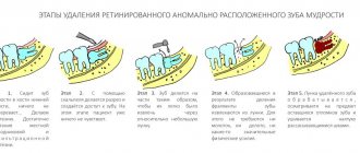
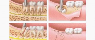
- Based on the characteristics of the localization of fragments and their sizes, the tactics for their extraction are determined. If the particles are close to the surface of the gum, they are removed using a special tool. If they are deep, you have to cut through the soft tissue. In any case, the dental surgeon uses anesthesia so that the patient does not feel pain.
- Removes root particles and treats the wound with an antiseptic solution. Puts an antibacterial drug in it.
Soon after the operation the situation returns to normal. Now the tissues can calmly heal and recover.
How to remove a tooth root without an apex
Stages of treatment:
- the use of anesthesia selected individually, taking into account contraindications and characteristics of the body;
- separation of the circular ligament from the neck and edge of the alveolus (in the absence of inflammatory processes of soft tissues);
- cutting the gums, drilling out bone structures to access the root part;
- direct removal with forceps or an elevator, use of a drill (if necessary);
- treatment of the hole with a special composition to accelerate healing.
In the absence of a crown, the procedure becomes more complicated and cannot be performed at home. An experienced doctor will remove all the fragments along with the base in an average of half an hour.
Recovery after surgery
When the hole is cleaned, you need to create optimal conditions for its speedy healing. To do this you need:
- Take an antibacterial drug prescribed by a dental surgeon. The standard course duration is 5-7 days.
- Carry out therapeutic rinses. For this purpose, you should use Chlorhexidine, Miramistin, etc.
- Drink painkillers. Its use is indicated only if the gums hurt very badly.
- Do not touch the wound with your hands. A blood clot will form on its surface. Under no circumstances should you remove it, lift it, or feel it. If it falls off prematurely, an infection may enter the gum tissue.
- Apply cold compresses to your cheek for the first 24 hours. You can keep them for 10-15 minutes. Warming your face is prohibited!
- During the entire rehabilitation period, do not eat anything spicy, sour, or salty. Such products irritate damaged mucosal tissues and have a bad effect on the course of recovery processes.
- In the first three days, refrain from performing intense sports exercises and heavy physical work, and do not drink alcohol.
Preparatory stage
To reduce risks as much as possible, you must:
- Brush your teeth;
- Rinse your mouth with balm, calendula tincture or chlorhexidine.
- Take a painkiller and wait half an hour.
Prepare:
- Antiseptics;
- Spitting container;
- Tampons and sterile gauze pads;
- Mirror.
The use of pliers and similar tools is prohibited.
Sequence of actions if you need to pull out a tooth at home:
- Cover the tooth with sterile gauze, grasp it tightly and slowly loosen it to remove it from the socket;
- Use tampons to remove accumulated blood;
- Act slowly and forcefully so that no root fragments remain in the hole;
- After extracting the tooth, apply a tampon to the wound and bite it firmly;
- After half an hour, carefully remove the tampon so as not to provoke bleeding.
Do not drink or eat for the next 4 hours. Apply cooling compresses for several minutes. In the next few days, eliminate bad habits and do not use physical activity.
Complications
The human body always tries to cleanse itself of dangerous substances and particles, so it is possible that over time the unremoved root element will “come out” on its own. But it is unreasonable to hope that this will happen. If the image confirms the presence of a fragment in the hole, it is necessary to remove it surgically. Otherwise, you may encounter dangerous complications, including:
- Acute inflammatory process in the root area. A large amount of pus is released, the tissues become severely inflamed. The roots of neighboring units or even the entire dentition may be damaged. It is possible that the abscess will spread to the periosteum.
- Osteomyelitis. Damage to the jaw of an infectious nature. Pathogenic microorganisms actively multiply in the unhealed hole. As a result, osteonecrosis develops - the jaw tissue begins to die. Pathology can occur in acute, subacute and chronic form. The patient feels weakness and headaches. The oral mucosa turns red, the lymph nodes become enlarged. If osteomyelitis is not stopped in time, it can develop into meningitis, meningoencephalitis, sepsis, or brain abscess.
- Inflammation of the periosteum. The gums swell, and when pressed, a sharp pain appears. The roots of units located on both sides of the inflamed hole may be affected. Treatment involves eliminating the root cause of the disorder and carrying out long-term antibiotic therapy.
- Sharp pain radiating to the neck. It is neurological in nature. It is the result of an inflammatory lesion of the jaw.
- Phlegmon. The gums swell and hurt. Purulent exudate is released from it. The neck and cheek become swollen. The patient's general well-being worsens.
All consequences of a root element not removed from the hole are dangerous. Therefore, if you suspect that your tooth has not been completely removed, be sure to take a photo and consult with an experienced dentist. This way you will avoid many health problems.
Price
The total cost of removing the left fragment from the hole consists of the price for the following manipulations:
- radiography (orthopantomography or visiography) - about 1 thousand rubles;
- anesthesia - up to 500 rubles;
- hole cleaning - about 800 rub.
Several other factors influence the final cost: the complexity of the manipulation, scheduling subsequent treatment of the complication, the pricing policy of the clinic, its status and location, and the qualifications of the specialist.
