Acute pulpitis is one of the most common dental diseases. The cause of acute pulpitis in most cases is developing caries. First, it affects the tooth enamel, and if measures are not taken in time, the infectious process reaches deep structures - dentin and subsequently the neurovascular bundle of the tooth or, in other words, the pulp. It becomes inflamed, and in the vast majority of cases the process is acute.
However, other causes of acute pulpitis include:
- trauma - fracture of the crown or root of a tooth, bruise, etc.;
- chemical burn - exposure of the tooth cavity to phosphoric acid, violation of the technology for applying pastes containing arsenic, etc.;
- thermal burn - due to grinding of a tooth to install a prosthesis;
- violation of filling technology - incomplete adherence of the material to the walls of the tooth, improper use of antiseptics, as a result of which bacteria multiply in the voids.
Regardless of the cause of pulpitis, it has quite characteristic symptoms.
Symptoms of acute tooth pulpitis
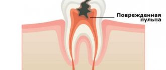
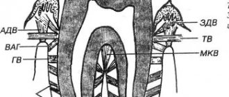
All types of pulpitis have one common feature: damage to the pulp - connective tissue with a large number of microvessels and nerves. It is the presence of a large number of nerve endings that explains the fact that the disease is almost always accompanied by severe pain. Acute pulpitis is considered a signal that pathogenic bacteria have entered the tooth cavity and caused inflammation. Despite the fact that this form is often the initial stage of pulp damage, it causes a number of very unpleasant sensations in the patient from the very beginning.
List of characteristic features
- Sudden attacks of acute pain (on average 10–20 minutes), worsening at night and usually subsiding during the day.
- Reaction to stimuli (usually hot and cold).
- Painful sensations can be localized both within the tooth and can be felt in the temples, ears, and even in healthy teeth (in the second case, these are symptoms of diffuse pulpitis).
The average duration of acute pulpitis is about two weeks. After this period, the disease usually acquires a chronic stage. Due to the gradual destruction of nerve fibers, the pain becomes aching, less pronounced, but longer lasting. Of course, acute pulpitis does not occur on its own, but is a consequence of bacterial activity.
What is pulpitis and how does the disease progress?
Pulpitis develops when carious destruction of the integrity of the tooth occurs when food and microorganisms penetrate the tooth cavity (pulp). Another way of developing the disease is infection from neighboring affected teeth through blood vessels, which occurs much less frequently and is called retrograde pulpitis. There are other, rare pulpitis:
- Traumatic, develops as a result of a crack, break, or chip of a tooth.
- Concrete occurs when there is excessive deposition of mineral formations that begin to replace the internal tissues of the tooth.
- Inflammation develops due to incorrect treatment, the use of low-quality materials, and accidental opening of the pulp.
Regardless of the type of infection, inflammation requires immediate treatment. Among the factors that increase the likelihood of developing pulpitis are: insufficient hygiene, osteoporosis and diabetes.
There are three stages of pulpitis: acute, chronic and aggravated chronic pulpitis. According to the ICD-10 classification used when making a diagnosis, the disease is divided into several categories.
Pulpitis acute
- K04.00 - initial inflammation of the pulp.
- K04.01 - acute focal pulpitis.
- K04.02 - purulent abscess.
The diagnosis is made when the pulp first comes into contact with infection. This includes the serous form of the disease, focal and purulent abscess.
First, the tooth becomes sensitive to temperature: hot or cold. Unlike caries, pain does not subside when the irritant is removed. Then comes the diffuse phase - the pain intensifies at night, is periodic, and often radiates to the temple or other parts of the face. With the transition to the chronic phase, the pain disappears, or is characterized by the patient simply as an unpleasant sensation.
Chronic pulpitis
- K04.03 - fibrous.
- K04.04 - gangrenous.
- K04.05 - pulp polyp.
Chronic pulpitis occurs after the acute phase of the disease. It is characterized by (irreversible) destructive changes in tissues, divided into three types according to the type of changes:
- Fibrous
- the most common sluggish form of chronic pulpitis. It is characterized by the growth of connective, fibrous-granulation tissue in the pulp.
- Gangrenous
- This is a putrefactive inflammation, which is marked by a characteristic odor from the mouth. The gums often swell, and fistulas may form through which exudate flows out.
- Hypertrophic
- the most rare pulpitis, characterized by the growth and exit of the pulp into the carious cavity.
The chronic form is not painful, but severe pain may occur periodically.
Note! In the acute stage, the infected tooth mainly reacts to cold; in the chronic form, the source of pain is exposure to high temperature.
Complicated forms
- K04.1 - pulp necrosis.
- K04.2 - pulp degeneration.
By complicated course of the disease we mean both inflammation of the entire pulp and simultaneous damage to several canals. When the pulp degenerates, denticles are formed - dense mineral formations (stones). They cause pain when there is a sudden change in body position, as this causes them to shift. For example, such patients experience pain when flying, going up in an elevator, or playing sports.
Necrosis refers to the death of cells in the neurovascular bundle of the dental root. In fact, this is the final stage of the disease. Necrosis can occur due to pulpitis, damage to the blood supply to the tooth, or under the influence of toxic components of the filling material.
Causes of acute pulpitis
- Complications of caries. Neglected or incompletely cured caries leads to the penetration of streptococci into the pulp through the dentin tubules - bacteria that have an acid-forming function and thereby create the foundation for the development of infection.
- Injuries of a mechanical or chemical nature that violate the integrity of the tooth. Without a protective layer of enamel and dentin, the pulp becomes extremely vulnerable to bacteria and the effects of the external environment.
- Gum disease in which infection spreads through open periodontal pockets.
- Unsuccessful dental treatment (infection, damage to tooth tissue, etc.).
Stages of treatment
Most of the operations, the main task of which is to heal the inflamed pulp, can be divided into a list of conditional stages:
- Excision of dental tissue.
- Extraction of the neurovascular bundle itself.
- Channel cleaning.
- Filling.
The question of how pulpitis is treated in 2 visits with the removal of teeth and nerves can clearly be answered by any more or less qualified dental specialist. However, not everyone, even experienced doctors, had to deal with the most severe forms of this disease. It is not difficult to guess that the key to the success of the entire therapy as a whole is a visit to a specialized specialist who knows how to handle modern equipment and the advanced range of today’s pharmacological drugs. Such professionals, of course, include dental staff.
Acute diffuse pulpitis
Usually it is the next stage of focal pulpitis (sometimes the peak form of chronic) and appears on the third or fourth day. With diffuse pulpitis, inflammation affects both the coronal and root parts of the pulp. The pain is paroxysmal and long-lasting, with periods of relief being rare. The pain is often radiating and radiates to various parts of the head. The fabric begins to become saturated with exudate.
Some sources also highlight acute fibrous pulpitis, but this is not entirely true. Fibrous pulpitis is a form of chronic; acute fibrous pulpitis is also not a completely correct term. It is a chronic complication of diffuse pulpitis. When gangrenous, extensive tissue necrosis occurs and the tooth often takes on a characteristic gray tint.
What might you encounter after treatment?
Some patients report discomfort after filling. It is important to distinguish whether this pain is correct (post-filling) or one that requires immediate consultation with a doctor.
Mild pain is almost normal after pulpitis treatment. This is due to the fact that a serious intervention was performed, especially if inflammatory and pathological processes had previously occurred. Temporary pain occurs during the first couple of days and occurs when chewing. There is no need to go to the dentist, wait a while, the pain will go away.
The pain is sharp, intensifying and occurring throughout the week may indicate the continuation of the inflammatory process. Swelling of the gums indicates the same thing. In this case, you need to visit your dentist.
Diagnosis of acute pulpitis
Diagnosis of acute pulpitis includes several stages. First, the doctor conducts a visual examination of the oral cavity: if the tooth has a carious cavity or is injured, and the patient complains of acute pain, then there is a high probability that we are talking about pulpitis. Acute and chronic pulpitis have quite significant differences, so visual diagnosis alone is impossible.
Each of the forms of acute pulpitis (they will be discussed later) has its own manifestations and characteristics. To identify the type and stage of the disease, specialists resort to x-rays and electroodontic diagnostics (EDD), which determines the reaction of the pulp to electric current. Rheodentography (assessment of the blood supply to the pulp) and a thermal test are also quite often performed. In addition, differential diagnosis of acute pulpitis is necessary in order to make an accurate diagnosis and not confuse pulpitis with other diseases with similar symptoms.
Prevention of gangrenous pulpitis
Due to the fact that gangrenous pulpitis is a consequence of an acute inflammatory process, a mandatory condition for its prevention is a visit to the doctor for symptoms of pulp damage - the appearance of throbbing pain in the tooth. It is fair to note that a visit to a specialist must be made even before pain appears - it is important to treat caries in a timely manner, without waiting for it to reach the internal structures of the tooth.
In order to prevent gangrenous pulpitis, like any other form of it, it is important to be attentive to oral hygiene, visit a dental hygienist twice a year, and undergo professional teeth cleaning.
Treatment of acute tooth pulpitis
Treatment of caries, pulpitis and acute periodontitis forms the basis of the activities of any dental clinic. It is important for the patient to find a doctor whom he can trust with his health. Below is a table that describes the main types and methods of treating acute dental pulpitis.
Treatment methods for acute pulpitis
✔
Classical technique: tooth depulpation, removal of affected tissue and filling.
✔
Biological method of treating acute forms of pulpitis with partial preservation of the pulp. It is carried out in two visits. The first stage involves preparing the tooth, applying therapeutic and insulating spacers, and installing a temporary filling. At the second appointment, a visual inspection and fixation of the permanent filling is performed. This type of treatment is possible only with minimal inflammation of the pulp and at a young age (up to 26-27 years).
Treatment of acute diffuse pulpitis in the vast majority of cases requires complete removal of the pulp. A biological method with the possibility of preserving part of it is possible in a minimal percentage of cases.
Other treatments
In 2022, there are additional ways for doctors to save people from troubles associated with pulp inflammation:
- laser therapy;
- complex of physiotherapeutic processes.
Both mentioned operations, in principle, can also answer the question of how to cure pulpitis without removing the nerve. True, they can only be used in the early stages, when the pathology has not yet had time to develop and acquire serious symptoms. In addition, to use such funds, a person will need to go to a truly modern and advanced clinic with an experienced medical team. For example, a set of works related to the use of a laser is carried out exclusively in the presence of high-quality, proven equipment. Such installations, of course, are present in dental departments.
Treatment of complicated stages of acute pulpitis
Focal and diffuse pulpitis have their own varieties. The first has serous and purulent forms. Focal acute serous pulpitis often occurs as a complication of caries or during improper treatment of caries. It affects the root part of the pulp, which causes sharp attacks of pain. Diffuse serous acute pulpitis spreads to both the coronal and root parts of the pulp, causing severe swelling, which can develop into phlegmon.
Acute purulent pulpitis (pulp abscess), as the name suggests, is characterized by the accumulation of pus in the pulp chamber, throbbing pain, which can radiate to various parts of the jaw and head (which is why the patient often cannot identify the diseased tooth). For successful treatment, it is necessary to completely remove the pus from the canals, and in most cases the tooth is subsequently killed. Treatment of acute traumatic pulpitis is usually more difficult.
Removal of enamel and dentin destroyed by caries
The doctor must carry out this stage of work carefully and competently: it is important to efficiently remove all tissue damaged and destroyed by caries. Errors in the process are fraught with complications, which will be very difficult to cure. To remove tissue, the dentist will use a drill: with this tool he will remove enamel and damaged dentin, as well as some of the healthy tooth tissue. Healthy tissues are affected as necessary: the specialist must gain access to the pulp chamber and canals.
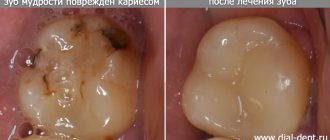
Is it possible to treat deep caries of a wisdom tooth?
The patient can make the decision to remove a wisdom tooth if pain occurs on his own. But only a highly qualified dentist can make a verdict on its treatment.
Treatment of a carious "eight" is considered rational if it has a partial covering in the form of a hood of gum, as well as an antagonist - the third molar located on the opposite side.
Removal shown:
- with a horizontal tooth position;
- presence or absence of 1st and 2nd molars;
- impossibility of treatment due to incomplete opening of the mouth;
- displacement of teeth in a row due to wisdom teeth.
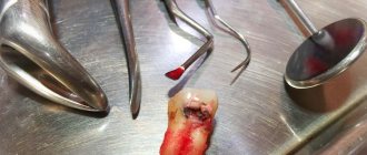
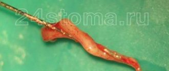
Removal of nerve (pulp)
Before this stage of treatment, the doctor carefully isolates the manipulation area from moisture. It is important to prevent saliva from getting into the canal cavities, because pathogenic microorganisms can also get inside the tooth along with it. Moisture insulation of a tooth can be carried out using different methods, but the most modern and effective method of protection is the installation of a rubber dam - a special lining made of latex material.
Next, the nerve is removed. To do this, the dentist takes a special tool with which he winds and then removes the pulp. The treatment of pulpitis does not end there - the next step in it will be the process of treating the canals, before which the doctor must accurately determine their length and get an idea of the shape. For these purposes, an x-ray is taken, and a specialized tool is used: an apex locator.
Definition of the concept in dentistry
Deep caries is the last stage of the carious process occurring in the tooth, which is characterized by extensive damage not only to hard tissues, but also to the deep layers of dentin. In general, the concept of “deep caries” reflects the depth of the lesion.
Caries can be:
- primary - a consequence of untreated secondary caries;
- secondary (recurrent) - the occurrence of a carious process under a filling in a previously treated tooth.
Based on the sensation of pain and clinical examination, the disease is divided into 3 forms:
- Acute form. It is determined by the narrow entrance opening of the carious cavity and the wide base (during examination), as well as by descriptions of the patient’s sensations - reaction to food temperature and chemical irritants.
- Chronic form. It is characterized by a wide (funnel-shaped) inlet and a narrow base. The patient feels pain when the “hollow” is clogged with food and during examination with a probe by the dentist.
- Deep cervical caries. It is characterized by the development of demineralization of enamel tissue and the carious process in the cervical area of the tooth (near the gums).
According to the clinical course, caries is also divided into several forms:
- compensated,
- subcompensated,
- decompensated.
Clinical researches
Repeated clinical studies have proven that regular use of preventive toothpaste ASEPTA ACTIVE for a month can reduce bleeding gums by 60%, improve the overall condition of the oral cavity by 44% and reduce inflammation by 33%.
Sources:
- Clinical and laboratory assessment of the influence of domestic therapeutic and prophylactic toothpaste based on plant extracts on the condition of the oral cavity in patients with simple marginal gingivitis. Doctor of Medical Sciences, Professor Elovikova T.M.1, Candidate of Chemical Sciences, Associate Professor Ermishina E.Yu. 2, Doctor of Technical Sciences Associate Professor Belokonova N.A. 2 Department of Therapeutic Dentistry USMU1, Department of General Chemistry USMU2
- The problem of treating inflammatory periodontal diseases in patients suffering from type 2 diabetes mellitus E.A. KHROMOVA* Ph.D., Associate Professor of the Department I.V. KULIK* Ph.D., Associate Professor of the Department N.A. UDALTSOVA**, ***candidate of medical sciences, associate professor of the department; Deputy Chief Physician for Organizational and Methodological Work A.K. IORDANISHVILI****,*****Dr.Med.Sci., Professor, Professor of the Department *Department of General Dentistry, North-Western State Medical University named after. I.I. Mechnikov" of the Ministry of Health of Russia; ** St. Petersburg State Budgetary Healthcare Institution “Dental Clinic No. 29”, Frunzensky District, St. Petersburg; ***Department of Maxillofacial Surgery and Surgical Dentistry of the Federal State Budgetary Educational Institution of Higher Education "St. Petersburg State University" of the Government of Russia ****Department of Orthopedic Dentistry of the Federal State Budgetary Educational Institution of Higher Education "North-Western State Medical University named after. I.I. Mechnikov" of the Ministry of Health of Russia; *****Department of Maxillofacial Surgery and Surgical Dentistry of the Federal State Budgetary Educational Institution of Higher Education "Military Medical Academy named after S.M. Kirov" of the Russian Ministry of Defense;
- Clinical studies of antisensitive toothpaste “Asepta Sensitive” (A.A. Leontyev, O.V. Kalinina, S.B. Ulitovsky) A.A. LEONTIEV, dentist O.V. KALININA, dentist S.B. ULITOVSKY, Doctor of Medical Sciences, Prof. Department of Therapeutic Dentistry, St. Petersburg State Medical University named after. acad. I.P. Pavlova
Deep caries of the front teeth. What are the differences in treatment?
Deep caries of the front teeth should be treated as carefully as possible so as not to burn the pulp. The main difficulty in treatment is:
- minimum thickness of all tooth tissues;
- inconvenient location of the cavity, if we are talking about caries located in the interdental space.
When infection penetrates into the pulp, pulpitis develops rapidly. A few hours is enough. The disease is accompanied by increasing pain. It needs to be treated immediately.