A panoramic photograph of teeth or orthopantomogram (OPTG) is a radiographic examination technique that makes it possible to study in detail the anatomical features of both jaws, the condition of soft and bone tissue, dental roots, maxillary sinuses and joints. The study is carried out using equipment with reduced radiation exposure and does not pose any threat to the health of the body. Today we’ll talk about what this is, why a panoramic shot is needed and how often it can be taken.
What is OPTG and why is it performed?
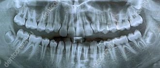
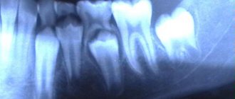
Unlike a standard targeted photograph, an orthopantomogram allows you to obtain a fairly clear and detailed image of both jaws at once with the ability to study not only the condition of the teeth and their root system, but also the jaw bones, paranasal sinuses and other important anatomical structures. To understand what this diagnostic technique is, you first need to answer the questions: what does it do and why is it carried out. So, here's what the x-ray shows:
- deviations in the formation of occlusion - defects in bite, position, shape and size of teeth;
- early stages of caries and hidden foci of infection;
- periodontal pockets;
- neoplasms: cysts, granulomas, fibromas;
- hidden inflammatory processes and the extent of their damage;
- condition of the temporomandibular joint;
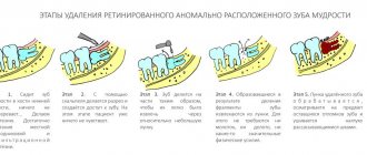
- impacted teeth that have not yet erupted - including wisdom teeth;
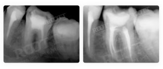
- condition of the root canals after treatment;
- diseases of the paranasal sinuses, for example, sinusitis.
The procedure is mandatory before orthodontic treatment, prosthetics and, of course, implantation. In the latter case, the image gives the doctor the opportunity to carefully examine the condition of the jawbone, select suitable locations for implants, or prescribe a bone tissue augmentation procedure. To understand what the result of OPTG looks like, take a look at the example image below. Important! It should be noted that today, within the framework of one-stage implantation techniques, the preparatory stage involves undergoing a computed tomography scan. This is a more modern X-ray method that allows you to obtain a three-dimensional image of the patient’s jaws with the possibility of layer-by-layer study of tissues, without distorting the actual sizes and shapes of the teeth. OPTG is also performed before treatment or removal of wisdom teeth or “eights”. The image will allow you to identify the exact location and position of the elements, detect hidden carious lesions and make a decision on the need for removal. In general, an orthopantomogram helps to carefully examine the clinical picture, make an accurate diagnosis and prescribe adequate treatment.
Orthopantomogram: description of the procedure
An orthopantomogram, or OPTG, is an overview of a circular photograph of the teeth, in which the entire jaw is visible, unfolded in a plane. In addition to the teeth, their roots, canals, the presence of fillings and crowns, the panoramic image shows the condition of the periodontal tissues, maxillary sinuses, and sinuses.
A panoramic photograph of the teeth, or orthopantomogram, allows you to detect hidden carious cavities, inflammation, cysts, abscesses, the presence of periodontal pockets, identify the exact condition of the roots and periodontal tissues, and assess the condition of the maxillary sinus and temporomandibular joint. It is also necessary to take an orthopantomogram when planning orthodontic treatment with braces or aligners.
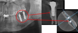
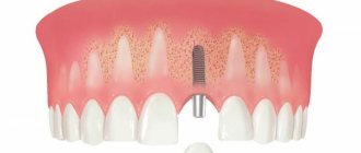
In the process of preparing for dental implantation, using an orthopantomogram, a specialist can determine whether there is enough bone tissue for implantation or a sinus lift is needed, how many implants will be needed and what size they should be, and also predict the dynamics of their engraftment.
It is very important to take a panoramic photo to determine the position and condition of your wisdom teeth. Only an orthopantomogram can give an adequate idea of the “eights”; no targeted photograph will help here. Therefore, competent dentists decide whether to treat or remove wisdom teeth only after x-raying the entire jaw.
What equipment is used as part of the study?
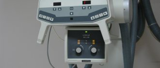
This is not a new method - by now, OPTG has been actively used in dentistry for decades. And if earlier film cameras were used to obtain a panoramic image, today they have been replaced by more modern digital devices - orthopantomographs. The image is generated in a special computer program and, if necessary, can be printed on film. Images taken using digital equipment are more accurate and detailed, which virtually eliminates the risk of making an incorrect diagnosis. Answering the question about how long a panoramic photograph of teeth is valid, it must be said that the information received will remain relevant for several months - a maximum of six months. If more time has passed since the examination, the procedure must be repeated.
Indications and restrictions
The procedure is completely painless, carried out as quickly as possible and without any discomfort for the patient. OPTG is prescribed in the following cases:
- as part of implantation – carried out to study bone tissue, select implant models and places for their implantation. Based on the data obtained, the doctor will be able to draw conclusions about the advisability of building up the jawbone;
- to assess the quality of dental canal treatment and the condition of the roots - before prosthetics;
- before starting orthodontic treatment - before installing braces and other corrective structures;
- before an upcoming operation, complex removal, including wisdom teeth;
- to examine the dental system in children, assess the correctness of its formation and identify possible deviations;
- in order to determine the extent of damage to periodontitis and periodontal disease;
- to identify neoplasms in hard and soft tissues.
This procedure may be required in various clinical cases. X-ray diagnostics is not advisable in the first and last trimester of pregnancy - it is carried out only in the presence of emergency indications and with the permission of a gynecologist.
Features of conducting OPTG for children
We figured out what is visible in panoramic photographs and why they are taken, and found out that this is a completely safe diagnostic method. Now it should be noted that an orthopantomogram for children is carried out taking into account some nuances. A radiation exposure of 55 μSv (1 shot) is completely safe for a child, so a similar procedure is allowed from 5-6 years of age. The examination assumes that the patient must stand still for some time, but for young children this task can be quite difficult . When it comes to small patients, the procedure is carried out in exposure mode - the area of exposure is reduced, and the sensor is adjusted to a trajectory of movement corresponding to the small size of the jaws.
“My son also recently had a panoramic photo taken. He's 10, everything went great. Now there are new technologies everywhere, everything is safe. The procedure took less than a minute, and the doctors assured me that there was nothing wrong with it. How else can you start correcting your bite? The orthodontist prescribed OPTG for us. So far I’ve made the plate, but in a couple of years we’ll get braces. There’s no place here without pictures!”
In pediatric dentistry, OPTG is used to detect the rudiments of permanent teeth, assess the correctness of their formation, to diagnose adentia and identify malocclusions. Most often, this procedure is prescribed by an orthodontist before starting to correct defects in bite and teeth position. The procedure is completely safe for the child
Harm and degree of radiation exposure
Let's start with the fact that radiation exposure is measured in micro- or millisieverts - μSv or mSv, respectively. According to the resolution of the Ministry of Health of the Russian Federation, the permissible radiation dose within the framework of OPTG for an adult patient should not exceed 55 μSv, and for children under 15 years of age - 24 μSv 1 . It should be noted right away that only digital X-ray equipment meets these requirements, while film devices provide a slightly higher load. To determine how many times you can take a panoramic photo, you need to take into account that the maximum permissible radiation dose for an adult patient per year corresponds to 1000 μSv. However, it is still not worthwhile to partake in such procedures, even despite their obvious safety and harmlessness . X-ray irradiation has a negative effect on a weakened body, especially with repeated examinations. The children's body is more susceptible to this kind of influence, therefore it is strictly forbidden to exceed the permissible dosages when examining small patients. When contacting the clinic for OPTG, be sure to clarify what type of equipment is used and what the degree of radiation exposure from one image will be. Some private dentistry purchases cheap, outdated equipment, which can produce 2-3 times the recommended dose. The best option where you can take a panoramic photograph of your teeth is a specialized diagnostic center equipped with modern high-precision equipment.
Orthopantomogram on film or digital: which one to choose?
Today, electronic devices have almost replaced film devices. Their cost is higher, but they do not require a large amount of consumables, as for film orthopantomographs. To develop film, you need a darkroom, which is prohibited by law from being placed in residential buildings, which can become a problem for small private clinics. In addition, the irradiation exposure of film devices is several times higher than that of electronic devices. An orthopantomogram of the jaws, made on a digital device, is immediately transferred to the doctor’s computer, who can be located anywhere. It is much easier to store such an image: it will simply remain in the patient’s electronic record and can be viewed at any time. If desired or necessary, you will receive the result of the orthopantomogram on disk or printed on transparent film.
Unlike a panoramic photograph on film, a digital orthopantomogram will not be lost, which means that the patient will not have to undergo radiation again, which is present during dental x-rays, albeit in very small doses: only 0.002 - 0.003x mSv, whereas, according to Sanitary Regulations , a person can receive up to 1 mSv per year for research purposes, and from the environment we receive 2 - 3 mSv over the same period. In addition, the image clarity and image resolution on digital cameras is much higher than on film cameras.
Digital OPTG - description of the process
Now let's move on to consider the question of how a panoramic photograph of teeth is taken. To begin with, the patient is asked to remove all metal products from himself so that they do not affect the operation of the equipment and the final result. Next, the patient stands in the place indicated by the specialist, bites down on a special plate that ensures the immobility of the jaws, and after turning on the orthopantomograph, its moving part begins to rotate around the head. The procedure takes literally half a minute - the exact duration will depend on the type and newness of the equipment. Next, the resulting image is formed in digital form, that is, in a special computer program, or on film media (outdated version). A digital photograph can also be printed onto film if necessary.
Orthopantomogram 3D
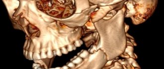
A three-dimensional panoramic x-ray of the jaw can be performed with a computed tomograph. He takes many sequential photographs in different projections and then completes the image in a computer program. The result is a three-dimensional image of the jaw without distortions, which are inevitably present on a conventional otopantomogram, since it conveys a curved object in a flat form. Only with the help of computed tomography does it become possible to look at an object from a different angle or in a different projection without taking new pictures. Also, this method of examination allows the doctor, in the absence of the patient, to see an image of his jaw and examine it from any side and even at any depth.
Information content of the image and reading (deciphering) technique
If you undergo an examination at a specialized diagnostic institution, the finished image is usually accompanied by an explanation from a specialist with a list of detected problems. Many people are interested in how to decipher an image on their own, how to read it correctly, and whether it can be understood at all. It should be noted here that in order to correctly explain the results of OPTG, you need to have a good understanding of the anatomy of the maxillofacial apparatus, and without the appropriate education, little can be understood. It is better to entrust this task to an experienced, qualified doctor.
Orthopantomogram during implantation
As part of implant treatment, a panoramic image is taken more than once. First, an X-ray examination is carried out at the preparatory stage - the doctor carefully examines the resulting image, assesses the condition of the bone tissue, selects places for implants, and makes a verdict on the need for bone grafting. If we are talking about one-stage immediate loading protocols, then patients are usually sent for a computed tomography scan of the jaw - a 3D image provides more detailed information about the clinical picture.
3D diagnostics makes it possible to study a more detailed three-dimensional image
You will have to undergo a repeat examination after implantation. In this case, OPTG will give the doctor the opportunity to assess the quality of the operation performed, monitor the process of engraftment of artificial roots, and recognize possible complications in the later stages of their development. Several photographs may be required during the osseointegration period of the implants.
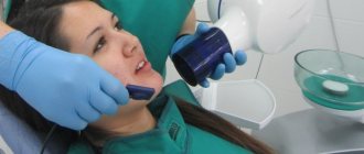
How is an orthopantomogram done?
No special preparation is required for the study. You will need to remove glasses, earrings, hair clips, other metal objects, and dentures. An orthopantomogram is usually done in a standing position. A special X-ray protective apron-cape is put on the chest. The position of the head is fixed with special clamps. As directed by the doctor, you will need to bite a special plastic mark (which is located in a disposable cover) - this will allow the image to be correctly centered. The tongue should be kept pressed to the palate. During an orthopantomogram, the module will move around the head, this will take 15-20 seconds, and during this time you must stand strictly still in order for the image to be of the proper quality.
You can get an orthopantomogram in Moscow at Family Doctor JSC.
Sign up for diagnostics Do not self-medicate. Contact our specialists who will correctly diagnose and prescribe treatment.
What are the advantages and disadvantages of OPTG
An orthopantogram can significantly improve the quality of diagnosis, and today dental treatment is practically not carried out without radiography, except for minor problems. Among the undeniable advantages of the technique, experts highlight the following points:
- speed of the procedure - literally in a couple of minutes the image is ready for study;
- modern equipment makes it possible to adjust the height and trajectory of the emitter, which made diagnostics possible for children and wheelchair users;
- minimal radiation exposure and absolute safety for the body;
- high quality and accuracy of the obtained images;
- the ability to zoom in on the area under study and study it in detail on a computer monitor.
With an orthopantomogram, minimal radiation exposure is used. If we talk about the disadvantages, the main disadvantage of OPTG is obtaining a flat two-dimensional image. As a result, the image may slightly distort the dimensions of teeth, root canals and bone tissue. Such deviations can vary from 15 to 30%. To obtain a more accurate image, it is better to resort to computed tomography.
Approximate prices
How much a panoramic dental photograph will cost directly depends on whether the study will be carried out using film or digital equipment - in the latter case, the cost will be an order of magnitude higher. Approximate prices are given below:
- a photograph on a film camera – about 700 rubles;
- digital photo - from 1000 rubles and 150-200 rubles for printing on film.
We have already mentioned above that today, for a more detailed layer-by-layer study of the tissues of the jaw apparatus, computed tomography (CT) is performed. Essentially, this is the same panoramic photo, but much more detailed and in 3D format. An important advantage of this technique is the accurate transmission of the sizes and shapes of all elements of the dental system.
Where can I take a panoramic photograph of my teeth or an orthopantomogram?
Today, an orthopantomogram is included in the list of services in almost every dentistry. You can use it in the same place where you decided to undergo treatment, or you can take a panoramic photo at a better price in another place. Some clinics offer regular clients a panoramic dental photo at a significant discount, which means it can be quite inexpensive. Prices for an orthopantomogram in Moscow start from 750 rubles and reach 2500 rubles. On average, dentistry in Moscow offers a high-quality panoramic photograph of teeth for 1000 - 1300 rubles.