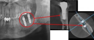
An x-ray is the dentist’s main tool in making the correct diagnosis. However, a conventional orthopantomogram or targeted photograph has limited diagnostic potential and does not provide complete data on the condition of the teeth and maxillofacial area. But technologies are constantly being modernized and today, more informative technology has come to the aid of conventional radiography - dental computed tomography (CT).
What does a 3D dental x-ray show?
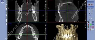
3D dental tomography is a highly accurate diagnostic method that makes it possible to obtain a three-dimensional image of the dental system in different projections. Volumetric images obtained with CT allow the specialist to enlarge, rotate and examine the area of interest from all sides and at different depths:
- The entire maxillofacial apparatus.
- A dentition or an individual tooth.
- Paranasal sinuses.
- Bone and periodontal tissues.
Dental CT allows you to detect inflammation, assess the homogeneity of the filling material and check the quality of installation of a filling, crown or implant, see the number of dental roots and their fragments, identify neoplasms, assess the degree of curvature of teeth, determine the exact parameters of bone tissue (height, width, density, etc.). d.). The information obtained allows the doctor to optimize treatment measures and predict the result.
Why is this necessary?
An image during dental implantation plays an important role from a diagnostic point of view. With its help, the doctor can objectively assess the condition of your bone tissue and, based on this, select the most suitable implantation option. For a more complete picture, several photographs are therefore taken using different methods.
As you know, implants, if professionally installed, can last you for decades. It is a competent examination before the operation that guarantees such a result. And without a photo, an examination is impossible. With its help, the specialist studies:
- Height and condition of bone tissue.
- The presence of remnants of roots that must be removed without fail.
- How are the chewing elements located?
- What condition are the canals of the teeth?
- Distance between teeth.
Dental implantation in Voronezh, carried out at the Dentika clinic, always ends successfully. This is a common practice thanks to accurate examination and diagnosis using modern equipment, as well as the use of high-quality implants.
Why are dental x-rays prescribed?
A 3D photograph of teeth is performed if the following indications exist:
- Injuries of the maxillofacial area.
- Preparation for endodontic treatment (structure of root canals, pathological processes in the periodontium, degree of pulp damage, etc.).
- Diagnosis of neoplasms (cysts, abscesses, granulomas, tumors).
- Anomalies of development and deformation of the maxillofacial apparatus.
- Quality control of filling and implant installation.
- Planning of orthodontic treatment (identification of impacted and dystopic teeth, analysis of the condition of the tissues around each tooth, etc.).
- Detection of hidden periodontal cavities and pockets.
- Implantation planning (assessment of jaw bone parameters, indications for sinus lift or osteoplasty, modeling the result of implantation).
- Endogenous pathologies of the maxillary sinuses.
Three-dimensional x-ray examination is the gold standard when planning any complex dental procedure or surgery. CT allows you to quickly make an accurate diagnosis, competently plan treatment or dental prosthetics, and monitor the results.
Why is a CT scan prescribed before dental implantation?
Installing implants to replace a lost tooth or group of teeth is not as complicated as planning an operation. The dentist must take into account many different factors: the thickness of the alveolar ridge, the proximity of the nasal sinus or infraorbital foramen, the location and inclination of implantation of the artificial root, and other features. This cannot be done without special equipment.
When performing surgery using conventional x-rays, the risk of errors is very high. Bone deficiency will not allow the implant to anchor well, so it may fall out. If a pin is installed too small, the chewing load will be too strong, which will ultimately lead to peri-implantitis. If you make a mistake with the location, then during the operation the bottom of the maxillary sinus, nerves, sinuses may be damaged, and the artificial tooth itself will cause a pathological change in the bite.
What equipment is used
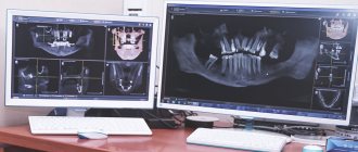
To carry out 3D diagnostics, a three-dimensional computed tomograph SOREDEX Scanora 3D with advanced functionality is used. This is the latest generation equipment, which allows you to obtain three-dimensional images of the anatomical structures of the maxillofacial region in a few seconds, with the least radiation exposure for the patient.
The program analyzes the obtained multiplanar sections and builds them into a 3D model, thanks to which the specialist is able to accurately assess the condition of the dental system, detect all pathological processes occurring in this area and competently plan a treatment regimen.
A virtual 3-dimensional model of the scanned area can be recorded on any digital media (CD, flash drive), which allows the attending physician, if necessary, to view diagnostic data or involve related specialists in the analysis of the received information.
Sight shot
Targeted dental radiography is a rapid diagnosis. A 2D image allows the doctor to see the condition of 1-3 teeth located nearby. To carry out this type of diagnosis, the ROOTT clinic uses a digital radiovisiograph “SCANORA”. The examination is carried out directly in the dental chair.
Targeted photographs of teeth
The purpose of a targeted photograph before implantation is to assess the condition of neighboring teeth. If they require treatment, the patient is referred to a general practitioner. This is necessary so that infection of neighboring teeth does not get into the socket when installing the implant.
On a targeted image you can find:
- inflammatory process in the area of the apexes of the roots of the teeth;
- quality of sealed canals;
- possible fractures and root injuries.
Advantages:
- obtaining a clear image of 1 to 3 teeth in a row
- carrying out several procedures in a row without the risk of adverse effects on the body
- To assess the clinical picture, the image can be enlarged several times
- In the clinic, targeted X-rays are performed free of charge
There are no absolute restrictions for conducting targeted imaging, but contraindications include early pregnancy and infancy (up to 2 years). The lactation period is not considered a contraindication.
Possible harm
Cone beam dental computed tomography is the safest and fastest diagnostic method. Thanks to the use of a conical X-ray beam, the radiation dose received during the study is 10 times less than when using spiral CT. And the pulsating mode of the X-ray beam further reduces the radiation dose. The three-dimensional computed tomograph SOREDEX Scanora 3D is one of the safest devices in terms of X-ray radiation dose - only 0.035 m3v.
However, despite the safety of the study, CT also has contraindications. If we just talk about dental tomography, it is not performed during pregnancy (in the 1st trimester). 3D dental x-rays with contrast are prohibited for pregnant and lactating women, patients with endocrine disorders (diabetes mellitus, thyroid pathologies), renal failure and intolerance to iodine-containing drugs.
Contraindications for CT examination
Computed tomography is not recommended more than twice a year. If the procedure was recently performed in another clinic, the patient can show the doctor the finished CT result of the teeth. In some cases it is contraindicated.
Restrictions on CT scanning:
- period of pregnancy, breastfeeding, as the body is exposed to x-rays;
- claustrophobia - the patient is in a confined space, so panic may occur;
- high motor activity of children under 14 years of age due to the need to remain completely still during scanning.
During breastfeeding, you can temporarily transfer the baby to artificial feeding. The milk needs to be expressed and discarded. Breastfeeding is resumed the next day.
Online consultation with a doctor
If you are concerned about the condition of your teeth. Painful sensations arise, gums bleed for a long time, and seals have appeared on the jaw. You are concerned about previously installed dental implants. And there was a need to take a 3D photo of the teeth. Then, after receiving three-dimensional visualization, it is better to go for an examination or consultation with a dentist to interpret the images and compare the results with your current complaints. It is impossible to independently understand the nuances of a 3D image, much less make a diagnosis. The specialist will explain the situation and give recommendations before the in-person appointment.
Computed tomography after implantation
In some cases, a CT scan may be required after implant placement. It is prescribed to determine the reasons for the instability of the artificial root, identify complications, and confirm the engraftment of the rod.
If you need to do a CT scan of the brain, you must inform your doctor about the presence of implants. Metal objects can distort the image, but doctors with many years of experience can correctly interpret CT results if a patient has titanium roots or metal pins.
Advantages of the method
- The ability to rotate, enlarge, and examine images in any projection and section, which is impossible with conventional 2-dimensional scanning.
- The examination lasts only a few seconds (8-20 seconds).
- Complete diagnostic information.
- Maximum security.
- Digital information format.
- Detection of any pathological processes at an early stage.
- No prior preparation required.
- 3D reconstruction without distortion or artifacts.
- A wide range of purposes - from endodontic dental treatment and implantation to maxillofacial operations.
Initial examination
Before dental implantation, you need to visit the dentist's office for an initial consultation. During this procedure, the doctor will carefully examine the oral cavity, assess its condition and give a referral for x-ray diagnostics and tests. Already at this stage, the dentist can:
- determine indications and contraindications for implantation;
- inform the patient about the features of the upcoming operation.
In addition, during the initial examination, the patient must fill out a questionnaire (survey card), in which the presence of bad habits, chronic diseases and surgical interventions should be noted.
| We strongly recommend that you fill out the survey card honestly. Then our doctors will know better how your body will behave during implantation. |
Is there an alternative to CT
There are many other diagnostic imaging methods (x-ray, orthopantomogram, ultrasound, etc.), but only CT provides the possibility of highly accurate, separate images of all types of tissue at different angles and to different depths. Although a panoramic dental photograph remains an equally important diagnostic tool for a dentist today, it can only provide a general overview. In turn, a 3D tomogram allows you to obtain not a single flat image of the jaw, but a whole series of sequential multiplanar images in different projections and without the distortions inherent in a panoramic image.
Example:
due to the different density of bone structures exposed to X-ray radiation, it is impossible to see less dense bone in a 2-dimensional image; accurate information is provided by a 3- D image of the teeth.
Possible consequences in the absence of CT diagnostics
Dentists strongly recommend that patients undergo a CT scan. It not only minimizes the risks of complications, but also helps doctors perform quality work in accordance with the clinical picture.
When performing an operation without CT detail, the patient may encounter the following problems:
- nerve damage;
- perforation of the maxillary sinus and the development of chronic runny nose, sinusitis;
- the need for subsequent orthodontic treatment;
- reinstallation of the implant;
- increased sensitivity, inflammation of the gums due to improper distribution of the chewing load;
- reducing the service life of the titanium root.
To avoid such consequences, you need to go through all the preparatory procedures prescribed by the doctor.
How does the procedure and decoding work?
To take a 3D photograph of teeth, a standing or sitting patient needs to bite a special plate and fix his position in the device using a fixing stand. During the entire scanning time, you must remain absolutely still.
The tomograph sensor makes a series of revolutions around the patient’s head for 8-20 seconds, producing about 200 images in different projections. Processing digital data takes 5-15 minutes, after which the information is written to a disk or flash drive. No preparation is required, you just need to remove all metal jewelry from your neck, ears, and hair before the procedure.
Read also
A new way to restore teeth - implantation without surgery
Modern dental implantation: methods and their features
Why do you need to prepare for implantation?
It allows you to divide patients into those who are indicated for implantation and those who are suitable for traditional methods of orthopedic care. Not only negligent attitude towards health is a contraindication. Excessive coffee consumption and smoking are social contraindications.
What if the implant doesn’t take root?
The likelihood of rejection is minimal. It does not exceed 1% out of 100. Sometimes rejection is a consequence of an incorrect approach to treatment, when the material and dimensions of the implant were chosen incorrectly. Sometimes the patient himself is to blame for such developments. Let’s say that during a dental implantation operation he did not maintain sufficient hygiene, as a result of which inflammation of the tissues of an infectious nature developed.
Preparation before dental implantation
will disappear if the patient’s chronic diseases worsen. Individual intolerance to individual components will also interfere with engraftment.
Here's Why Preparing for Dental Implants
. The presence of an experienced doctor nearby in combination with the patient’s compliance with all recommendations leads to successful implantation.
What to do before implantation?
- Do not eat or drink 2 hours before implantation. But eliminating breakfast completely is not recommended. Strength will definitely come in handy, so a light snack will even do you good.
- You are allowed to take sedatives an hour before surgery. The doctor chooses the drug individually when the patient experiences strong emotional anxiety before the manipulation. What you shouldn't do is use alcohol as a stress reliever. It is prohibited to use it.
- Teeth brushing is required 30 minutes before implantation.
- Good sleep and a positive attitude are almost half the success, because in this way you protect yourself from dark thoughts. Go to bed early the night before surgery, preferably the day before.
Speaking about preparation for implantation, a specialist should definitely pay attention to the oral mucosa. The following parameters are important:
- Thickness;
- Mobility.
The anatomical formations of the dental system and their location relative to each other are also important. In addition, the implantologist looks at the features of the bone tissue, its structure and size, and the anatomical features of the jaw.
Preparation for dental implantation. Video.
How to avoid pain?
Individual selection of painkillers is the prerogative of the attending physician. If dental implantation is planned, preparation for surgery
makes it possible to select anesthesia to eliminate pain. Patients today have access to high-quality painkillers. In combination with modern equipment, they allow dental implantation to be performed without pain.
Age restrictions
There are no strict restrictions. Even in elderly patients whose age exceeds 70 years, good results are observed. As for adolescents, the success of engraftment is determined by the characteristics of the bone tissue. For boys, it is recommended to have implants from 19 years of age, while for girls the procedure can be performed from 17 years of age.
Dental implant design and choice
It is defined:
- Condition of the dentition;
- Width of jaw bone tissue;
- Heights of the jaw bone tissue.
First of all, the outcome of implantation is influenced by two factors:
- Surgical technique;
- Proper preparation of the material.
The next step is the doctor’s recommendations and how the patient implements them.
How long will implants last?
There is no service life as such for dental implants. But this is fair to say in relation to those of them that were installed correctly. If the implant takes root well, the person will use it throughout his life. He will not have to worry about the service life of the implants.
How to achieve this:
- Adhere to general hygiene rules;
- Reduce jaw injuries to a minimum;
- Avoid mechanical factors;
- Professional cleaning combined with regular home cleaning.
All this will extend the life of the implants and guarantee oral health.
Features of treatment in early and late stages
Obstetricians and gynecologists say that dental treatment during pregnancy is safest in the 2nd trimester. And the maximum danger from dental interventions is in the first trimester. And at the same time, doctors are convinced that the risk of harm to the fetus from progressive dental diseases is much higher than the risks from treatment. Therefore, in case of deep caries or pulpitis, you must contact the dentist immediately, regardless of the stage of pregnancy. If symptoms of early caries appear, you can delay the visit to the doctor until 20 weeks. But at 36-38 weeks (if there is no severe pain and purulent inflammation), it is better to refuse dental interventions. Treatment during this period can provoke premature birth.
Important: if the pregnancy is short and the pregnancy is not yet visually noticeable, be sure to inform the dentist before starting treatment that you are expecting a baby. Otherwise, he will not be aware of the need to take extra precautions during pain relief and X-ray diagnostics.