We always pay a lot of attention to tooth enamel; this is what we clean, treat it with care and try not to destroy it under any circumstances. But what is hidden under this shell, which protects the inner body of the tooth from bacteria and all sorts of negative factors? This is dentin - the inner shell, which is located both in the crown and in the root, and protects the pulp and nerve fibers. Dentin is essential to the functioning of each tooth and affects its appearance and health.
Let's try to highlight a few interesting facts to understand what dentin is and exactly what function it performs.
Tooth dentin: what is it, composition and structure
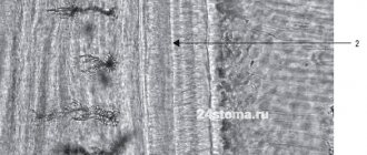
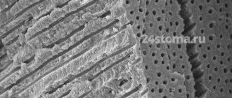
Dentin is a hard tissue located under the cement and enamel of teeth, respectively, in the roots and crown. It has a special structure and is second only to enamel in strength. Such properties are provided by a unique biochemical composition, which is represented by organic (20% by weight) and inorganic (70%) compounds, water (10%). The thickness of the dentin layer ranges from 2 to 6 mm. In the cervical region it is smallest. Its structure is presented in the following table:
| Types of fabric | Layer Features | Functionality |
| Dentinal tubules and tubules | Permeate the entire structure | The fibers that fill these elements are responsible for nutrition and metabolic processes in all dental tissues. Thanks to the tubules, dentin has high permeability |
| Peritubular dentin | Covers the surface of tubules and tubules | Minerals contained in this layer in large quantities nourish all dental tissues. |
| Interglobular dentin | Located between the dentinal tubules. There are two types: peripulpal, or predentin (surrounds the pulp), mantle (in contact with the enamel) | The processes of pear-shaped odontoblast cells, which make up predentin, deliver nutrients to the dental epithelium |
Basic diagnostic methods
Independent determination of dentin caries is possible based on pain and other unpleasant symptoms. However, only a doctor can make an accurate diagnosis after conducting a full dental diagnosis. It includes basic and additional research methods.
The main diagnostic methods for dentin caries include the following:
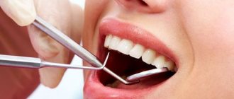
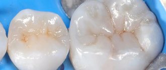
- Probing - using a sharp probe, the doctor determines the presence and depth of carious cavities. If the bottom of the cavity has a soft structure and a light brown color, this may indicate an acute course of caries. Dense pigmented dentin in the bottom area indicates a chronic form of the disease. Also, with the help of probing, you can detect hanging edges of tooth enamel, chips on it, and by the localization of pain during probing, determine the depth of the carious lesion.
- Percussion is tapping on the teeth with tweezers or the back of a dental probe.
- Palpation - feeling.
These methods are used to determine the viability of the pulp, which is especially important in differential diagnosis.
Additional research methods include:
- Thermometry - measuring the temperature of dental tissues.
- Electroodontometry is the study of pulp using electric current.
And the following methods are used to assess the condition of the pulp:
- X-ray - an X-ray of the tooth helps determine the exact location of the carious cavity and its depth.
- Transillumination - the transillumination method is widely used in the diagnosis of hidden dentin caries, for example, with an isolated, atypical form of progression. When transilluminated, healthy tissues appear transparent, while those affected by caries create a shadow effect.
When the doctor has made a preliminary diagnosis based on the above methods, differential diagnosis of dentin caries with other types of dental diseases - enamel caries, pulpitis, periodontitis may be required.
Types of dentin
Dentin is constantly changing. The following types are distinguished:
- Primary. It is formed during intrauterine development of the fetus. It is preserved until the tooth erupts.
- Secondary (replacement). Its formation begins when a tooth appears above the gum. Lasts for life. It differs from the primary one in its slower pace of development and in a large amount of organic matter. Has high permeability.
- Tertiary (irregular). Appears in case of injury, disease and tooth preparation. Its occurrence is a response to an inflammatory process or external irritants. Tertiary dentin performs a protective function. During its formation, the structure of the tissue changes: the tubules have a chaotic arrangement or are absent altogether (usually due to severe inflammation). But the process of formation of tertiary tissue is not endless.
There is also sclerosed (transparent) dentin, which appears as a result of the deposition of the peritubular layer in the tubules. This leads to their gradual narrowing. Similar changes can occur with age. As lime salts are deposited on the walls of the tubules, permeability is reduced, thereby prolonging the viability of the pulp.
Types, location areas
From an anatomical point of view, the mantle and peripulpar layers of dentin are distinguished. The first is located immediately under the enamel, the second - above the pulp. Histologically, dance and radial layers are distinguished, the differences of which relate to the location of collagen fibers.
The following types of fabrics are distinguished:
- primary, beginning to develop before the unit erupts;
- secondary, the development of which begins after the tooth has erupted;
- tertiary, also called reparative, irregular or replacement, appearing as a response to injuries and other damage.
The predentin zone is also distinguished, where secondary and tertiary species begin to form. This region belongs to the germinal region, where the process of formation of new layers begins.
Diseases
Dentin, like other tissues, is subject to age-related changes. The volume of minerals decreases, and degenerative processes begin. The tubules become narrower, sclerosis occurs, and the trophic function practically stops. Metabolic and other processes slow down, which negatively affects the strength and condition of tissues. Resistance to the development of caries decreases, teeth become thinned by various negative factors.
Caries is the main disease diagnosed most often. This is due to various nuances, for example, a local decrease in acidity, demineralization, an increase in the value of carbohydrates and others. With the death of odontoblasts and narrowing of the tubules, tissue nutrition deteriorates, which also makes the layer vulnerable to pathogenic bacteria.
Recovery methods
For treatment, it is recommended to remove the affected tissues followed by filling. After cleaning, the cavity is treated with antiseptics, which protects it from further problems. Composite fillings restore the shape and size of the tooth and allow for proper load distribution. The color is selected in accordance with the shade of the surrounding fabrics.
To prevent the development of diseases, it is recommended to regularly visit the dentist. During a routine examination, problems will be identified in a timely manner, which will allow treatment to begin while preserving tissue.
Functionality and regenerative qualities of dentin
Dentin consists of several layers. They, as already mentioned, protect the pulp from bacterial infections. In addition, this elastic fabric acts as a shock absorber for the enamel. It is also responsible for tooth sensitivity. Thanks to dentin, the pulp responds in a timely manner to infection, injury, and thermal irritants.
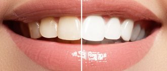
Many people believe that the color of teeth is determined by the enamel. In fact, it is translucent, and the snow-whiteness of a smile depends on dentin, which comes in different shades - from white to gray and yellowish. Often yellowness indicates high mineralization of tissues, and therefore the health of the teeth.
Dentin has the ability to regenerate thanks to monoblast cells. Recovery is possible only if the innervation of the dental tissues is preserved. After depulpation, regeneration processes stop.
Hypophosphatasia
The peculiarity of this pathology is a reduced level of alkaline phosphadase in the blood, as well as impaired bone mineralization. The disease is inherited in an autosomal recessive manner. It is characterized by skeletal deformities and changes in the structure of the lower extremities.
Due to pathological root resorption, temporary incisors fall out prematurely. Abnormalities of calcification are observed in primary teeth, while the crowns retain their normal size and shape. Uneven calcification is observed only in permanent molars and incisors. Signs of hypoplasia are obvious on the enamel.
X-ray shows:
- osteoporosis of bone and underdevelopment of the alveolar process;
- root resorption too early;
- resorption of the root tips of intact teeth;
- W-shaped roots;
- sometimes - taurodontism.
Caries: causes of development, diagnosis, therapy
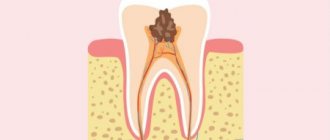
Caries is the most common disease of dentin. Develops with insufficient oral care, low levels of acidity in the mouth, abuse of sweets, and genetic predisposition. The onset of caries development is the stage of a white or pigmented spot. At this stage, dentin is not yet affected. The internal tissues of the tooth are involved in the painful process if left untreated. Depending on the degree of their damage, they distinguish between medium caries, when the middle layers of tooth tissue are affected, and deep caries, in which the pathogenic process almost reaches the pulp.
With caries, the degree of mineralization of tissues decreases and their structure changes. The processes of some odontoblasts are destroyed. The inflammatory process begins. If it is not eliminated in time, it affects the pulp. Diagnosis of the disease is not a problem for the dentist. Upon examination, a carious cavity filled with soft dentin is noticeable. Therefore, it is important to consult a doctor in time. When caries is diagnosed, treatment involves the following:
- administration of anesthesia (if necessary);
- removal of infected tissue;
- cavity disinfection;
- installation of a seal.
In case of deep caries, when a pathological process has developed in the root canals, treatment is mainly carried out in several stages. First, a temporary filling is installed. Various materials can be used for it, including artificial dentin - powder made from kaolin and zinc sulfate. It is diluted with water. Oil dentin is also used in dental practice. The powder is similar in composition to aqueous powder, but is mixed with a mixture of clove and peach oils.
Such components will protect the tooth cavity from bacteria and food debris getting inside. With the help of a sealing material, the medicine will remain in the canal for the required time. Among the properties of artificial dentin, it is worth highlighting its low strength. Therefore, the final stage of treatment should be replacing the material with a permanent filling.
To strengthen the enamel and dentin, remineralization is carried out. To do this, fluoride-containing varnish is applied to the teeth and special toothpastes are used. After the procedure, the enamel becomes less permeable. Carious cement has a layered structure. It is little permeable or impenetrable. Beneficial substances actively penetrate into demineralized dentin to a great depth and react with its inorganic component.
How does a tooth hurt with dentin caries?
Different types of pain are characteristic of different stages of caries. When dentin is damaged, the stages of medium and deep caries are distinguished. In the first case, the carious cavity is located closer to the enamel; in the deep stage, it is as close as possible to the area of the dental nerves.
The middle stage of dentin caries is characterized by a painful reaction in response to temperature or taste stimuli, the pain quickly passes, and the intervals without pain are quite long.
If caries is in the stage of deep damage to dentin, it is characterized by intense long-term pain with short intervals without pain.
Deep caries is the most dangerous, because at any moment it can be complicated by pulpitis, periodontitis, purulent inflammation, and the spread of infection beyond the dental tissues. But even the moderate one can turn into the deep stage at any moment, so if you have a toothache of any nature, you should consult a dentist as soon as possible.
Pathological abrasion of teeth
Abrasion occurs when the bite is incorrect, when the chewing load is distributed unevenly, in the absence of some teeth, constant exposure of the enamel to aggressive substances, and vitamin deficiency. Among the main causes, it is also worth mentioning bruxism - involuntary grinding of teeth during sleep. In these cases, the enamel is erased first, followed by the dentin.
The choice of treatment methods depends on the provoking factor. They may include wearing mouthguards, inlays, and temporary dentures. This allows you to adjust the height of the teeth. When the patient gets used to it, prosthetics are performed (usually with non-removable structures).
Isolated dentin caries: what are its features
Sometimes lesions and extensive carious cavities form in the dentin of a permanent tooth while maintaining the integrity of the enamel. This is how isolated, atypical caries develops. Its difference from the classical form is that the carious lesion does not affect the enamel, but immediately passes to the dentin.
This happens, for example, in cases where a person uses fluoride preparations for a long time for preventive purposes. This leads to strengthening of the enamel located on top of the affected dentin, and, as it were, “seals” the pathological process inside the tooth.
Cost of services
If treatment is started on the day of treatment, consultation and treatment plan are free
| Treatment of superficial caries with the installation of a light-curing filling | RUB 3,800 |
| Treatment of medium caries with light filling | 4,200 rub. |
| Treatment of deep caries with light filling | RUB 5,900 |
| Complex treatment of 1 tooth canal | 1,300 rub. |
| Filling 1 tooth canal | 1,400 rub. |
| Consultation | 550 rub. |
| Titanium anchor pin | 1,300 rub. |
| Anchor pin (carbohydrate, fiberglass) | RUB 1,850 |
