Summary
Reconstruction and rehabilitation of patients with defects of the upper jaw are the most difficult in reconstructive maxillofacial surgery, occupying the minds of oncological surgeons working in this area, microsurgeons, plastic surgeons and orthopedic dentists involved in maxillofacial and somatotropic prosthetics.
The fundamental difference between patients who have undergone anatomical reconstruction of the alveolar process of the upper jaw, as well as nasal support with microsurgical grafts and patients who have undergone maxillofacial prosthetics with obturation structures is an increase in the volume of the respiratory space, improved speech, the absence of atresia, frequent acute respiratory viral infections and, fundamentally, in the absence of regular relocation of definitive orthopedic work [1]. An important difference is also the lack of mobility of dentures on dental implants, which avoids chronic trauma to surrounding tissues.
This article describes the experience of rehabilitation of patients with defects of the upper jaw using fibular and Chinese skin-bone flaps with further plastic surgery of bone and soft tissues in order to create conditions for dental implantation, orthopedic conditions and features of fixed prosthetics for this group of patients.
The developed protocols for the use of a particular graft on a vascular pedicle are described in terms of the type of defect and further restoration of bone joints, as well as measures for the formation of anatomically close to normal conditions for implantation and prosthetic dentistry in patients with a reconstructed upper jaw.
Prosthetics using implants
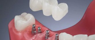
Today, implantation is the most effective method of restoring teeth. Implants can be installed for both partial and complete edentia.
The implant plays the role of a durable artificial root. It is implanted into bone tissue. The support necessary to put on the crown is attached to it. With proper care, the implant will serve the patient throughout his life. Replacements may require solely a crown.
The huge advantage of this method is that the bone tissue does not atrophy after tooth extraction. In modern dentistry, implantation can be performed in one operation - immediately after the removal of a dental unit, an implant is implanted in its place.
Introduction
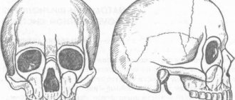
The upper jaw is the most complex area in reconstructive maxillofacial surgery, being more responsible for appearance than others, and due to the presence of such organs and anatomical formations as the eyes, zygomatico-orbital complex, and teeth.
The upper jaw is a support for the lower jaw from the point of view of the stationary container of antagonists, also a zone of concentration of air cavities and a complex of buttresses. Defects and deformations in this area lead to disfigurement and limit or make impossible vital body functions such as chewing, swallowing, breathing, and vision. Restoration of buttresses is necessary from the point of view of optimizing the transfer of stress of the masticatory load, as well as to strengthen the reconstructed alveolar process of the upper jaw. [3]
Despite the positive results of tissue autotransplantation using microsurgical techniques, creating conditions for fixed and conditionally removable dental prosthetics remains a serious problem. The presence of a skin area with an array of subcutaneous fat makes the creation of an orthopedic bed and the attachment of soft tissues around the completed orthopedic structures almost impossible and reduces the functional significance of the work performed aimed at the rehabilitation of the chewing function. [2]
The article is devoted to the synergistic efforts of a maxillofacial surgeon and an orthopedic dentist to create functionally significant conditions and long-term results in patients with reconstructed upper jaws using skin-bone autografts on a vascular pedicle.
In the world literature available to us on the rehabilitation of chewing function in patients with jaw defects, we did not find any descriptions of the formation of a prosthetic bed, features of the choice of orthopedic structures, or long-term results after dental prosthetics.
Key words: jaw defect, lack of occlusion, obstructive prostheses, dental implantation, fixed prosthetics, integrated implants, micro-surgical autotransplantation, bone grafts, fibula, parietal bone, three-dimensional bone augmentation.
Contraindications for prosthetics
The method of prosthetics is always chosen by the attending physician, taking into account many factors, including the general condition of the patient and an assessment of his psychological comfort. Among the contraindications that may cause this procedure to be postponed for some time are the following:
- various blood clotting disorders in the postoperative period or those of a chronic or temporary nature;
- first and last trimester of pregnancy;
- menstruation in women;
- presence of chronic infectious diseases;
- the presence of acute inflammatory processes;
- decreased immunity of various origins, etc.
Even ARVI can make it impossible to carry out prosthetic procedures. Before visiting the dentist, it is recommended to measure your body temperature, and if there are indicators different from the physiological norm, you should inform your doctor. Prosthetics for loose teeth
is carried out only after consultation with a doctor, since it can be difficult due to the lack of supporting teeth necessary for installing a crown. You may be asked to remove loose teeth.
Material and methods
From 2008 to 2014 in the reconstruction of the upper jaw, we used a fibular autograft for the reconstruction of total defects (5 cases) and a radial autograft for the reconstruction of subtotal defects (10 cases). When using a radial skin-bone autograft in the subsequent placement of dental implants, there is a need to recreate the second cortical-spongy layer of the alveolar process on the lingual side, which can be achieved using parietal or mandibular free autoblocks.
With VRGN, relatively small defects of the alveolar process of the frontal part of the upper jaw are noted; more often the problem is the elimination of the defect of the palatal plate. Bone reconstruction requires the alveolar process, represented by genetically modified bone tissue in the projection of the bone cleft. A palatal bone defect does not require repair, since functional loss does not occur in the absence of reconstruction of the bone component. It should be noted that with complete clefts the main problem is communication with the nasal cavity. To eliminate defects of the upper jaw, we have developed a technique for using a free split chin graft and transplanting a fasciocutaneous graft [Application: 2010118686/14, 05/12/2010. Patent No. 2435537 ](4 cases).
In cases where the patient's health condition did not allow microsurgical transplantation, we installed Zygomatic system implants and fixed prosthetics (6 cases).
After a clinical assessment, instrumental diagnostic methods were used to study the pathology of the integrity of the jaw/s: OPTG, TRG, CT (with angiocontrast and soft modes).
If a tumor was present, the extent of resection was assessed and the optimal graft was selected according to the developed algorithm. If the X-ray picture of the tumor was detected, a biopsy was performed and, when the diagnosis was verified, resection of part or all of the jaw was performed. For jaw defects, after clinical and instrumental analyses, treatment planning began.
Preoperative planning was carried out using 3D visualization programs that made it possible to simulate the sizes, shapes, and position of the revascularized autografts relative to the bone structures, taking into account the positioning of the condylar processes of the mandible in the temporal fossae (anterior-superior position in the articular cavities) according to CT studies and the formation of anatomical buttresses for the upper jaw. A special feature of the programs is the absence of distortions in the individual dimensions of the patient’s skull.
We used fibular and radial autografts, since only they allow us to perform 3D modeling of the bone component congruent with the defect in the maxillary defect. Subsequently, when using a radial graft, the technique of transplanting free cortical-cancellous grafts was used to thicken the alveolar process for further dental implantation. Thus, a comprehensive and step-by-step reconstruction of the jaw defect was carried out to create conditions for restoring chewing function with fixed and conditionally removable orthopedic structures, which is important for any jaw defects.
After engraftment and activation of the dental implants, the subcutaneous fat pad of the fibular and radial flaps and the removal of excess skin pad were removed.
Orthopedic rehabilitation consisted of choosing the design of the prosthesis and precise adherence to the accuracy of taking impressions to transfer the complex relief of soft tissues and the position of the implants onto precise plaster models. The usual occlusion and central relationship of the jaws were recorded for the subsequent selection of prosthetic tactics for patients with reconstructed upper jaws. Plaster models were installed into the articulator using a facebow. The existing occlusion and centric relation were assessed. Depending on the type of defect, we selected the desired position of the lower jaw to create optimal chewing function and aesthetic results.
Particular attention was paid to the creation of a prosthetic bed for a future orthopedic design supported by dental implants. In these clinical situations, there is no attached keratinized mucous membrane around the implants in the oral cavity, which makes it difficult to accurately transfer the relief of the soft tissues of the prosthetic bed to create adapted non-removable orthopedic structures.
After the manufacture and fixation of temporary crowns on the implants, the presence of tumor-like growths of the mucous membrane around the installed orthopedic superstructures, which were histologically described as polyps, was noted. Patients complained of bleeding and discomfort around the installed crowns. Attempts to change the eruption profile of structures from implant shafts did not produce a positive result.
We used a method of simultaneous surgical correction of the subcutaneous fat area of autografts with the installation of provisional structures supported by dental implants. In a number of cases, we used a removable compression acrylic plate on the gum formers, which held the surgically created profile until permanent prosthetics or installation of provisional fixed and conditionally removable structures.
After 3 months, we made permanent orthopedic fixed or conditionally removable structures.
For the manufacture of orthopedic structures, we used cobalt-chrome alloy, titanium and zirconium dioxide.
As a rule, after the integration of implants and placement of gum formers at the prosthetic stage, we were faced with the need to modernize the prosthetic elements of the implantation system for specific clinical cases.
The gum formers were needed to be longer and predominantly conical in shape. The cylindrical shape of the gum former created parallel walls of the crater of the mucous membrane, which caused rapid collapse of the walls during the placement of the impression coping and caused severe discomfort to patients during manipulations. We attribute this to the presence of excess connective tissue layer, which is located on the autograft bone.
It is important to note that immediately after corrective operations on the skin-fat part of the flaps, orthopedic compression plates were fixed to prevent further growth of the soft tissue component.
Patients were monitored every seven days and appropriate compression plate adjustments were made.
Depending on the chosen permanent structure, a set of further orthopedic manipulations was carried out to make prostheses.
The results were recorded using a Canon D 60 camera, 100 mm lens, and MR-100 ring flash.
Description of clinical observations
Patient U. , 18 years old, was admitted to the clinic on April 1, 2008 with a diagnosis of osteoblastoma of the upper jaw, a condition after subtotal resection of the upper jaw on the left. From the anamnesis: a neoplasm was discovered and removed in early childhood at the Federal State Institution Central Research Institute of Infectious Diseases and Maxillofacial Surgery. The patient was sent to the clinic of the Russian Scientific Center for Surgery named after. acad. B.V. Petrovsky.
Status localis: the configuration of the face in front and profile is changed, the upper lip is retracted on the left. On palpation, a defect in the alveolar process of the upper jaw on the left is noted.
Mouth opening is not limited, there is a through defect in the alveolar process of the upper jaw with teeth 21-28, the patient wears an obturating denture, the denture teeth are in the bite.
Bite according to the second class according to Angyu.
On OPTG and CT: there is a defect of the zygomatic tubercle, alveolar process on the left, absence of the spine of the upper jaw, the base of the pyriform foramen on the left, defect of the tubercle of the upper jaw on the left. An impacted 28th tooth is noted in the remains of the pterygomaxillary articulation.
Treatment tactics: since the defect of the upper jaw was subtotal and through, a radial skin-bone graft on a vascular pedicle was used. When transplanting a radial cortico-periosteal-skin graft, a zygomatic-maxillary buttress was formed using a free split graft from the branch of the mandible on the right. The second stage involved the formation of the alveolar process using parietal grafts.
Rice. 1. Appearance of the patient, existing defect of the upper jaw, bite, obturating prosthesis, CT scanThe patient was noted to have no nasal lining, and to anatomically restore aeration, before transplanting a microsurgical graft, the nasal lining was formed with local tissues.
Rice. 2. Stages of planning the bone and soft tissue components of the graft Rice. 3. Stages of surgery Rice. 4. Computed tomography of the patient after surgery and condition on the third day
Rice. 5. Scintigram after surgery
In the postoperative period, there was a divergence of the sutures and a reduction in the volume of the flap, and therefore a protective mouth guard was made to protect the soft tissue component of the flap from food getting into the graft. Kappa also pressed the flap into the defect area to allow healing by secondary intention.
Rice. 6. Postoperative mouth guard2 months later, the patient, being a cycling athlete, fell from the “saddle” and received a fracture of the upper limb at the site of collection of the radial skin-cortical-periosteal graft on the vascular pedicle. Therefore, the patient underwent osteosynthesis with the Ilizarov apparatus.
Rice. 7. Condition after osteosynthesis of the upper limb on the left, condition in the oral cavity Rice. 8. 3D reconstruction of the alveolar process using parietal grafts, CT scans after the second operationAfter 5 months, we installed three dental implants in the area of the reconstructed alveolar process using parietal bone autografts. Thanks to this technique for reconstructing the alveolar process, it was possible to obtain adequate bone thickness and height for placing dental implants.
Rice. 9. Implantation in the area of 3D reconstruction with parietal blocksAfter 5 months, we opened the implants and applied a method of one-stage surgical correction of the subcutaneous fat pad of the autograft with the installation of elongated gum formers in dental implants, which simplified the formation of soft tissues during surgery.
After completion of the surgical stage, the alginate impression was removed from the upper jaw and a compression plate was made using cold polymerization at a pressure of 3 atmospheres from acrylic plastic.
The patient was advised to wear the plate constantly, removing it only for hygiene procedures. Inspection and correction of the plate fit were carried out every seven days for a month.
Thus, we were able to form a stable mucosal contour around the gingival formers.
The next stage was the production of a conditionally removable prosthesis supported on a beam structure on implants.
Rice. 10. Correction of the subcutaneous fat area of the autograftConsidering the fact that microsurgical elimination of the jaw defect and reconstruction of the alveolar process was carried out by transplantation of free parietal cortical autografts, the further stage of orthopedic rehabilitation is practically no different from classical prosthetics for patients with an extended terminal defect of the dentition.
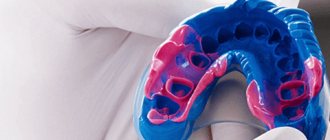
In order to be able to produce a conditionally removable beam-type prosthesis supported by implants, it was necessary to produce an accurate plaster model that displayed the entire relief of the prosthetic bed and accurately reproduced the position of the implants. To do this, we took primary impressions of the upper jaw using Clip-transfers for implants. The primary plaster model gave us the opportunity to make a custom impression tray with prepared transfer checks for precise transfer of the position of the implants.
Having made an accurate working model of the upper jaw and a model of the antagonists, we installed them in the articulator according to the average parameters. Registration of the central ratio was carried out using an occlusal hard wax plate with refinement on ALUWAX (soft wax with the addition of aluminum filings for long-term heat retention and elasticity).
Rice. 11. Impressions and plaster modelsAfter analyzing the relationship of the plaster models in the articulator, we manufactured a beam frame supported by three implants. The beam frame was made by vacuum casting from cobalt-chrome alloy. To achieve a passive fit of the frame, we used adapters for external connection with the implants.
We fixed titanium ball locks into the cast part of the frame. Then a model of the upper jaw was made from a fire-resistant mass for modeling and casting the mating part of the prosthesis itself.
Rice. 12. Beam frame and mating part of the prosthesisTaking into account the possibility of changing the relief of the soft tissue component of the autograft, the frame of the response part was modeled in such a way that it was possible to change the acrylic base part, changing the fit of the prosthesis to the bed.
After fitting the beam and frame in the oral cavity, we manufactured the base part of the conditionally removable denture with the installation of acrylic artificial teeth, using the method of cold polymerization of plastic under a pressure of 3 atmospheres and a temperature of 50 degrees Celsius. We took into account all possible characteristics of the patient at rest and when smiling, creating the most natural appearance of the entire orthopedic restoration.
The final fixation of the prosthesis was carried out in a certain sequence:
- Cleaning and disinfection of all components of the prosthetic restoration.
- Removing the gum formers, irrigating the internal shafts of the implants with a solution of 3% hydrogen peroxide and 0.05% chlorhexidine solution, installing adapters on the external connection.
- Installation of the beam structure on three implants, tightening the fixing screws with a force of 30 N/cm2.
- Fixing the actual removable denture on the beam frame, checking the occlusion, teaching the patient about hygienic measures.
Surgical preparation of the oral cavity for prosthetics
Surgical preparation of the oral cavity for prosthetics
80% of patients who use dentures do not have good support for their fixation in the oral cavity.
The task of surgical preparation of the oral cavity for prosthetics is the creation of a reliable supporting structure from bone and soft tissues for the subsequent manufacture and optimal functioning of dentures. Reasons for the lack of support for fixing dentures in the oral cavity:
1. Atrophy of the alveolar processes of the jaws after tooth extraction.
2. Trauma during tooth extraction and frequent loss of one of the walls of the alveoli.
3. Progression of atrophy due to systemic diseases and involutive processes (osteoporosis of bones in menopause and postmenopause).
4. Progression of atrophy due to wearing prostheses, especially if they are poorly fixed.
5. Atrophy of the alveolar process in diseases of marginal periodontium.
6. Disproportion of the alveolar processes during atrophic processes of the jaws.
7. Individual anatomical features of the jaws (severity of the torus, malocclusion).
8. Reduction of the arches of the vestibule of the oral cavity, the severity of the frenulum of the lips and tongue, mucous and muscle cords due to atrophy of the alveolar processes.
9. Cicatricial changes in the mucous membrane after tooth extraction, wearing dentures, injuries and operations.
Preparing a patient for oral surgery
1. Referral from an orthopedic doctor.
2. The patient’s psychological readiness to use prostheses, especially removable ones, as well as for surgical interventions in this regard.
3. Conducting a general examination and determining the absence of general contraindications to surgical interventions.
4. A thorough examination of the oral cavity (assessment of changes in soft tissues and bone formations that impede prosthetics).
5. Assessment of jaw models and x-ray examination.
There are: • Operations on the bone tissues of the jaws. • Surgeries on soft tissues (oral mucosa, muscle bundles, periosteum) • Surgeries on the peripheral branches of the trigeminal nerve. • Raising the bottom of the maxillary sinus (sinus lift), nose.
Operations on the bone tissues of the jaws
Alveoloplasty
Indications:
Detection of deformation of the alveolar process during treatment of a postoperative wound after the removal of one or more teeth.
Operation technique:
1. Peeling off the mucoperiosteal flap to expose the affected area of bone.
2. Elimination of deformation along the outer and inner surface of the alveolar arch using bone nippers, a bone file, bur or cutter.
3. Placement of the mucoperiosteal flap in place and suturing.
Intraseptal alveoloplasty
Indications:
Protruding interalveolar septum, displacement of the lateral plate of the alveolar process, discovered during tooth extraction surgery.
Operation technique:
1. The protruding or inadequate interalveolar septum is removed.
2. Reposition the lateral plate of the alveolar process of the upper jaw or the alveolar part of the lower jaw with strong finger pressure.
Reduction and correction of the uneven bone surface of the alveolar process of the upper jaw, the alveolar part of the lower jaw
Indications:
Bone tuberosity, which prevents normal prosthetics, which is caused by bone protrusions, as well as excess, hypertrophy of the soft tissue covering it.
Operation technique:
1. The mucoperiosteal flap is peeled off and the alveolar process or alveolar part of the jaw is exposed on both sides.
2. Areas of protrusions, irregularities and other bone deformations are removed with bone nippers, burs, and cutters.
3. If there is excess soft tissue, they are excised, and the wound is sutured with knotted catgut sutures or sutures made of polyamide thread.
When operating on the upper jaw, it is necessary to take into account the boundaries of the maxillary sinus in order to avoid damage to its bottom. On the lower jaw - you should pay attention to the location of the mental foramen and the neurovascular bundle emerging from it.
Removal of exostoses on the upper and lower jaws
Indications:
The presence of pronounced exostoses in the area of the upper and lower jaws, contributing to the balancing of the dentures and traumatization of the mucous membrane.
Operation technique:
1. A linear incision is made along the alveolar arch or supplemented with vertical incisions, folding back an angular or trapezoidal flap.
2. Each section of deformed bone is exposed.
3. Exostoses are removed with bone nippers. Smooth the surface of the bone with a bur or cutter.
4. The mucoperiosteal flap is placed in place and fixed with an interrupted or continuous suture.
Resection of the alveolar process of the upper jaw, alveolar part of the lower jaw
Indications:
Excess tissue, bone deformations, lack of space for antagonist teeth.
Operation technique:
1. Using models, the required amount of bone resection is determined.
2. The location of the nasal and maxillary cavities is assessed radiologically in order to avoid their damage during surgery.
3. A linear incision is made along the alveolar arch, then additional vertical incisions are made, separating angular or trapezoidal flaps.
4. Excess alveolar part is removed with bone nippers, a chisel, as well as burs and cutters, which allow smoothing the surface of the bone. In accordance with the occlusal planes of the alveolar arches necessary for prosthetics, the operated area is given the desired shape.
5. Excess soft tissue is removed in such a way that the edges of the wound come together without tension.
Removal of exostoses in the area of the palatine ridge of the hard palate
Indications:
Exostoses of the torus - palatine ridge, deforming the palatine vault.
Operation technique:
1. Incisions are made along the midline of the palate with releasing incisions at an angle of 30-45 degrees at the anterior and distal ends.
2. The mucoperiosteal flap is peeled off to the sides, taken along the edges with ligatures, exposing the base of the bony protrusion.
The bony protrusion is removed using a chisel and hammer, bur or milling cutter.
3. The surface of the bone is smoothed, and the mucoperiosteal flap is placed in place, pressing the soft tissue with a finger to the surface of the bone.
4. Excess soft tissue is excised and knotted sutures are applied to the wound without tension on its edges.
Reduction and removal of the mylohyoid line
Indications:
- sharp ridge of the mylohyoid line;
- ulceration of the thin mucous membrane covering the ridge of the mylohyoid line;
- an obstacle to the fixation of the orthopedic structure due to the muscle fibers attached to this area.
Operation technique:
1. Linear incisions are made along the top of the ridge on both sides at the level of premolars, the mucous membrane and periosteum are peeled off. The incision and removal of soft tissues are made so as not to damage the lingual nerve.
2. The attaching muscle is cut off at the point of protrusion or the sharp surface of the line, leaving part of the muscles, the fascia, in the middle section. Using bone cutters, a bur and a dental pin, remove the protruding part of the ridge and smooth the bone.
3. It is advisable to put on a prosthesis or splint immediately after suturing the wound with knotted sutures and, in accordance with the necessary reduction of the floor of the mouth, increase its oral edge.
Reduction of the mental tubercle and mental protuberance
Indications:
The presence of a protruding mental tubercle or protrusion, which is an obstacle to adequate fixation of the denture in case of atrophy of the lower jaw.
Operation technique:
1. An incision is made along the alveolar arch at the level of the incisors.
2. The mucoperiosteal flap is peeled off from the lingual side, the genioglossus muscle is cut off, and the exposed area of the mental tubercle or protrusion is carefully removed with a chisel or bone nippers, and the surface of the bone is smoothed with a bur.
3. The muscle is sutured or left without fixation so that the floor of the mouth is lowered.
Removal of the mandibular ridge
Indications:
The presence of protruding ridges on the lower jaw, located on the inner surface of the bone corresponding to the small molars. The tori on both sides are often enlarged.
Operation technique:
1. An incision is made along the crest of the alveolar part, 1-1.5 cm long, on both sides of the jaw at the level of the premolars.
2. Carefully peel off the mucous membrane with the periosteum, since they are often very thin.
3. A bur is used to make a groove at the top of the torus, which is then removed using a chisel and hammer.
4. Smooth the bone and, having laid the mucous membrane and periosteum, run a finger along their surface, assessing the result.
5. The wound is closed with knotted or continuous sutures.
6. A gauze swab soaked in iodoform liquid, sea buckthorn and rosehip oil is applied to the lingual surface at the operation site and the sublingual area for 12-24 hours.
Surgical interventions for leaving tooth roots in the alveoli
Indications:
Prevention of jaw atrophy and maintaining optimal conditions for prosthetics
Operation technique:
1. A thorough clinical and x-ray examination is carried out, well-filled teeth and roots are cut down to the surface of the bone so that the depth of the pocket at the gingival margin is no more than 3 mm. 2. If there is a deeper pocket and gum hypertrophy, a gingivectomy is performed. 3. Mobilizing the tissue, the roots are covered with a flap of the mucous membrane and periosteum and sutured tightly.
Operations on soft tissues of the oral cavity
Reduction of tuberosity of the mucous membrane and periosteum covering the alveolar process of the upper jaw and the alveolar part of the lower jaw
Operation technique:
1. Ellipse-shaped converging incisions are made bordering the pathological area.
2. Mobilize the mucoperiosteal flaps from the vestibular and oral sides until they touch without tension.
3. The wound is closed with knotted or continuous sutures.
Reduction of retromolar tissue
In the retromolar region, excess tissue is usually associated with its hypertrophy.
Operation technique:
1. Ellipse-shaped incisions are made.
2. Thinner tissue along the edges of the defect.
3. The wound is sutured with knotted or continuous sutures.
Removal of excess soft tissue in the distal palate
Excess tissue in the distal part of the palatine vault causes its narrowing and creates difficulties in prosthetics.
Operation technique:
1. Excess soft tissue is excised with a sharp thin scalpel along the tangential surface to the depth of the mucosa and submucosal layer.
2. The edges of the wound are brought together and sutures are applied.
3. A protective plate is placed on the wound surface.
Complications:
Shallow tissue excision is recommended, as damage to the anterior palatine artery and loops of the pterygoid venous plexus is possible.
Removal of excess soft tissue of the alveolar arch
With bone atrophy and wearing inadequately fixed dentures, an excess of soft tissue is created that does not have bone support. The tissue is removed by two parallel incisions converging at the ends to the periosteum along the alveolar arch, and the wound is sutured using the usual method.
Removal of excess inflammatory tissue
• Excess inflammatory tissue is formed when wearing poorly fixed dentures, their inadequacy. • The simplest method is electrocoagulation or laser excision followed by wound healing by secondary intention under a tampon. • If the area of excess inflamed tissue is significant, conventional excision to the periosteum is performed with suturing the wound with an interrupted or continuous suture.
Operations for shortened frenulum of the tongue
To lengthen the frenulum of the tongue, a midline incision is made through the frenulum, two triangular flaps are formed, which are mutually moved and fixed with thin catgut or synthetic thread. During surgery, it is necessary to remember the location of the sublingual papillae to avoid injury.
If the frenulum of the tongue is significantly shortened, it is more advisable to perform the operation by horizontal dissection of the frenulum.
Excision of the frenulum of the lip (frenectomy of the lip), removal of scar muscle cords of the vestibule of the mouth
With a shortened frenulum of the upper and lower lips, difficulties are created in fixing dentures.
Operation methods:
- Excision of the frenulum - when attaching the frenulum of the lip to the alveolar arch with a wide base. The mucous membrane is sutured to the periosteum, preferably to the entire depth of the gingival sulcus. The resulting wound is sutured along its entire length along with the periosteum.
- Plastic surgery with opposing triangular flaps is used to lengthen the labial frenulum.