Description of fungi of the genus Candida
Candida are yeast-like single-celled fungi. In the external environment, they are found in soil and water, on food products (raw meat, fruits, juices, cottage cheese, milk), and on household items. They are opportunistic microorganisms - this means that they can live on the human body for a long time without causing concern, and manifest themselves only when immunity decreases. In people without immune system disorders, the fungus is found in the mouth and nose, on the mucous membrane of the digestive tract, and in the vagina [1]. In a suppressed state, it even brings benefits, participating in the formation of the body’s immunobiological reactivity [2].
Two decades ago, Candida fungi as opportunistic pathogens in the oral cavity were found in only 5.7% of healthy individuals.
This figure has increased and today is about 50%, which can also be explained by improving diagnostic methods aimed at identifying fungi. Sakharuk N. A., Ph.D., Associate Professor of the Department of Therapeutic Dentistry of VSMU [1]
Yeast fungi are resistant to external factors and can adapt to conditions of limited nutrition and oxygen access [2].
Treatment and prevention
Traditional antibiotic therapy for thrush not only does not bring results, but can also intensify the manifestation of the disease.- It is necessary to diagnose and treat the underlying process that led to decreased immunity. Without this, fungi in the child’s throat will persist for a long time, regardless of the therapy performed.
- Mushrooms of the genus Candida are afraid of an alkaline environment, so lubrication and gargling with a soda solution are successfully used locally.
- Depending on the severity of the process, the pediatrician prescribes antiseptic solutions.
- In children, at the first symptoms of candidiasis, local treatment is usually sufficient. Only chronic forms, when the spread of fungi throughout the body is proven, require the use of general antifungal drugs.
The determination of treatment tactics and the choice of drugs in the treatment of thrush in children should be carried out only by a pediatrician.
Prevention of fungal infections of the throat is aimed at maintaining hygiene standards and increasing the protective properties of the body. Children should eat a balanced diet (deficiency of vitamins and protein reduces immunity), follow a daily routine, and avoid excessive physical activity and stress. When caring for a newborn, it is important to follow all hygiene rules.
When children become ill, you should not self-medicate, especially through the unjustified use of antibiotics. If long courses of antibiotics or chemotherapy are necessary, antifungal drugs are taken as prescribed by a doctor.
Stages of infection development
The fungus can penetrate into the oral cavity in three ways: airborne, with food, and through contact with a contaminated surface (for example, cutlery) [2]. Microorganisms then go through the following stages:
- attachment to the surface of the mucous membrane;
- emergence of a colony;
- violation of the barrier properties of the mucous membrane;
- penetration into the underlying tissue.
This process occurs most simply with mucous membranes, the epithelium of which is not prone to keratinization—the oral cavity is covered with these [1, 2].
Patients and methods
In the Department of ENT Pathology of Children, State Budgetary Healthcare Institution “NIKIO named after. L.I. Sverzhevsky" DZM on the basis of the State Budgetary Institution "DGKB No. 9 named after. G.N. Speransky" DZM examined and treated 306 children with chronic tonsillitis and 402 children with chronic adenoiditis aged 2 to 16 years.
The diagnostic algorithm for examining children with fungal infections of the pharynx included: collection of complaints and medical history, otorhinolaryngological examination, endoscopic examination of the nose, nasopharynx and oropharynx, microbiological diagnostics (bacteriological and mycological).
When collecting an anamnesis of the disease, attention was paid to the child’s complaints, previous viral infections, and the presence of chronic diseases, in particular diabetes mellitus; features of the course and frequency of recurrence of the inflammatory process of the pharyngeal and palatine tonsils. The collection of medical history included anamnesis of antibacterial, corticosteroid and immunosuppressive therapy, since with long-term use of these drugs immunosuppression occurs, leading to the activation of a fungal infection.
During an objective examination, attention was paid to the condition of the nasal mucosa; for the presence and nature of pathological discharge in the common nasal passage; for the presence or absence of changes in the mucous membrane of the oropharynx, palatine tonsils, regional lymph nodes (submandibular, anterior cervical, posterior cervical).
When conducting an endoscopic examination of the nasopharynx, we assessed not only the shape, size and location of the pharyngeal tonsil in relation to the paranasal structures, but also the absence or presence of signs of inflammation of the pharyngeal tonsil (swelling, hyperemia, smoothness of the grooves, the presence of secretions, the nature and extent of pathological overlays).
When collecting pathological material from the surface of the pharyngeal tonsil in children, we used a method developed by us, which allows us to take material directly from the pharyngeal tonsil, excluding contamination from adjacent paranasal structures (priority certificate for the issuance of a RF patent for an invention No. 2011107242). The use of endoscopic control during the selection of pathological material made it possible to visualize and select the exact localization of the pharyngeal tonsil site. When carrying out this method, a rigid endoscope with 0° optics was used.
To determine the prevalence of fungal inflammation, samples were taken from different loci (from the palatine tonsils, from the nasopharynx and from the surface of the mucous membrane of the posterior pharyngeal wall).
Laboratory microbiological diagnostics were carried out in two directions: microscopy of pathological discharge from various loci of the pharynx and nasopharynx, as well as cultural research methods - inoculation of pathological discharge on liquid and solid nutrient media, followed by counting fungal colonies and determining the growth rate. The preparations were stained using the Gram method, and the calcofluor white staining method was also used. When conducting a cultural study, Sabouraud's medium was used to identify the fungus. Species identification was carried out using standard API 20 test systems (bioMerieux, France).
The sensitivity of fungi to antifungal drugs was determined using the disk diffusion method to modern antifungal drugs: fluconazole, ketoconazole, clotrimazole, itraconazole, voriconazole and amphotericin.
To identify the bacterial flora, 5% blood agar, 10% yolk-salt agar, and Endo medium were used as nutrient media. After isolating a pure culture, the sensitivity of bacteria to antibiotics was determined using the disk diffusion method.
To exclude side effects of systemic antifungal drugs, on the 5th day of taking antifungal therapy, a biochemical blood test was performed (monitoring liver enzymes: ALT, AST, alkaline phosphatase).
Why are Candida mushrooms dangerous?
The fungus contains several substances that are harmful to our body:
- endotoxins - poisons released during the breakdown of a microorganism after its death;
- aspartyl proteinases and phospholipases are enzymes that cause tissue necrosis;
- oligosaccharides in the cell wall that suppress immune reactions [1].
In addition, the fungus is capable of transforming into a thread-like form (the so-called pseudomycelium), which penetrates into the intercellular spaces and destroys tissue.
Reasons for the development of candidiasis
Sources of infection for candidiasis are previously infected individuals. In this case, the main routes of transmission of infection to a child are:
- intrauterine;
- aerosol;
- infection during childbirth when moving through the birth canal of mothers suffering from urogenital candidiasis;
- contact.
Factors contributing to the development of the disease are:
- violation of requirements regarding hygienic care of the child’s oral tissues and teeth;
- weakened immunity or the presence of severe disturbances in the functioning of the immune system;
- allergization of the body;
- gastrointestinal diseases;
- anomalies in the structure of the tongue;
- oral acidosis;
- deficiency of vitamin C, B vitamins;
- long-term treatment using antibacterial agents;
- the presence of severe systemic diseases;
- morphological and functional immaturity of epithelial tissues in the mouth;
- undergoing a course of therapy using hormone-containing drugs.
When does a fungus become pathogenic?
Candida fungi provoke disease only in the presence of factors that contribute to their increased reproduction and penetration from the surface of tissues into the deep layers. These factors can be exogenous and endogenous - that is, caused by external causes or internal problems of the body, respectively.
Exogenous factors
- Occupational hazards: working with acids, alkalis, cements, solvents. Work in confectionery and canning shops can be dangerous, where high levels of fungal spores are recorded.
- Damage to the mucous membranes of the mouth, which is caused by poorly fitting and heavily worn dentures, sharp edges of fillings, and poor oral hygiene.
- Chemical burns with resorcinol, formaldehyde, electrical burns during electrophoresis, radiation therapy in the head and neck area.
- Local inflammatory processes in the oral cavity (recurrent aphthae, lichen planus, leukoplakia).
- Irrational and too long course of antibiotics, treatment with corticosteroids, simultaneous use of drugs from these two groups.
- Smoking and alcohol abuse.
- Stressful situations.
- Unfavorable environmental conditions.
Endogenous factors
- Acquired or congenital immune deficiency.
- Metabolic disorders: protein, fat, carbohydrate, vitamin, mineral.
- Endocrine diseases: diabetes mellitus, amenorrhea, dysmenorrhea, hypothyroidism.
- Chronic diseases of the digestive tract - especially those in which the acidity of gastric juice decreases.
- Iron deficiency.
- Serious diseases: tuberculosis, blood diseases, oncology [1, 2, 3].
Risk group and causes
In order for a fungus of the genus Candida to trigger symptoms of inflammation, it is necessary to reduce the protective properties of the body. Therefore, children with reduced immunity are at risk.
- Premature newborns.
- Children with intrauterine growth retardation and congenital defects.
- Birth trauma helps to reduce defense mechanisms.
- Severe infectious diseases.
- An unbalanced, monotonous diet leads to hypovitaminosis and decreased immune status.
- Long-term antibiotic therapy and chemotherapy directly suppress the immune system.
- Metabolic disorders.
- Endocrine diseases.
- Poor hygiene of newborns. The fungus reaches the baby from the mother through dirty nipples, hands, and care items.
The listed reasons for decreased immunity are the most studied. In reality, there are many more such factors. That is why the diagnosis of oral thrush is very common in pediatric practice.
Classification of oral candidiasis
The infection can occur in several forms with different symptoms. All of them are collected in a pivot table.
| Form of candidiasis | Preferential localization | External signs | Patient's feelings | At-risk groups |
| Acute pseudomembranous (thrush) | Cheeks, palate, tongue | White grains appear on the mucous membranes, which can merge into a cheesy film. | Burning and dryness in the mouth, pain while eating. | Usually affects children, almost never occurs in adults. |
| Acute atrophic | Cheeks, tongue | The lesions are redness with a smooth, varnished surface without plaque. | Severe pain, the mucous membrane becomes extremely sensitive to any irritants. | After thrush, a course of antibiotics or corticosteroids. |
| Chronic hyperplastic | Cheeks, tongue | White spots and plaques that, without treatment, turn into rough gray films. | As a rule, the course is painless, only some patients complain of pain when eating spicy and sour foods. | Smokers, patients with tuberculosis or blood pathology. |
| Chronic atrophic | Tongue, areas under or adjacent to dentures. | Redness and swelling of the mucous membranes. In later stages, the papillae of the tongue atrophy, and its surface becomes smooth. | Dry mouth, burning, secretion of viscous viscous saliva. | Patients with gastritis or diabetes, elderly people with dentures [2]. |
How to recognize?
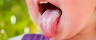
The most characteristic sign of the disease from which the word “thrush” comes is a white, cheesy coating. Fungi in a child’s throat multiply on the tonsils and oral mucosa. Therefore, plaque during candidiasis is localized in these places. At first it is scattered, white, and can be removed well. After some time, the whitish deposits turn into a grayish film, which is difficult to remove. The surface of the mucous membrane underneath bleeds.- Before plaque forms, it is difficult to diagnose candidiasis. Symptoms at the first stage of the disease are nonspecific - swelling, redness, dry mouth, sore throat.
- Painful swallowing accompanies thrush at all stages. This is due to the fact that when fungi reproduce, they release toxins that dissolve areas of the mucous membrane. Children refuse food and become capricious. If the fungus is not treated, ulcers will form in the child’s throat under the influence of toxins.
- An increase in body temperature is not a characteristic sign of candidiasis. The severity of the child’s condition depends on the severity of the pain syndrome.
Read also: Barking cough in a child: everything you need to know
Prevention of candidiasis
A few simple rules can significantly reduce the risk of developing an infection:
- use of antibiotics strictly according to indications;
- regular sanitization of children's pacifiers, toys, household items;
- brushing your teeth 2 times a day with high-quality toothpaste - for example, Colgate Total 12;
- sanitation of the oral cavity: timely treatment of caries and periodontal diseases, removal of the roots of damaged teeth;
- dental assistance: rational prosthetics, correction of occlusion pathology;
- careful adherence to the rules of care for removable dentures [1].
Candidiasis can cause serious harm to the oral cavity if the pathological process is not eliminated at an early stage. If you notice any symptoms of the disease, you must consult a doctor, who will make an accurate diagnosis and prescribe effective treatment.
List of sources:
- Sakharuk N. A. Candidiasis: etiology, clinical picture, diagnosis, treatment: Monograph. Vitebsk: VSMU, 2010. // URL: https://elib.vsmu.by/bitstream/123/10746/1/Sakharuk-NA_Kandidoz%20etiologiia%20klinika%20diagnostika%20lechenie_2010.pdf (access date 12/10/2020) .
- Molokov V.D., Galchenko V.M. Oral candidiasis. Irkutsk: Irkutsk State Medical University, 2009. // URL: https://ismu.baikal.ru/src/downloads/5eb06a90_kandidoz_polosti_rta.pdf (access date 12/10/2020).
- Weisgeim L. D., Dubacheva S. M., Gavrikova L. M. Complex treatment of oral candidiasis // International Journal of Applied and Fundamental Research. 2014. No. 2. pp. 48-51. // URL: https://applied-research.ru/ru/article/view?id=4692 (accessed 12/10/2020).
Candidiasis (thrush) in children
The following forms of thrush are distinguished:
- Thrush of the mucous membranes (oral cavity, tongue, gums, pharynx, tonsils, trachea, larynx, red border of the lips, corners of the mouth, teeth, vagina, vulva).
- Thrush of the skin and its appendages.
- Thrush is visceral, systemic.
- Allergic manifestations of thrush in children.
Candida infection most often manifests itself as thrush . It most often affects newborns, infants and preschoolers. The main symptom is curdled white deposits on the mucous membrane of the cheeks, gums, and palate. The overlays are initially located pointwise and then merged. Easy to remove. If treatment is not carried out in time, the overlays become denser and become grayish-dirty in color, they are increasingly difficult to remove, and the mucous membrane bleeds after removing the overlays. The general condition is not significantly disturbed if the children in the first days of life are not sick with anything else.
If the infection affects the mucous membrane of the tongue, then not only fungal overlays appear, but also areas without papillae. Swelling of the tongue, focal hyperemia and striations with furrows are recorded. The tongue becomes more sensitive to spicy and hot foods. Patients may complain of a burning sensation and dry mouth. It is difficult for infants to suckle milk, which is why they have difficulty eating.
Candidiasis tonsillitis , as a rule, appears against the background of candidiasis of the oral mucosa. It manifests itself by the appearance of loose whitish deposits on the surface of the tonsils, which can be removed with a spatula without effort. The tissue of the tonsils is practically unchanged. The child's general condition is almost normal. The temperature is elevated only if candidiasis of the tonsils occurs against the background of ARVI.
When diagnosed, fungal tonsillitis is distinguished from localized diphtheria of the pharynx by the absence of fever, the absence of hyperemia of the tonsils, and the normal size of the regional lymph nodes.
Candidiasis infection of the mucous membranes of the oral cavity and tonsils can spread to the mucous membrane of the larynx, trachea, and esophagus. In such cases, symptoms such as hoarseness and difficulty breathing appear.
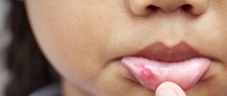
Candidiasis of the corners of the mouth (jam) in children occurs in rare cases. It is recognized by cracks in the corners of the mouth and erosion with perifocal infiltration. Usually both corners of the mouth are affected. The disease is distinguished from streptococcal infection - with it the inflammatory reaction is more pronounced.
Candidiasis of the red border of the lips (in the literature referred to as cheilitis) is often combined with candidiasis of the oral mucosa and erosions of the corners of the mouth. The red border of the lips swells and becomes hyperemic. The patient complains of dryness and burning of the lips. The disease lasts a long time.
Vulvovaginal candidiasis is symptomatically manifested by white discharge. The mucous membrane of the genital organs is moderately hyperemic, with loose cheesy deposits of a whitish or grayish tint visible on it. Rarely does it have superficial erosions. Overlays can be found in some cases on the vaginal mucosa and cervix. The external genitalia are very itchy and there is a burning sensation.
Intertriginous candidiasis occurs most often in infants in the area of large skin folds. The skin is hyperemic or eroded, and maceration of the stratum corneum is observed on it.
Smooth skin candidiasis in infants occurs mainly as a result of the spread of intertriginous candidiasis from skin folds. Candidiasis of the scalp is rare in children .
Chronic generalized granulomatous candidiasis is typical for children who are malnourished, suffer from gastrointestinal disorders, and bronchitis. The disease begins with oral thrush, then the process causes glossitis, cheilitis and seizures, which are treated poorly and ineffectively. In many cases, deep dental caries develops. The process subsequently affects the skin on the face and under the hair, and later on the torso, arms and legs. The appearance of hyperemic spots with a bluish tint, with infiltration and superficial peeling is recorded. These epidermal lesions gradually become granulomatous. Papules and tubercles appear, many of which are covered with a yellow-brown crust, under which papillomatous growths form. In almost all cases, the disease affects the nails and nail folds.
Microscopy results show the presence of yeast-like fungi in the urine and feces, and in some cases in the blood. Serological reactions in all sick children are positive.
In recent years, visceral candidiasis most often manifests itself as pulmonary candidiasis, which appears as a result of long-term therapy with antibiotics that were prescribed incorrectly. Pulmonary candidiasis manifests itself with a variety of symptoms. The disease can be acute or take a protracted, chronic nature. Relapses and exacerbations are possible.
Abscessing and cavernous forms of candidiasis pneumonia, pleurisy, which are difficult to distinguish from tuberculosis based on symptoms and radiographs, have been recorded in the literature.
Gastrointestinal candidiasis is distinguished by the fact that the deposits are abundant, sometimes completely fungal, they can cover the entire mucous membrane of the esophagus. Symptoms such as progressive dysphagia and the inability to swallow food are noted. Babies stop reaching for their mother's breasts and vomit. In especially severe cases of the disease, due to the massiveness of the deposits on the esophagus, its lumen may narrow, or even obstruction may occur. Histologically, deep destruction of the esophagus is revealed. In most cases, damage to the esophagus is also accompanied by thrush of the oral mucosa, which provides clues to diagnosticians.
Gastric candidiasis is a rare disease in children. It can only be detected by obtaining histological examination data. On the affected part of the stomach, hyperemia of the mucous membrane and small erosions are noted. Typical overlays, as with thrush, are quite rare.
With intestinal candidiasis, symptoms of enterocolitis or colitis, intestinal colic, and bloating are recorded. The stool is watery and may contain blood. The disease lasts a long time, there are relapses.
Damage to the urinary tract by Candida fungi manifests itself as urethritis, pyelitis, cysts, nephritis.
Generalized candidiasis in children can lead to endocarditis with damage to the heart valves or meningitis and meningoencephalitis, which is typical mainly for infants and preschoolers. With candidiasis meningitis, mild meningeal symptoms and a slight increase in temperature are observed. The disease progresses sluggishly, torpidly, and relapses are possible.
The most severe manifestation of candidal infection is candidal sepsis . This form of the disease occurs mainly in babies from 0 to 6 months. Before the disease, as a rule, there is another serious illness or microbial sepsis, which is complicated by an associated superinfection with the Candida fungus.
Candidiasis can spread through the oral mucosa to the esophagus, intestines or to the larynx, bronchi and lungs and end in sepsis . Also from the oral mucosa, Candida can spread through the blood. The symptoms of candidal sepsis are almost similar to ordinary bacterial sepsis. Diagnosis is made by isolating Candida from the blood of a sick child.
Prevention of candidiasis in childhood
Measures aimed at preventing the occurrence and development of the disease in childhood include:
- limiting contacts between children and carriers of this disease in preschool educational institutions, schools, sections and clubs;
- maintaining the baby’s personal hygiene;
- competent organization of the process of feeding the baby;
- maintaining a healthy diet;
- inclusion in the child’s diet of foods rich in all essential vitamins;
- sterilization of bottles, pacifiers and nipples;
- systematic visits to the pediatric dentist to assess the state of oral health.
It is important to understand that self-medication for oral candidiasis not only does not promote recovery, but can also lead to consequences dangerous to the health and life of the baby. That is why, if any symptoms indicating the development of this disease are detected, it is necessary to contact a dental clinic as soon as possible and undergo a course of treatment according to a regimen drawn up by an experienced dentist.