A scary story about terrible killer bacteria is one of the most effective ways to influence people's consciousness and try to make them more careful about maintaining hygiene rules, as well as draw attention to vaccination issues. But without visual material to show, dry data on microbiology research, interspersed with scientific terms, most often does not gain even a tenth of the necessary attention. Often such information remains unclaimed. To convince the listener or reader that bacteria are indeed literally everywhere and can play a decisive role in human destiny, these bacteria need to be shown and this display must be made as colorful as possible.
General information about microbes
These tiny organisms get their name from the abbreviation “microscopic object.” It was proposed back in 1878 by the French Emile Littre and Charles Sedilot.
Billions of such small organisms inhabit the air, the earth's surface and water everywhere. They are also found in human, animal and plant organisms. The first time microbes were seen was by the Dutch optician Antonio Leeuwenhoek, looking at a drop of ordinary water through a magnifying glass.
Microbes are the most ancient inhabitants of the planet. Their earthly history is more than 3.5 billion years. And for the first billion years they were the only living organisms on Earth. In the human body, the ratio of microbes and our own cells is 50/50. Most microorganisms benefit the body.
The 2 largest groups of microbes are nuclear-free prokaryotes and eukaryotes, which contain a nucleus in their cells.
The total number of microbes inhabiting the Earth and their ubiquitous distribution make them the most important link in maintaining the balance of the earth's biosphere.
Microbes vary in shape:
| Microbes | Form |
| Staphylococcus or streptococcus | Round cocci |
| Spirochetes | Spiral-shaped organisms |
| bacilli | Similar to sticks |
| Bifidobacteria | They live in fermented milk products and look like a two-toothed saw. |
There are also multifaceted microbes in the shape of stars, triangles and polyhedra. Some microbes are immobile, but mostly these microorganisms have flagella and are capable of movement.
The following classification:
- bacteria;
- the simplest single-celled organisms, for example amoebas;
- microscopic fungi.
Wear gloves
In some professions, this is not just a desire or request, but a requirement. For example, dentists must put on clean, disposable, sterile gloves before beginning a procedure. The same goes for chefs who serve food in a public place.
Try this rule in your family, it will help prevent the spread of germs. Surgical gloves are not such an expensive product that an ordinary person cannot buy them. However, this is how you can stop Campylobacter from getting into your food - a bacteria that can cause serious poisoning.
Wear surgical or latex gloves when handling raw poultry and discard them immediately after you have handled the product and placed it in the pan or pan. This reduces the risk of spreading the virus to the kitchen faucet when you go to wash your hands. It is also possible that when working with raw meat you can contaminate other food products.
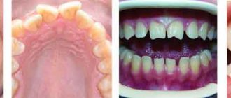
Bacteria in the mouth
The oral cavity is the most bacteria-populated area of the body. This is due to the fact that this place has suitable conditions for the development of microbes: it is warm, constant humidity, and acidity and oxygen saturation are the best for microorganisms. There is a lot in the mouth and all kinds of nutrients.
About 30 species of bacteria live here permanently. Interestingly, the species composition of bacteria in humans does not change, but the number of each specific type of microbe may change throughout life. The basis of the microflora is bacteria, of which 90% can live without oxygen.
Most bacteria are found on the back of the tongue. Here the smallest plaque is formed, in 1 g of which almost 300 billion bacteria can be found. The second favorite place to live for most bacteria is the surface of the teeth. Microbes under a microscope (photo of streptococci that cause sore throat) allow you to see their structure.
Those microbes that are constantly in the oral cavity cause periodontitis and caries. Today, 95% of people suffer from caries.
The following bacteria are constantly found in the human mouth:
- Veillonella alkasenses. This is a microbe that leads to the development of infectious diseases.
- Candida mushrooms. More than 100 species of such fungi can be pathogenic or normal, not causing disease.
- Leptothrix bacteria , very similar to curved rods with sharp or round edges. They can be found in the cervical area of the teeth. These bacteria form tartar and cause bad breath.
- Corynebacteria, leading to the development of purulent inflammation in diseases of the gums or teeth.
- Lactobacillus casei. These microorganisms produce fatty acids. They are the ones who create bad breath.
- Treponema dental. This bacterium takes part in the destruction of gums and is directly dependent on oral hygiene.
- Solobacteria Moore cause a severe putrid odor in the mouth: they produce hydrogen sulfide, which evaporates very quickly and enters the exhaled air.
- Streptococci, forming chains of spherical anaerobic bacteria.
- Staphylococci that can live in the air and cause purulent infections. They can lead to pneumonia, severe pneumonia and general sepsis.
Nobody cares about hygiene
An American microbiologist conducted research and found out that every fifth person does not wash their hands with soap after using the toilet, especially a public one. This means that even if these four people from the statistics thoroughly clean their hands, apply antiseptic gel and dry the surface of the skin, they can still “pick up” pathogenic microorganisms.
Streptococci and staphylococci
The following is a description of the most numerous bacteria that inhabit the human oral cavity and skin.
Streptococci in the mouth
The very first of the bacteria that enter the baby’s oral cavity as the child moves through the birth canal. To date, scientists have identified 17 species of streptococci containing the C-antigen and streptococci lacking this antigen. It is microbes without such an antigen that cause caries. Such streptococci are found in the saliva and in the gum pockets of all people without exception.
Streptococci can grow rapidly: this requires a temperature of 37 degrees and glucose with carbohydrates. Therefore, during a sore throat, giving sweets to sick children is not recommended.
Streptococci on the skin
These microorganisms enter human skin in several ways:
- With inhaled air or dust particles.
- Upon contact with contaminated objects: children's toys, bus handrails, banknotes, books, other people's clothing.
- When shaking hands, touching.
- During medical procedures performed with non-sterile instruments.
- From soil or unwashed fruit or when eating with dirty hands.
It is easier for infections to gain a foothold in the body if it is weakened. This happens, for example, in the following cases:
- The child’s defenses have not yet been formed, the immunity is weak from birth or weakened for some external reasons.
- The acid-base balance of the skin is greatly changed, which is normally 5.2 - 5.5 units.
- There are hormonal imbalances in the body: it is hormones that are responsible for the normal course of metabolic processes and cell life.
- The skin is damaged by wounds, abrasions, sunburn, and insect bites.
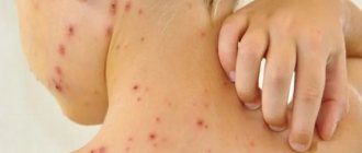
Streptococcus causes 2 serious health problems: erysipelas and streptoderma. Both of these diseases can lead to blood poisoning and death. Streptoderma has 3 types: superficial, ulcerative and deep.
Staphylococcus in the mouth
These elongated bacteria are the second most numerous inhabitants of the oral cavity. In the microscope photo, their clusters resemble a bunch of grapes. The favorite place for such bacteria to accumulate is the area at the base of the gums and plaque on the teeth. Such microorganisms are capable of multiplying at temperatures from 7 to 46 degrees. But 35-39 degrees Celsius is most suitable for them.
In a simple environment, the population of staphylococci grows very quickly. When breaking down carbohydrates, they produce acid. They process staphylococci and proteins. The result of such processing is hydrogen sulfide. Staphylococci lead to the rapid development of dental plaque.
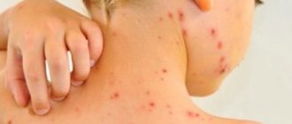
Staphylococcus on the skin
Such bacteria live on the skin and mucous membranes of living beings, on food, and household items. They cause illness only when the human body is weakened and does not find the strength to resist microbes. The ways in which staphylococcus spreads are standard: through the air or through direct contact with the patient’s skin and objects that contain these microbes.
Photo of staphylococcus microbe under a microscope
Contact with staphylococcus on human skin is always painful. Swelling, suppuration, pain and redness of the skin develop at the site of infection. The main indicator is all kinds of rashes on the skin.
This rash may look like this:
- abscesses;
- large red spots;
- blisters with fluid inside;
- inflammation of the nail phalanges;
- acne;
On the face, Staphylococcus aureus causes a huge number of acne. When such skin inflammations appear, they look like bright red, painful bumps. After a short time, white pus appears in the center of the acne. Even later, such an abscess bursts, and in its place a small pit-scar remains. On the hands, staphylococci most often infect the nail phalanges.
Signs of the disease are:
- suppuration at the site of infection;
- swelling and swelling of the skin;
- severe throbbing pain;
- change in color of the skin and nail plate.
Staphylococcus can form on any part of the body, but in most cases the back, chest and abdomen are affected. Basically, these are the same acne with pus in the center, but there is also a more severe form: boils.
In common parlance they are called boils. In the center of the boil there is a long purulent rod. This rod goes into the deep layers of the skin. Boils most often occur at the site of sebaceous, sweat or hair glands. Another form of staphylococcal disease is erysipelas.
It looks like this:
- at the site of inflammation, swelling begins and body temperature rises;
- a large red spot appears on the skin;
- sometimes transparent blisters appear filled with purulent mass;
- many small pinpoint hemorrhages appear.
Where to look for reliable images
Photos in which you can see the main vital centers of a bacterium (circular DNA, ribosomes, etc.), cell division and its external structure can be easily found either in professional medical albums on microbiology or in textbooks for higher educational institutions specializing in the study of microbiology .
The photos and pictures published in these publications are not as colorful as in popular literature, but more informative. The bacteria imprinted on them live their natural lives, and do not float in multi-colored clouds that resemble intergalactic space rather than a nutrient medium on a microscope slide.
Yulia Pyatirubleva
Fungi on the skin
On the body, fungal infections can develop anywhere: on the head, feet, buttocks or palms.
The most common forms are:
- Dermatophytosis: damage to the deep layers of the skin, which is caused by both yeast and mold fungi.
The most common fungal disease. This infection triggers an inflammatory process and leads to the formation of pink or red spots or plaques. The size of such plaques is very diverse. Fungi that cause dermatophytosis are permanent residents of hair and skin. They are able to perfectly absorb and assimilate keratin from hair and nails. - Keratomycosis: a disease that affects the top layer of skin. Symptoms of the disease are small, hard nodules on the skin that are filled with pus. The forms of the disease vary and include pityriasis versicolor, axillary trichomycosis and erythrasma.
- Deep mycosis: the disease affects the subcutaneous tissue, muscles, joints and bones, mucous membrane, tissues of internal organs and parts of the nervous system. With such a lesion, warts and deep fistulas appear on the skin.
- Candidiasis: The infection is caused by yeast. The most common sites of lesions on the body are in the groin area, in the armpits and under the female breast.
The manifestation of the disease depends on the location of the lesion and the type of fungus.
Candida and dermatophytes cause itching, discoloration of the skin to red-bluish, peeling of infected areas, dandruff and dry hair, and discoloration of nails.
With mycosis of the foot, in addition to peeling of the skin, blisters up to 2 mm in diameter appear, filled with liquid. These bubbles collect between the toes. The smell that appears when the fungus decays also causes trouble for the patient. Often a person gets such diseases in a sauna or in the summer, when wearing tight shoes for many hours.
When fungi get on the skin of the face, they first attack dead epithelial cells. If treatment is not carried out, the skin begins to peel, become rough, and red or yellow spots appear. During the transition to the severe stage, purulent foci and ulcers appear. Fungal infections of internal organs begin.
On the head, fungal infections lead to hair loss, flaking, dandruff, and the formation of bald patches.
Studying Cipollino
A microscope will help your child learn that all living things are made up of cells. Under a microscope you can see not only a cell, but also examine its structure. To do this, together with your child, prepare a simple and visual preparation from ordinary onions. Why onions? This plant has very large cells, and they are clearly visible under relatively low magnification. So, cut the onion into several parts and separate one juicy layer. Cut a small piece from it, and then use tweezers to separate the thin film from the concave side of the piece. Drop distilled water onto a glass slide, place a film in it and carefully straighten it with a needle. Then add a couple of drops of an aqueous solution of methylene blue or an aqueous solution of iodine. This should be done so that the colorless cells become colored and become better visible. If you can find a red-violet onion, you don’t need to add any dye. Cover the resulting “beauty” with a cover glass and blot away any escaping liquid. Try to view the drug first at low and then at high magnification. Tell your child that both plants and animals are made up of tiny cells. These are the ones that are visible through a microscope, like little bricks. Why were they called cells? This name was invented by the English botanist R. Hooke. Examining a section of the cork under a microscope, he noticed that it consisted of “many boxes.” He also called these “boxes” chambers and... cells. After all, it really looks like someone drew the onion film into squares.
At high magnification, the cell wall, nucleus, and vacuole are clearly visible. Explain to your child that a cell wall is a partition, a wall between cells. It protects the cell and helps maintain the desired shape. Thanks to the nucleus, the cell grows and reproduces. And inside the vacuole there is cell sap. The one that splashes in different directions and brings tears to our eyes when we cut onions.
Microbes on your hands: what they look like under a microscope
It is recommended to show photos of microbes under a microscope to children who forget to wash their hands. This photograph of the palm of her 8-year-old son was taken by Californian microbiologist Tasha Sturm, who works at Cabrillo College.
Microbes under a microscope (the photograph reflects them in huge quantities) exist even in a healthy child. The baby whose hand is in the photo only played a little in the house and with the dog.
Photo of a child's palm covered in germs:
The transcript of the photo is as follows:
- white colonies of microbes around the fingertips - staphylococci;
- micrococci are marked in yellow;
- Colonies colored pink are serratia.
Staphylococci and micrococci are often harmless and are part of the natural microflora of humans. Serratia, on the contrary, can cause infections, especially in weakened people.
On the hands, in addition to the already mentioned streptococcus, staphylococcus and fungal bacteria, you can find the following microbes:
- salmonella, which have the shape of rods up to 7 microns in length and cause intestinal infections, including severe typhoid fever;
- Escherichia coli measuring 0.4-0.8 × 1-3 microns, sometimes leading to death in elderly people and small children due to general poisoning of the body;
- Shigella, which causes dysentery and seizures in children;
- spheroid-shaped brucellae without flagella that infect internal organs.
Use an antibacterial spray
Despite the fact that the main ingredient in an antibacterial gel or spray is ethyl alcohol, it effectively kills many bacteria, disinfecting the skin.
Stock up on this product when you are traveling on public transport or going on a trip. Even if you want to have a snack on the way, you don’t have to worry about poisoning. Just apply a little gel to the surface of your hands, rub it in and wait until it dries completely. Do the same when you visit a public restroom. While at home, treat frequently used surfaces (faucets, door handles, sinks) with an antiseptic that contains bleach or alcohol.
Found a violation? Report content
Microbes that live under your nails
The area under the nails contains all types of bacteria that live on the hands. But the concentration of such microbes is hundreds of times higher. This is due to the inaccessibility of the space under the nails for treatment with disinfectants and anti-inflammatory agents.
Interestingly, there are many times more such microbes under artificial false nails than under natural ones.
Permanent residents of the body inhabiting this zone:
- fungal bacteria;
- staphylococci,
- streptococci.
The difficulty of disinfecting fingers is that the nail plate reliably protects this part of the body from the action of the most effective antibacterial agents.
Choosing the right model
Many novice researchers are interested in which device to choose in order to examine fermented milk, as well as other common categories of bacteria.
The budget segment of microscopes demonstrating 640x magnification will not give the same effect that can be appreciated in a video made with a more powerful microscope. Bacteria in urine, for example, can only be seen under equipment lenses that magnify 1000x or more.
The phase-contrast type of device works by detecting different particle densities. This microscope, which allows observation and magnification of bacteria, colors the elements in a light gray or dark gray shade. In this video you can see a multiple increase in bacteria in the urine.
A dark-field microscope allows you to see lactic acid bacteria (you can also see what they look like in the photo). Its advantage is the scattering of light coming not directly through the lens, but from the side. The device also allows you to understand the actual movement patterns of bacteria.
Bacteria living in the intestines
Scientists believe that more than 2,000 species of bacteria live in the human intestine. This number includes both beneficial microorganisms and microbes that lead to diseases. It is difficult to calculate the total number, but science puts the figure up to 100 trillion. If we translate these numbers into weight, then from 200 g to 1 kg of our body weight is accounted for by intestinal bacteria.
The small intestine, compared to the large intestine, contains a smaller set of bacteria: in this part of the body there is a more acidic environment, more oxygen and antimicrobial agents. Therefore, fast-growing organisms develop in the small intestine that can aggressively attach to its walls. The bacteria of the large intestine are excellent feeders: they get carbohydrates that are not broken down in the small intestine.
The main functions of beneficial intestinal bacteria:
- produce vitamins B and K;
- directly affect the functioning of the brain, internal organs and psyche;
- help to obtain more energy from the food consumed;
- increase the intestinal barrier;
- help the production of bile acids;
- decompose toxic substances and carcinogenic cells.
The bulk of intestinal bacteria are Firmicutes and Bacteroides.
In addition to bacteria from these 2 groups, which make up up to 90% of all intestinal microflora, several other populations live in the intestine:
- Actinobacteria, shaped like branching threads.
- Bifidobacteria, which look like curved rods up to 5 microns long. They provide the fight against harmful putrefactive microbes.
- Lactobacilli. These organisms produce lactic acid from lactose and carbohydrates. The acidic environment obtained in this way prevents the development of harmful bacteria.
- Proteobacteria. This group includes almost a third of all known bacteria, and is diverse in shape, ability to breathe and move. There are proteobacteria necessary for humans, and those that cause infections.
In children, the intestinal microflora differs at different periods of life. Those who are bottle-fed contain equal proportions of bacteroides and bifidobacteria. The set is complemented by staphylococci and clostridia. In this case, E. coli is also required.
In those who feed on breast milk, the microflora consists of lactobacilli and streptococci. Microbes under a microscope (photo of E. coli, which causes bacterial diarrhea) allow you to understand how numerous they are.
Presentations for children contain photographs of microbes visible on hands and mucous membranes under a microscope. They help the child understand why it is necessary to wash their hands, bathe or brush their teeth, eat washed apples or not touch stray kittens. Photos of these microorganisms are supplemented with text fragments and a sound sequence.
Article design: Vladimir the Great
Development of the disease
Nail mycoses, or onychomycosis, affect every tenth representative of the globe. The main pathogens are dermatophytes, such as T. rubrum. But there are forms caused by mold or yeast-like fungi. Factors that provoke onychomycosis: wearing uncomfortable, tight shoes and neglecting hygienic care of hands and feet.
Onychomycosis occurs in several stages:
- Spots of a yellowish or gray-white hue appear on the platinum.
- The plate becomes brittle, flakes, crumbles, and some areas thicken.
- The bed takes on a brownish tint, and the nail plate comes off.
Pathological changes can be observed both in a local area and on the entire surface. The fungus gradually spreads to neighboring fingers.
Pasteurization of milk
This is also an interesting experiment that can be done at home, only aimed at destroying bacteria.
The world owes the appearance of shelf-stable milk (pasteurized) to the Frenchman Louis Pasteur. This scientist developed a process to kill microorganisms found in liquids. True, Pasteur processed wine and beer, not milk.
In a regular kitchen you can easily pasteurize milk. To do this, place the container with milk in a steam bath (in a pan with hot water) and, with constant stirring, bring it to a temperature of 63 - 65⁰C. After half an hour, the container with milk is transferred to cold water to quickly reduce the temperature.
What are the treatment methods for caries in children?
Treatment methods depend on the stage of caries development. The more serious the problem, the longer it will take to fix it.
Preventive
At the initial stage, a delicate intervention is sufficient, which can stop or reverse the process of destruction that has begun. In this case, for example, remineralization (treatment of teeth with a solution of calcium and fluoride) or deep fluoridation (treatment with fluoride) is used. Such procedures are carried out in courses until the condition of the teeth is stabilized.
There is also a treatment method without drilling, which allows you to quickly and completely painlessly prevent the development of inflammation. The procedure is suitable for children from 4–5 years of age and causes them minimal discomfort.
Medicinal
For medium and deep caries, classical filling with preliminary preparation of teeth for the procedure is already necessary. To ensure that the treatment is calm and painless, the most modern and effective anesthesia is used. Your baby doesn’t even have to endure the pain of the needle because the injection site is first numbed. The drill is used to a minimum in the treatment of childhood caries - if something can be done manually, then the doctor will do so.
If a child has a complex case of caries, or is very restless in the dentist's chair, treatment is carried out under anesthesia. After consulting with an anesthesiologist and passing all the necessary tests, the baby will be selected the appropriate drug and dose, he will be put into deep sleep, during which the doctor will carefully monitor the condition of the body. Time will fly by in a dream - the child will wake up with healthy teeth, a clear head and an unspoiled mood.
Why is bottle caries dangerous?
Since baby teeth are temporary, many adults do not understand why they need to be treated. A simple explanation would be an example of possible complications.
If bottle caries is not treated, then within 2-3 months it develops into pulpitis - acute or chronic. In the chronic stage, pulpitis of primary teeth is often painless and asymptomatic and gradually turns into periodontitis. With purulent inflammation in the area of the root of the diseased tooth, fistulas with purulent discharge appear, or an abscess begins. This is already an indication for the removal of a baby tooth.
Untreated bottle caries of baby teeth is a threat:
- formation of the rudiments of permanent units;
- development of bite pathologies - with early tooth extraction;
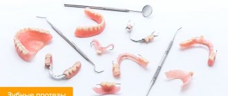
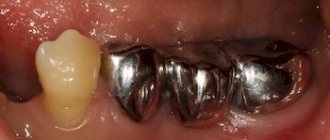
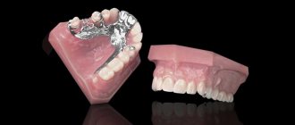
- prosthetics of baby teeth – incisors and molars;
- decreased general immunity;
- the appearance of gastrointestinal diseases - due to a violation of a balanced diet.