Classification of fluorosis
The disease is divided into degrees of severity:
- The first is doubtful fluorosis. These are barely noticeable white spots on the enamel that become better visible when the surface dries.
- The second is very weak. Opaque white spots occupy up to 25% of the enamel surface.
- The third is weak. The lesion covers from 25 to 50%.
- The fourth is moderate. The surface of the tooth is completely damaged, abrasion and brown spots are noticeable on the enamel, and the relief is modified.
- Fifth – heavy defeat. There are significant brown areas and serious enamel destruction.
How does dental fluorosis manifest?
Dental fluorosis is primarily manifested by external changes in the tooth enamel - spots of various sizes and colors (chalky, dirty yellow, dirty brown) appear on the tooth surface. The stains are located on the entire outer surface of the tooth, but in different quantities. As a rule, fluorosis affects adjacent or symmetrical teeth - central incisors, front teeth.
At the beginning of the development of enamel fluorosis, the patient most often discovers small and white dots on the teeth. Instead of dots, stripes may also form. The first teeth affected by fluorosis and the ones that react most sensitively to fluoride are the incisors. As the disease develops, other teeth in the mouth are also affected.
Over time, the initially formed spots begin to darken and acquire a dirty hue. This is evidence that the disease is entering a chronic stage.
Forms
The streak form is characterized by barely noticeable stripes in the subsurface enamel layer. They are hardly noticeable and form on the vestibular surface of the incisors.
In the spotted form, chalky spots alternate with areas of healthy enamel. The incisors are most often affected. Sometimes the formation takes on a yellowish-brown tint. But the enamel always remains smooth and dense, with an intact structure.
The chalky mottled form has various symptoms. Distinct pigmented spots are visible on the enamel. The enamel may be yellow, with spots and dots, and other minor defects. It quickly wears away, exposing pigmented dark brown dentin.
The erosive form is accompanied by unaesthetic erosions on which there is no enamel. Destructive is characterized by a violation of the shape of the crowns of the teeth due to abrasion of hard tissues. They become brittle and break off easily, but due to the formation of replacement dentin, the tooth cavity is not opened.
Reasons for development
Fluoride is necessary for dental health and the formation of strong tooth enamel, but when it is in excess, the opposite processes occur in the body. Microelement intoxication does not occur without a reason. Fluoride is not produced inside the body, so the provoking factor is always external.
Causes of fluorosis include:
- regular consumption of fluoridated water;
- long-term use of toothpaste and rinses containing fluoride;
- professional activities requiring constant contact with high concentrations of fluoride.
However, even under the influence of provoking factors, fluorosis rarely develops in adults. A weakened immune system, osteosarcoma, osteoporosis, cardiovascular disease and osteosclerosis may increase the risk of dental damage.
Subtleties of differential diagnosis of fluorosis
It is very important to distinguish fluorosis from enamel hypoplasia, especially from its patchy form. Chalky spots in both diseases are located symmetrically, in areas of the crown that are not typical for caries. As a rule, these are the labial and lingual surfaces, cusps and cutting edge. With fluorosis, they have a pearly white tint, are shiny, do not cause pain during probing, and gradually turn into healthy enamel.
With hypoplasia, the spots are white and dense, also shiny, but with clear boundaries. Under the influence of UV rays, with fluorosis, chalky spots give a light blue glow (pigmented ones - red-brown), with hypoplasia - light yellow. Fluorosis is not prone to changes in spots, while formations with hypoplasia often require treatment for caries in children, since they change and progress.
Dental restoration after fluorosis
For initial or spotty forms of dental fluorosis, whitening can be performed. The result will be that the shade of the white spots on the teeth and the natural shade of the teeth will be equal, and the spots will no longer be noticeable. Minor aesthetic defects can also be sanded down.
Remineralization of enamel is very helpful during the treatment of fluorosis. Beneficial substances - calcium and fluorine, penetrate into hard dental tissues during remineralization, enriching and restoring teeth, strengthening them.
For severe forms of enamel fluorosis, as well as for serious aesthetic defects (dark, dirty spots), direct dental restoration using light-curing composites, as well as prosthetics using veneers, lumineers, and dental crowns, are indicated.
Treatment of fluorosis
The treatment regimen depends on the severity of the disease, general health and the influence of endemic factors. Therapy solves the following problems:
- reduce excessive intake of fluoride into children’s bodies from drinking water and food products;
- weaken the toxic effect of increased concentrations of fluoride on the body (the child is prescribed calcium preparations that bind fluoride and remove it).
Topical treatment for fluorosis depends on the severity. For any clinical picture, it is recommended to brush your teeth twice with toothpastes that contain calcium glycerophosphate, for example “Pearls” or “Calcium Complex”. At the first stage, no further action is required.
In the presence of pigmented spots, whitening is sometimes carried out followed by remineralization - only after 16 years under the supervision of a dentist. There are other methods of combating age spots, for example, removing pigmented enamel using microabrasion.
In erosive and destructive forms, when there is loss of hard tissue, the anatomical shape of the tooth can be restored with composite materials. Orthopedic treatment is sometimes indicated.
Diagnostic methods
Diagnosing dental fluorosis is not difficult. The doctor relies on the clinical picture characteristic of each degree of damage. Differential diagnosis includes:
- Mild opacity of the enamel is detected by drying the surface to assess the condition of the protective layer in the area with altered pigmentation.
- Vital staining method - in case of fluorosis, the affected areas of the enamel are not subject to staining with methylene blue, unlike a carious defect.
- Luminescence study - mild types of the disease glow weakly in ultraviolet light; in moderate and severe forms, partial quenching of fluorescence occurs.
- Microradiography is aimed at determining the depth of damage to dentin tissue.
Drinking water must be sent to the laboratory for analysis to determine the percentage of fluoride compounds.
Prevention of fluorosis
Prevention of fluorosis should be carried out in geographic areas where the fluoride content in drinking water exceeds 2 mg/l. Ideally, it is desirable to solve this problem collectively - to eliminate the etiological factor. If this is not possible, drinking water is defluoridated using a reagent or filtration method. Such water is also mixed with water from artesian wells or mountain rivers to reduce the concentration of fluoride. You can also remove fluoride from water at home by freezing, boiling and settling, and filtering through a layer of magnesium oxide.
Individual prevention involves the use of imported clean water for cooking. It is very important to limit the consumption of foods high in fluoride - strong tea, sea fish, seafood. In the winter-spring period, children from 2-3 years of age at risk are prescribed calcium supplements for a month, as well as ascorbic acid.
Features of the disease
Fluorosis is a non-infectious and non-inflammatory chronic disease that develops when the level of fluoride in the body increases. With fluorosis, all teeth can be damaged, but intensive destruction of the enamel of the upper incisors is predominantly observed.
A distinctive feature of fluorosis from caries is the formation of not only white spots on the enamel, but also stripes, specks, etc. Gradually, destruction occurs, and cracks and chips form on the affected teeth. In advanced cases, the tooth can completely collapse, exposing dentin.
The risk group includes children during the period of change from primary to permanent dentition. Fluorosis is less common in adults, but is no exception. The disease develops slowly. It is characterized by the onset of periods of exacerbation and remission.
FAQ
How to treat fluorosis in children?
If your child has fluorosis, treatment options are much the same as for adults. Yellow stains on children's teeth and other manifestations of the disease are eliminated using whitening procedures. Be sure to eliminate the source of increased fluoride.
In addition, children do not undergo restoration, as well as prosthetics and installation of veneers. If we are talking about baby teeth, then such events are pointless, because permanent teeth will replace them.
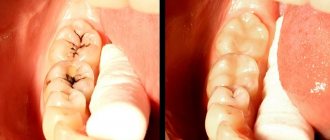
Fluorosis in children: photo
What happens if fluorosis is not treated?
Pigmentation on teeth is far from the only trouble with fluorosis. If the disease is not treated, it progresses, until the complete loss of the tooth. When the first symptoms of the disease appear, it is extremely important to consult a doctor and undergo treatment.
Is it possible to whiten teeth with fluorosis?
This procedure can be carried out in the initial stages of the disease, when no more than 25% of the enamel is affected. Whitening is carried out to restore the previous aesthetics. It must be remembered that without eliminating the cause of the disease, such an event is meaningless. This way you will only temporarily regain the lost whiteness of the enamel, because if you continue to take fluoride in large quantities (with water, food or medicine), the disease will not go away.
How to avoid dental fluorosis?
To ensure that bones and teeth do not experience problems associated with an excess of fluoride, it is important to monitor the composition of drinking water. In regions where water has high levels of fluoride, people should be informed about this. Don’t be lazy – take an interest in this question. If you buy drinking water, ask for certificates certified by experts that indicate the chemical composition. Normally, 1 liter of drinking water contains from 0.5 to 1 milligram of fluoride.
What is the danger of the disease?
If fluorosis treatment is not started in a timely manner and the root cause of the pathological process is not eliminated, this is fraught with serious consequences. In addition to aesthetics, teeth will become weak, crumbly, and ultimately this can lead to tooth loss. In addition, in advanced stages, any treatment is ineffective; the only way out of the situation will be prosthetics or dental implantation. In addition, high levels of fluoride are not only harmful to tooth enamel, but can also trigger more serious diseases such as osteoporosis.
Pathogenesis
During normal enamel formation, matrix proteins (ameloblastin and amelogenin) are gradually replaced by growing hydroxyapatite crystals. The space between adjacent enamel prisms is minimized.
When there is an excess of F- ions, they are more quickly incorporated into the crystal lattice than the larger and asymmetric OH- ions. Fluoridated enamel preserves matrix proteins. As a result of incomplete crystal growth, significant spaces remain along the periphery. The penetration of food pigments turns these cavities brown. Porosity of enamel in severe forms of fluorosis contributes to the spread of caries and increases the risk of chipping from mechanical stress.


