Removing a tooth is the last thing a dentist will suggest. Due to a number of subjective and objective reasons, it is impossible to maintain the dentition in ideal condition until old age.
Dental defects are the loss of one or more dental units, leading to a violation of the integrity of the dentition, deviation in the development of physiological occlusion, and an abnormal arrangement of individual teeth.
Reasons contributing to the development of dental defects
Dental defects arise due to:
- untimely treatment of caries;
- inflammatory processes in the oral cavity;
- presence of malocclusion;
- genetic conditioning;
- diseases leading to changes in metabolic processes;
- damage to the jaw apparatus;
- various neoplasms.
Classification of dentition defects
There are several types of classification. Dentists mainly use three of them:
- Kennedy classification:
- Group 1 - bilateral absence of chewing teeth;
- Group 2 - unilateral absence of a molar;
- Group 3 - unilateral absence of lateral teeth with preservation of distal support;
- Group 4 - defect of the anterior jaw.
- Beltman classification:
- Class 1 - the defect includes edentulous chewing teeth.
a) dentition with unilateral terminal defects;
b) dentition with bilateral terminal defects.
- Class 2 - there is one or more included defects (if the terminal units are present, there may be no teeth in other parts of the row).
a) the number of missing units does not exceed three;
b) the number of missing units is three or more.
- Classification according to the Gavrilov system:
- Group 1 - dentition with terminal one- and two-sided defects;
- Group 2 - dentition with anterior and lateral one- and two-sided defects;
- Group 3 - combined defects;
- Group 4 - single preserved dental units.
Diseases of hard dental tissues
Treatment for partial destruction of tooth crowns
The main goal of orthopedic treatment for partial defects of the hard tissues of the tooth crown is restoration of the crown through prosthetics in order to prevent further tooth destruction or relapse of the disease.
The important preventive value of orthopedic treatment of defects of hard dental tissues, which is one of the main directions of orthopedic dentistry, is that restoration of the crown allows you to prevent further destruction and loss of many teeth over time, and this in turn allows you to avoid serious morphological and functional disorders of various departments of the dental system.
The therapeutic effect of prosthetic restoration of crown defects is expressed in the elimination of disturbances in the act of chewing and speech, normalization of the function of the temporomandibular joint, and restoration of aesthetic standards. The odontopreparation used in this case as an act of impact on dental tissue also creates certain conditions for the activation of reparative processes in dentin, as a result of which a targeted restructuring is observed, expressed in the natural compaction of dentin and the formation of protective barriers at various levels.
As a treatment for defects in the coronal part of the tooth, mainly two types of prostheses are used: inlays and artificial crowns.
An inlay is a non-removable prosthesis of part of the tooth crown (microprosthesis). Used to restore the anatomical shape of a tooth. The insert is made from a special metal alloy. In some cases, the prosthesis can be lined with an aesthetic material (composite materials, porcelain).
An artificial crown is a permanent prosthesis that is used to restore the anatomical shape of a tooth and is fixed to the stump of a natural tooth. Made from metal alloys, porcelain, plastic. Can serve as a supporting element for other types of prostheses.
As with any medicinal product, there are indications and contraindications for the use of inlays and artificial crowns. When choosing a prosthesis, the disease that caused the destruction of the natural tooth crown and the degree (size and topography) of the destruction are taken into account.
Tabs
Inlays are used for caries, wedge-shaped defects, some forms of hypoplasia and fluorosis, and pathological abrasion.
Inlays are not indicated for circular caries, MOD cavities in combination with cervical caries or wedge-shaped defect, or for systemic caries. It is undesirable to use tabs for people taking gastric juice or hydrochloric acid for medicinal purposes, or working in acid shops. In these cases, artificial crowns are preferable.
It should be remembered that varying degrees of dental caries damage and a number of other diseases of hard tissues (hypoplasia, fluorosis, dysplasia) require complex treatment.
The question of a treatment method for partial defects in the coronal part of a vital tooth can be decided only after removal of all necrotic tissue.
Odontopreparation and treatment of inlays. Local treatment of defects in the coronal part of the tooth consists of surgical removal of necrotic tissue, surgical formation (by odontopreparation) of a corresponding cavity in the tooth and filling this cavity with an inlay in order to stop the pathological process, restore the anatomical shape of the tooth and connect it to the chewing function.
The clinical and laboratory stages of restoring the coronal part of a tooth with inlays include: forming a cavity for the inlay by means of appropriate odontopreparation, obtaining a wax model of it, making an inlay by replacing the wax with an appropriate material, processing the metal inlay and fitting it to the model, fitting and fixing the inlay in the tooth cavity.
The formation of a cavity in a tooth for the purpose of its subsequent filling with an inlay is subordinated to the task of creating optimal conditions for fixing the inlay, which does not have side effects on healthy tissue. The surgical technique of odontopreparation of cavities in a tooth is based on the principle of creating a cavity with walls that can perceive both pressure when a bolus of food of varying consistency and density directly hits them, and pressure transmitted from the prosthesis when it is loaded during the chewing process. The design features of the prosthesis should not contribute to the concentration of additional pressure on the remaining hard tissues: the pressure should be fairly evenly distributed over their entire thickness. In this case, the inlay material must be hard, but not brittle, not plastic in the hardened state, not corrode or swell in the oral environment, and have an expansion coefficient close to that of enamel and dentin.
The principle of the operational technique of forming a cavity and then filling it with an inlay is subject to the laws of redistribution of chewing pressure forces.
In case of caries, the cavity is formed in two stages. At the first stage, technical access is made to the carious cavity, its expansion and excision of pathologically altered enamel and dentin tissues. At the second stage of odontopreparation, a cavity is formed according to the configuration in order to create optimal conditions for fixing the inlay and optimal distribution of chewing pressure forces on the tissue.
To open a carious cavity, shaped carborundum and diamond heads, fissure or spherical burs of small diameter are used. The opening of a carious cavity on the contact surface presents a certain difficulty. In these cases, the cavity is formed towards the chewing or lingual surface, removing unchanged tooth tissue to facilitate access to the cavity. A free approach to the cavity from the chewing surface is also necessary to prevent the occurrence of secondary caries.
After expanding the carious cavity, necrotomy and formation of a cavity for the inlay begin. To facilitate further study of the topic, we will describe the main elements of the formed cavity. In each cavity, there are walls, a bottom, and the place where the walls connect with each other and the bottom - the corners. The walls of the cavity may converge with each other at an angle or have a smooth, rounded transition.
Depending on the topography of the damage to the tooth crown, it is possible to have two or three cavities combined with each other, or a main cavity (localization of the pathological process) and an additional one, created in healthy tissues and having a special purpose.
The nature and scope of surgical interventions on hard dental tissues are determined by the following interrelated factors:
- the relationship between the hard tissue defect and the topography of the tooth cavity and the preservation of the pulp;
- thickness and presence of dentin in the walls delimiting the defect;
- topography of the defect and its relationship to occlusal loads, taking into account the nature of the action of chewing pressure forces on the tooth tissue and the future prosthesis;
- the position of the tooth in the dentition and its inclination in relation to the vertical cavities;
- the relationship between the defect and the areas of greatest caries damage;
- the reason that caused the damage to hard tissues;
- the possibility of restoring the full anatomical shape of the tooth crown with the proposed design of the prosthesis.
The question of the effect of occlusal loads on tooth tissue and microprosthesis deserves special study. When eating food, chewing pressure forces of varying magnitude and direction act on the tooth tissue and denture. Their direction changes depending on the movement of the lower jaw and the food bolus. These forces, if there is an inlay on the occlusal surface, cause compressive or tensile stress in it and in the walls of the cavity.
Thus, with cavities of type 0 (class I according to Black) in a vertically standing tooth and a formed box-shaped cavity, force Q causes deformation - compression of the tissues of the bottom of the cavity. The forces R and P are transformed by the walls of the cavity, in which complex stress states arise. With thin walls, over time this can lead to their breaking. If the tooth axis is inclined, then the forces R and Q cause increased deformation of the wall on the inclined side. To avoid this and reduce wall deformation, you should change the direction of the walls and bottom of the cavity or create an additional cavity that allows you to redistribute part of the pressure to other walls.
Similar reasoning, which is based on the laws of deformation of a solid body under pressure and the rule of the parallelogram of forces, can be applied to cavities of the MO and OD types. Additionally, the effect of force P directed towards the missing wall should be considered. In this case, the horizontal component of the force tends to displace the inlay, especially if the bottom is formed with an inclination towards the missing wall. In such situations, the rule for forming the bottom also applies: it must be inclined away from the defect, if the thickness of the preserved contact wall allows, or a main cavity must be formed on the occlusal surface with retention points.
The patterns of redistribution of chewing pressure forces between the cavity wall microprosthesis system allow us to formulate the following pattern of cavity formation: the bottom of the cavity should be perpendicular to the vertically acting pressure forces, but not to the vertical axis of the tooth. In relation to this level, the walls of the cavity are formed at an angle of 90°. The pressure from the inlay on the tooth walls under occlusal forces depends on the degree of destruction of the occlusal surface.
As an indicator (index) of the degree of destruction of hard tissues of the crowns of chewing teeth in classes I-II of defects, V. Yu. Milikevich introduced the concept of IROPD - an index of destruction of the occlusal surface of the tooth. It represents the ratio of the size of the “cavity-filling” area to the chewing surface of the tooth.
The area of the cavity or filling is determined by applying a coordination grid with a division value of 1 mm2, applied to a transparent plexiglass plate 1 mm thick. The sides of the mesh square are aligned with the direction of the proximal surfaces of the teeth. The results are expressed in square millimeters with an accuracy of 0.5 mm2.
To quickly determine IROPD, V. Yu. Milikevich proposed a probe that has three main sizes of defects in the hard tissues of teeth for cavities of classes I and II according to Black.
If the value of IROPD a is from 0.2 to 0.6, treatment of chewing teeth with cast metal inlays with the following features is indicated. When cavities of type O are localized and the index value is 0.2 on premolars and 0.2 - 0.3 on molars, the cast inlay includes the body and the rebate. If the value of IROPD is 0.3 on premolars and 0.4 - 0.5 on molars, occlusal covering of the slopes of the tubercles is carried out. With values of IROPD a of 0.3 - 0.6 on premolars and 0.6 on molars, the entire occlusal surface and cusps are covered.
When the cavity is displaced towards the lingual or vestibular surface, it is necessary to cover the area of the corresponding tubercle with a cast inlay. On molars with IROPZ = 0.2 - 0.4, the slopes of the cusps should be covered; with IROPZ = 0.5 - 0.6 - completely cover the tubercles. The design of the inlays must include retention micropins.
When cavities of the MOD type are localized on premolars and the value of IROPD = 0.3 - 0.6, on molars and the value of IROPD = 0.5-0.6, it is necessary to completely cover the occlusal surface with the tubercles.
When odontopreparation for inlays, as well as when odontopreparation for other types of prostheses, it is necessary to know well the boundaries within which you can confidently excise the hard tissues of the tooth crown without fear of opening the tooth cavity. The hard tissues of the crowns of the upper and lower front teeth on the lingual side at the level of the equator and cervix can be excised to a greater extent. The most dangerous place for injury to the incisor pulp is the lingual concavity of the crown.
With age, in all teeth, the safe preparation zone expands at the cutting edge and at the level of the neck, since the coronal pulp cavity is obliterated due to the deposition of replacement dentin. This is most often observed in the lower central (2.2±4.3%) and upper lateral (18±3.8%) incisors in people aged 40 years and older.
When forming cavities for inlays, as with other types of prosthetics, in which it is necessary to excise the hard tissues of the tooth crown in order to avoid injury to the pulp, you should use data on the thickness of the walls of the tooth tissues. This data is obtained using radiographic examination.
An essential condition for preventing the development of secondary caries after treating an affected tooth with an inlay is the mandatory preventive expansion of the entrance cavity to the “immune” zones. An example of such preventive expansion is the connection between carious cavities located on the chewing and buccal surfaces of molars. It eliminates the possibility of the development of secondary caries in the groove present on the buccal surface of the molars and extending to their occlusal surface.
Another condition for preventing secondary caries is creating a tightness between the edge of the cavity formed in the tooth and the edge of the inlay. This is achieved by grinding down enamel prisms along the edge of the tooth defect.
The next important rule of odontopreparation is the creation of mutually parallel walls of the cavity, forming right angles with its bottom. This rule must be observed especially strictly when forming MO, MOD and other cavities, in which the walls of both cavities and the bridges must be strictly parallel.
During odontopreparation, a cavity is created under the inlays from which the simulated wax model can be removed without interference and then the finished inlay can also be freely inserted. This is achieved by creating weakly diverging walls while maintaining the overall box-like shape, i.e., the entrance to the cavity is slightly expanded compared to its bottom.
Let us consider the sequence of medical actions and reasoning using the example of the formation of cavities under the inlay for carious lesions of class I and II according to Black.
So, if, after removing necrotic tissue, medium caries is established in the center of the occlusal surface, in which the affected area does not exceed 50 - 60% of this surface, the use of metal inlays is indicated. The task of the surgical technique in this case is to form a cavity, the bottom of which is perpendicular to the long axis of the tooth (the direction of inclination is determined), and the walls are parallel to this axis and perpendicular to the bottom. If the inclination of the tooth axis to the vestibular side for the upper chewing teeth and to the lingual side for the lower ones exceeds 10-15°, and the thickness of the wall is insignificant (less than half the size from the fissure to the vestibular or lingual surface), the rule for forming the bottom changes. This is explained by the fact that occlusal forces directed at the inlay at an angle and even vertically have a displacement effect and can cause chipping of the tooth wall. Consequently, the bottom of the cavity, obliquely directed away from the thin walls that are not resistant to mechanical forces, prevents spalling of the thinned cavity wall.
With deep caries, the depth of the cavity increases the load on the tooth wall, and the increased size of the wall itself creates a moment of tearing force when a food bolus hits the occlusal surface of this wall. In other words, in these situations there is a danger of breaking off part of the tooth crown. This requires the creation of an additional cavity to distribute the forces of chewing pressure onto thicker, and therefore more mechanically strong, areas of tooth tissue. In this example, such a cavity can be created on the opposite (vestibular, lingual) wall along the transverse intertubercular groove. For an additional cavity, it is necessary to determine the optimal shape in which the greatest effect of redistribution of all components of chewing pressure can be achieved with minimal surgical removal of enamel and dentin and minimal pulp reaction.
The additional cavity should be formed somewhat deeper than the enamel-dentin border, but in vital teeth the optimal shape will be one in which the width is greater than the depth. Additional cavities are characterized by the presence of connecting and holding parts. The connecting part departs from the main part in the vestibular direction and connects with the retaining part, which is formed in the mediodistal direction parallel to the walls of the main cavity. The dimensions of the additional cavity depend on the strength of the material used for the insert. Thus, when using a cast inlay, a cavity is created that is smaller in both depth and width than when filling with amalgam.
The thinned wall, especially its occlusal part, also requires special treatment and protection from occlusal pressure in order to prevent partial spalls. To do this, the thinned sections of the wall are ground down by 1-3 mm in order to subsequently cover the inlays with material. In case of deep caries and cavities of class I according to Black, it is especially necessary to carefully determine the thickness of the remaining hard tissues above the pulp. Painful probing of the bottom of the cavity, discomfort when pressing with a blunt instrument on the bottom, a thin layer of tissue above the pulp (determined by x-ray) determine the specificity and purposefulness of odontopreparation of a carious cavity. In this case, it is necessary to take into account the redistribution of chewing pressure forces on the tooth tissue after insertion of the inlay. Chewing pressure acting on the inlay strictly along the axis of the cavity deforms the latter and is transmitted to the bottom of the cavity, which is also the roof of the dental pulp, which causes irritation of its neuroreceptor apparatus. Mechanical irritation of the pulp is accompanied by pain of varying intensity only during eating and can be regarded by a doctor as a symptom of periodontitis. In such cases, unjustified depulpation is often performed, although percussion of the tooth and x-ray examination do not confirm the diagnosis of periodontitis.
In order to prevent such a complication, which over time can lead to the development of pulpitis, after removing the softened dentin and creating parallelism of the walls, it is necessary to additionally excise healthy enamel and dentin at a level of 2.0 - 1.5 mm below the enamel-dentin border along the entire perimeter of the cavity. As a result, a ledge with a width of 1.0 - 1.5 mm is created, which makes it possible to relieve pressure from the bottom of the cavity and thereby the side effect of the inlay on the tooth tissue. This can be done with thick walls surrounding the main cavity (IROPZ = 0.2 - 0.3). With further destruction of the occlusal surface, the pressure on the bottom of the cavity decreases due to the sections of the inlay covering the occlusal surface.
For similar defects in the crowns of pulpless teeth, instead of an additional cavity, a pulp cavity and root canals with their thick walls are used. The canal (or canals) of the tooth root is expanded with a fissure bur to obtain a hole with a diameter of 0.5 - 1.5 mm and a depth of 2 - 3 mm. It is recommended to use clasp wire of the appropriate diameter as pins.
When making inlays, the pins are cast together with the body of the inlay, with which they form a single unit. This necessitates the need to obtain holes in the channel parallel to the walls of the main cavity.
In case of tooth crown defects of class II according to Black, it is necessary to surgically remove part of the healthy tissue and create an additional cavity on the occlusal surface. The main cavity is formed in the lesion. If two contact surfaces are simultaneously affected, it is necessary to combine the two main cavities into a single additional one, running along the center of the entire occlusal surface.
In the case of deep caries, when the occlusal and contact surfaces are simultaneously affected, the use of fillings is contraindicated. Odontopreparation for inlays in this case, in addition to creating the main (main) and additional cavities, involves removing tissue from the entire occlusal surface by 1-2 mm in order to cover this surface with a layer of metal.
In case of unilateral carious lesions within healthy tooth tissues, a rectangular main cavity is formed, with parallel vertical walls. The cervical wall of the cavity can be at different levels of the crown and must be perpendicular to the vertical walls. When using an inlay, protection of the enamel edges is achieved not by the formation of a bevel (rebate), but by an inlay that rests on part of the contact surface in the form of an armor-like or scaly coating. To create this type of bevel, a layer of enamel is removed along the plane using a one-sided separating disk after the formation of the main cavity. From the contact surface, the bevel has the shape of a circle. The lower part of its sphere is located 1.0-1.5 mm below the cervical edge of the cavity, and the upper part is at the level of the transition of the contact surface to the occlusal one.
In order to neutralize the horizontally acting forces that shift the tab towards the missing wall, it is necessary to create additional elements. An additional cavity is formed on the occlusal surface, most often in the shape of a dovetail or T-shaped with a center along the medio-distal fissure. This shape causes a redistribution of the angular component of chewing pressure directed towards the missing wall.
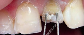
In case of extensive damage to the contact and occlusal surfaces by the carious process and thinning of the remaining tooth tissues (IROPZ = 0.8 or more), medical tactics consist of devitalization of the tooth, cutting off the coronal part to the level of the pulp chamber, and from the contact sides to the level of the carious lesion, making a stump inlay with pin. In the future, such a tooth should be covered with an artificial crown.
In class III and G cavities, the main cavities on the anterior and lateral teeth are formed in places of carious lesions, additional cavities are formed only on the occlusal surface, mainly in healthy enamel and dentin.
The optimal shape of the additional cavity is one that ensures sufficient stability of the inlay with minimal removal of tooth tissue and preservation of the pulp. However, cosmetic requirements for the restoration of anterior teeth, as well as their anatomical and functional differences, determine the characteristic features of the formation of cavities in these teeth.
When choosing a location for the formation of an additional cavity on the occlusal surface of the front tooth, it is necessary, along with other factors, to take into account the unique shape of this surface and the different location of its individual sections in relation to the vertical axis of the tooth and the main cavity.
A horizontally located bottom can be formed perpendicular to the long axis of the tooth in the cervical part of the contact sides. The specificity of the surgical technique for odontopreparation of anterior teeth for restoration with inlays is the formation of vertical walls and the bottom of the cavity, not only taking into account the redistribution of all components of chewing pressure (the leading component is the angular component), but also the route of insertion of the inlay.
There are two ways to insert the insert: vertical from the cutting edge and horizontal from the lingual side anteriorly. In the first case, vertical walls are formed along the contact surface; additional cavities are not created, but parapulpal retention pins are used. The pins are inserted into the tissue of the tooth in the cervical region and cutting edge, focusing on the safety zones, which are clearly visible on an x-ray. A recess for the retention pin is created along the cutting edge, grinding it down by 2-3 mm, but this is only feasible in cases where the cutting edge is of sufficient thickness. A pin only in the main cavity on the contact side cannot provide sufficient stability of the inlay, since the force directed towards the inlay from the palatal side and the cutting edge can rotate it. The use of an additional small pin on the cutting edge significantly increases the stability of the inlay.
If the carious cavity is localized in the middle part of the tooth and the angle of the cutting edge is preserved, then in teeth of significant and medium thickness the formation of a main cavity in the direction of the tooth axis is in principle excluded, since this would require cutting off the angle of the cutting edge, which must be preserved. Therefore, the cavity is created at an angle to the tooth axis. In such cases, an additional cavity on the occlusal surface is also formed at an angle to the tooth axis. This direction of formation of an additional cavity is also necessary because it ensures the stability of the inlay and prevents it from moving towards the missing vestibular wall.
An indispensable condition for the formation of a cavity in case of damage to the vestibular wall, as well as the cutting edge, is the complete removal of the enamel layer, which does not have a dentin sublayer. Preserving a thin layer of enamel in the future will certainly lead to its breaking off due to the redistribution of chewing pressure throughout the entire volume of the tooth.
With small transverse dimensions of the crown, i.e. in thin teeth, the use of retention pins is difficult. Therefore, an additional cavity is formed on the palatal side of such teeth, which should be shallow, but significant in area on the occlusal surface of the tooth. The location of the additional cavity is determined based on the fact that it should be in the middle of the vertical dimension of the main cavity. Retention pins must be placed along the edges of the vertical dimension of the main cavity.
The cavity formed under the inlay is cleaned of sawdust from the hard tissues of the tooth crown and modeling begins.
In the direct method of modeling an inlay, carried out directly in the patient’s mouth, heated wax is pressed into the formed cavity with a slight excess. If the chewing surface is being simulated, the patient is asked to close the dentition until the wax hardens in order to obtain impressions of the antagonist teeth. If there are none, modeling of the cutting edge and tubercles is carried out taking into account the anatomical structure of the tooth. In the case of modeling inlays on the contact surfaces of the teeth, contact points must be restored.
When making an inlay reinforced with pins, pins are first inserted into the corresponding recesses, after which the cavity is filled with heated wax.
An important element of prosthetics is the proper removal of the wax model, eliminating its deformation. If the insert is small, it is removed with one wire sprue-forming pin; if the inlay is large, parallel U-shaped pins are used. In a well-formed cavity, drawing out a model of the inlay is not difficult.
With the indirect method, modeling of a wax reproduction of an inlay is carried out on a pre-fabricated model. In order to obtain an impression, a metal ring is first selected or made from calcined and bleached copper. The ring is fitted to the tooth so that their diameters match. The edge of the ring along the buccal and lingual (palatal) surfaces should reach the equator. When making an inlay from the contact side of the tooth, the edge of the ring should reach the gingival edge.
The ring is filled with thermoplastic mass and immersed in the formed cavity. After the mass hardens, the ring is removed. The quality of the cast is assessed visually. If a good cast is obtained, it is filled with copper amalgam or superplaster. Copper amalgam is introduced in excess, which is used to form a base in the form of a pyramid, which is convenient when holding the model in your hands while modeling the wax insert. After modeling the wax inlays, the metal model is cast.
In the case of the presence of antagonists, as well as to create good contact points, an impression of the entire dentition is made, without removing the impression with the ring from the tooth. After receiving the general impression, the combined model is cast. To do this, the ring is filled with amalgam and a base up to 2 mm long is modeled, then the model is cast according to the usual rules. To remove the ring with thermoplastic mass, the model is immersed in hot water, the ring is removed and the thermoplastic mass is removed. This is how a combined model is obtained, in which all the teeth are cast from plaster, and the tooth prepared for the inlay is made from metal. A wax inlay is modeled on this tooth, taking into account occlusal relationships. Currently, two-layer impression materials are more often used to take impressions. The model can be made entirely from supergypsum.
To cast a metal inlay, the wax reproduction is placed in a refractory mass placed in a casting cuvette. The sprues are then removed, the wax is melted, and the mold is filled with metal. The resulting insert is carefully cleaned of plaque and transferred to the clinic for fitting. All inaccuracies in the fit of the inlay are corrected using appropriate techniques using thin fissure burs. The cement insert is fixed after thorough cleaning and drying of the cavity.
When making inlays from composites, odontopreparation is carried out without forming a bevel (rebate) along the edge of the cavity, since the thin and fragile layer covering the bevel will inevitably break. The simulated wax model of the inlay is covered with a liquid layer of cement, after which the model with the sprue (and cement) is immersed in plaster poured into a ditch, so that the cement is located below and the wax is on top. Replacing the wax with plastic of the appropriate color is carried out in the usual way. After fixing the inlay on the tooth, it undergoes final mechanical processing and polishing.
In rare cases, porcelain inlays are used. The formed cavity is crimped with platinum or gold foil with a thickness of 0.1 mm to obtain the shape of the cavity. The bottom and walls of the cavity are lined so that the edges of the foil overlap the edges of the cavity. The foil form (impression) must accurately copy the shape of the cavity and have a smooth surface. The resulting foil cast is placed on a ceramic or asbestos base and the cavity is filled with porcelain mass, which is fired 2-3 times in a special oven. The finished inlay obtained in this way is fixed with phosphate cement.
Artificial crowns
For defects in the hard tissues of the tooth crown that cannot be replaced by filling or with inlays, various types of artificial crowns are used. There are restorative crowns, which restore the damaged anatomical shape of the natural tooth crown, and support crowns, which provide fixation of bridges.
According to their design, crowns are divided into full, stump, half-crowns, equatorial, telescopic, crowns with a pin, jacket, fenestrated, etc.
Depending on the material, crowns are distinguished as metal (alloys of noble and base metals), non-metal (plastic, porcelain), combined (metal, lined with plastic or porcelain). In turn, metal crowns, according to the manufacturing method, are divided into poured ones, made by casting metal in pre-prepared forms, and stamped ones, obtained by stamping from disks or sleeves.
Since artificial crowns can have a negative impact on both the periodontium and the patient’s body as a whole, when choosing their type and material, it is necessary to carefully examine the patient. Indications for the use of artificial crowns:
- destruction of hard tissues of the natural crown as a result of caries, hypoplasia, pathological abrasion, wedge-shaped defects, fluorosis, etc., which cannot be eliminated by fillings or inlays;
- abnormalities in tooth shape, color and structure;
- restoration of the anatomical shape of the teeth and the height of the lower third of the face in case of pathological abrasion;
- fixation of bridges or removable dentures;
- splinting for periodontal disease and periodontitis;
- temporary fixation of orthopedic and orthodontic devices;
- convergence, divergence or protrusion of teeth when significant grinding is required.
In order to reduce the possible negative consequences of the use of artificial crowns on the periodontal tissue of supporting teeth and the patient’s body, crowns must meet the following basic requirements:
- do not overestimate central occlusion and do not block all types of occlusal movements of the jaw;
- fit tightly to the tooth tissues in the area of its neck;
- the length of the crown should not exceed the depth of the dentoalveolar groove, and the thickness of the edge should not exceed its volume;
- restore the anatomical shape and contact points with neighboring teeth;
- do not violate aesthetic standards.
The last circumstance, as shown by many years of practice in orthopedic dentistry, is essential in terms of creating a functional and aesthetic optimum. In this regard, porcelain, plastic or combined crowns are usually used on the front teeth.
Untreated foci of chronic inflammation of the marginal or apical periodontium, the presence of dental plaque are contraindications to the use of artificial crowns. An absolute contraindication is intact teeth, unless they are used as a support for fixed prosthetic structures, as well as the presence of pathological tooth mobility of the 3rd degree and baby teeth. The production of full metal crowns consists of the following clinical and laboratory stages:
- odontopreparation;
- taking impressions;
- model casting;
- plastering the model into an occluder;
- teeth modeling;
- receiving stamps;
- stamping;
- fitting of crowns;
- grinding and polishing;
- final fitting and fixation of crowns.
Odontopreparation for a metal crown consists of grinding the hard tissues of the tooth from all five of its surfaces so that the artificial crown fits snugly in the cervical area, and its gingival edge is immersed in the physiological gingival pocket (tooth sulcus) to the required depth without putting pressure on the gum. Violation of this condition can cause inflammation of the gums and other trophic changes, scarring and even atrophy.
There are different points of view on the sequence of odontopreparation. You can start it from the occlusal surface or from the contact surface.
Symptoms and consequences of dental defects
Dental defects appear:
- chewing function disorder;
- incorrect bite;
- articulation disorder;
- single displacement of teeth;
- violation of the integrity of the dentition;
- violation of the aesthetics of a smile.
Diagnosis of dental defects
The removal of one or more dental units requires consultation with a dentist and an orthodontist, especially when it comes to the anterior part of the dentition. Untimely correction of the defect does not have the best effect on the condition of adjacent teeth.
As a rule, a visual inspection is sufficient to determine how to correct the defect. In some cases, x-rays may be needed.
Types of cracks
This phenomenon occurs frequently. Treatment depends on how the crown cracks—the direction of the split matters. In medical practice, there are several types of cracks.
Vertical crack
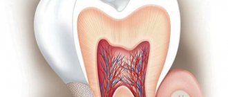
The vast majority are fixed on the front teeth. But it also happens on molars. It can stretch over the entire surface and, when neglected, affects the root. The integrity of the surface part is damaged, caries appears, quickly turning into pulpitis - such a crack opens the way to infection.
Horizontal crown crack
Statistics show: when the crown of a tooth is cracked like this, the destructive process is slower, and there is no accelerated danger to the pulp. If the crack is already large and there are prerequisites for chipping, then the tooth can be restored only by removing the damaged part and strengthening the tooth with a crown.
Oblique oblique
The pulp is often damaged. Treatment depends on the complexity of the specific situation. In an advanced state, only tooth extraction followed by implantation and prosthetics is possible.
Fracture of the tooth crown into pieces
A common option: separation - the crown of the tooth is cracked into 2 parts. The fragments put pressure on soft tissues and injure them. Food particles end up in the crack, causing pain. Treatment is urgently required; in case of serious pathologies, removal is practiced.
"Web"
Small cracks affect only the enamel of the tooth. At this stage, the treatment prognosis is favorable.
Damage to the masticatory tubercle
The pointed part of the tooth is injured. The result is a small chip. The pulp remains largely intact. It is possible to restore the tooth using modern filling materials or install a crown or veneer.
Methods for restoring a dental crown
The method of restoring the crown of a tooth is chosen by the dentist based on the size of the defect. Currently, reconstruction of the supragingival part is carried out in the following ways:
- Installation of a seal. The defect is filled layer by layer with composite material. The method is used for small damage (up to ⅓ volume). The advantages of the method are price, minimal time investment. Disadvantages - the filling changes color over time and stands out against the background of the enamel, and also requires replacement after 3 years.
- Installing a tab. For molar defects of more than ⅓ and less than ½ volume, ceramic inlays are used, which are made in a dental laboratory using an impression. The advantage of the method is efficiency and reliability (the inlay is much stronger than a filling and lasts up to 15 years). Disadvantages: complexity, price, need to wait for the product to be manufactured.
- Installation of veneers. The method is used for minor defects of teeth in the smile area (chips, cracks, stains). It involves removing enamel from the front surface of the tooth and fixing ceramic plates (veneers) with dental cement. The advantage of veneers is their multifunctionality; with their help, you can adjust the color, size, shape of units and at the same time eliminate minor defects. Disadvantages - restoration with veneers is irreversible and introduces some restrictions into the lifestyle.
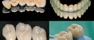
- Prosthetics. Doctors recommend installing artificial crowns (made of ceramic, metal, zirconium, or combined materials) in case of more than half tooth decay, since other methods are not reliable enough. The advantages of the method are that it allows you to quickly restore the functions and aesthetics of a row. The downside is the price.
In case of loss of the crown and root parts, the integrity of the row can be restored using a removable prosthesis (butterfly or dental bridge). Complete restoration of the functions of the lost unit is possible through implantation (installation of a root prosthesis and an artificial crown).
To quickly restore the crown in the optimal way for you, sign up for a consultation with our specialists!
Author: Parnikel Vitaly Evgenievich orthopedic dentist, 1st qualification category
Treatment of cracks in tooth crowns
If the damage does not cause inconvenience and was discovered by chance, then a visit to the doctor is required. The defect should not be viewed as purely cosmetic.
Treatment depends on the type of chip and its severity. For minor damage, the doctor may suggest restoring the tooth crown with durable materials. In more serious cases, canal cleaning or tooth extraction will be required. In each specific option, the decision is made individually.
In case of micro-manifestations, remineralization is recommended, followed by surface coating with composites and varnish. It is possible to install veneers, and in case of significant damage, crowns.