The development of dental pathologies, as a rule, is a consequence of the progression of pathological processes occurring inside the human body. Against the background of weakened immunity, the influence of external negative factors increases, which leads to the formation of problem areas. The causes of diseases can be different: by the symptoms manifested on the tongue, lips and gums, as well as by the results of clinical diagnostics, including the use of professional equipment, the source of concern can be determined. Classification of diseases of the oral mucosa helps to make a correct diagnosis and begin therapy in a timely manner, avoiding more serious negative consequences.
Classification of diseases of the oral mucosa
Stomatitis
Stomatitis is an inflammation of the mucous membrane, characteristic of children and adults. Most often, stomatitis is bacterial, viral or fungal in nature. A bad toothbrush with hard, scratchy bristles, poorly fitting braces or crowns, and biting the cheeks and lips can also cause canker sores.
Most often, stomatitis manifests itself in the form of itchy, bright red or whitish sores and erosions on the inner surface of the cheek, tongue or gums. A person may complain of burning and swelling, bad breath, pain when chewing and swallowing. In advanced cases, the temperature may rise, sleep may be disturbed, and the person becomes irritable.
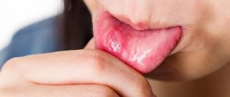
Glossitis
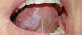
Glossitis is an inflammation of the tongue that can occur either as a result of injury (such as a burn), exposure to pathogens, or as a symptom of certain systemic diseases. Most often, glossitis is manifested by a burning sensation and discomfort in the mouth. The tongue becomes bright red and slightly swollen, and salivation may increase. The patient may complain of loss of taste or changes in the sense of taste, and eating or even just talking causes pain.
Highlit
Haylit (or cheilosis) is a disease in which the lips begin to peel, break, and “sticks” appear in the corners of the mouth. The reasons can be very different: exposure to wind and sun, allergic reaction, chronic diseases with skin lesions (dermatitis, psoriasis, etc.), endocrine pathologies or mycoses.
Oral leukoplakia
Oral leukoplakia is keratinization of the mucous membrane under the influence of aggressive factors, such as smoking. This condition is considered precancerous and therefore requires mandatory treatment.
Most often, oral leukoplakia appears as whitish, grayish, or red plaques that cannot be removed, rough or keratinized areas, or strange thickenings on the lining of the mouth. As a rule, the patient does not experience pain or discomfort, and therefore does not immediately consult a doctor.
Paradontosis
The periodontium is the complex of tissues that surround the tooth and hold it in place: the gums, periodontal ligament, periodontium, root cementum and bone tissue. Periodontal diseases include: gingivitis, periodontitis and periodontal disease.
Gingivitis
Gingivitis is an inflammation of the gums that most often occurs due to inadequate or irregular oral hygiene. Pathogens accumulate in plaque and tartar, causing inflammation.
With gingivitis, inflammation affects only the surface of the gums and may cause bleeding, swelling of the gums, mild pain or discomfort when pressing, and bad breath. If treatment is not started, the inflammation will go further and affect the periodontium.
Periodontitis and periodontal disease
Very often, patients confuse periodontitis and periodontal disease. Periodontitis is an inflammatory disease of periodontal tissues that causes bleeding gums and leads to the gradual exposure of tooth roots, their mobility and, as a result, their loss. Periodontal disease is a non-inflammatory periodontal disease in which the lining of the gums and jaw bone gradually decrease. Unlike periodontitis, in which tooth tissue is destroyed over several years, periodontal disease progresses very slowly and develops over decades. The patient may not even realize that he has gum disease. Periodontal disease is rare compared to other oral diseases.
| — Why are diseases of the oral mucosa dangerous? — Diagnosis of diseases of the oral mucosa — What provokes diseases of the oral mucosa? — Common diseases of the oral mucosa | — Herpetic stomatitis — Recurrent aphthous stomatitis — Leukoplakia — Lichen planus |
Why are diseases of the oral mucosa dangerous?
In the process of eating, food begins to break down in the mouth, under the action of enzymes contained in saliva. Inflammation of the soft tissues of the oral cavity leads to impaired fermentation, which causes problems in the gastrointestinal tract.
Diseases of the oral mucosa have the form of purulent formations that cause itching, burning, and aching pain. They are often accompanied by halitosis. Halitosis is bad breath that brushing your teeth cannot prevent.
The inflammatory process that damages the mucous membrane and soft tissues is caused by poor oral hygiene, smoking (due to the high tar content), abuse of alcohol, hot foods and sweets, as well as increased acidity in the oral cavity.
Such deviations and bad habits create favorable conditions for the development of pathogenic bacteria, and increased acidity irritates the mucous membrane due to the reaction of the acid to the alkaline environment of the oral cavity.
Diagnosis of diseases of the oral mucosa
The variety of types of diseases of the oral mucosa creates certain difficulties for diagnosis. Since the same symptoms can often serve as indicators of completely different diseases, an accurate diagnosis is often the result of long-term observations of the dynamics of the disease.
Methods for studying and diagnosing diseases of the oral mucosa are examination, observation, smears, scrapings. Bacterial cultures and allergy tests.
It is very important to take into account the fact that any inflammatory process in the body (and diseases of the respiratory system are nothing more than an inflammatory process) is a factor that provokes the development of cancer.
The patient can be advised to trust an experienced specialist, be patient and scrupulously follow all the recommendations and prescriptions of the attending physician while waiting for a correct diagnosis, and then take all possible measures to, with the help of a highly qualified dentist, fully and completely recover and avoid consequences.
What causes diseases of the mucous membranes?
An impressive list of reasons is involved in the emergence of diseases of the oral mucosa:
- diabetes mellitus: high blood sugar levels cause decay of mucous tissues, and low levels cause bleeding wounds;
- lack of calcium, phosphorus, fluorine makes tooth enamel and pulp capillaries fragile;
- colds of various etiologies;
- some types of bacteria, fungi and viruses;
- low hemoglobin due to iron deficiency;
- decreased immunity;
- lack of oxygen in soft tissues;
- vitamin deficiency, which destroys capillary walls and leads to the appearance of microthrombi in soft tissues;
- immune and autoimmune diseases (arthritis, HIV);
- vein diseases;
- cancer;
- allergic reactions;
- stress.
The most common diseases of the oral mucosa.
Among the variety of oral mucositis diseases, dentists highlight those that occur most frequently.
The most common are: acute and chronic herpetic stomatitis, recurrent aphthous stomatitis, leukoplakia, lichen planus, trauma to the oral mucosa.
The most common disease of the oral mucosa is herpetic stomatitis
. It progresses in the autumn-winter periods, when the body is susceptible to hypothermia and suffers from changes in temperature.
With herpetic stomatitis, the infection is transmitted by both non-contact airborne droplets and contact. The causative agent of the disease is the herpes virus. The disease is contagious and easily transmitted.
Visually, the disease manifests itself as rashes on the face, especially around the mouth, and on the oral mucosa in the form of single or grouped small blisters (papules) filled with exudate.
Over time, the contents of the vesicle become cloudy, it bursts and an aphtha is formed - a small ulceration of the mucous membrane. In the lip area, the blisters dry out, forming hemorrhagic crusts.
For treatment to be effective, patients must urgently consult a doctor who will conduct an examination and additional diagnostics and prescribe antiviral, antihistamines and vitamins. Additionally, it is necessary to treat the affected elements with leukocyte interferon and perform oral baths with antiseptic solutions. In case of severe pain, the oral cavity can be treated with local anesthetics as prescribed by the dentist. On the fifth day, it is recommended to apply an oil solution of vitamin A, rosehip oil or dental solcoseryl to the aphthae.
To relieve pain, prevent new rashes and shorten the healing time of already formed lesions, experts recommend the procedure of biostimulation with a diode laser at a low power therapeutic mode in the dentist's chair. The procedure is comfortable for the patient, does not require pain relief, and lasts four minutes.
It is also necessary for the first three days after the rash to exclude all planned manipulations in the oral cavity for dental treatment to prevent damage to adjacent areas of the patient’s mucosa and infection of medical personnel.
Erosion on the mucous membrane of the lower lip after opening of the vesicles.
Herpetic eruptions on the skin of the lower lip.
Recurrent aphthous stomatitis
accompanied by constant aching pain in the oral cavity, aggravated by speech and chewing loads. Aphthous stomatitis is an infectious-allergic, bacterial disease.
The causes contributing to the occurrence of aphthous stomatitis include atopy, trauma, endocrinopathy, diseases of the gastrointestinal tract, malnutrition, stress, and food allergies.
The formation of painful recurrent aphthae is preceded by the appearance of a pinhead-sized area of mucosal necrosis in the subcutaneous layer on a reddish base.
Gradually, the focus of necrosis increases, and the aphtha acquires a typical appearance: a round or oval epithelial defect measuring 0.3-0.5 cm, located in the inflamed area. The aphthae is covered with purulent plaque. Most often, aphthae are single and form once or twice a year. Small aphthae heal in two weeks on their own and do not leave scars.
It is impossible to effectively cure aphthous stomatitis, but some improvement is still observed when local anti-inflammatory drugs, Tantum Verde, Cholisal, as well as epithelium-restoring drugs, for example, dental solcoseryl, are applied to the lesion.
Good results are obtained by a single treatment of aphthae with a diode laser in therapeutic mode. Patients who underwent this procedure noted a decrease in pain the very next day after treatment, and the healing time of ulcers was reduced.
The positive effect of the laser is associated with its biostimulating properties. It consists in normalizing trophism and metabolic processes in the tissues of the working state of capillaries, improving microcirculation and regeneration.
Aphtha surrounded by a belt of hyperemia.
Afta before laser treatment.
Afta after laser treatment.
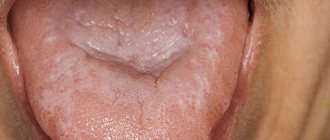
Leukoplakia
It is a white plaque on the oral mucosa that cannot be separated by friction. The disease is more common in men aged 45 to 65 years.
Leukoplakia develops against the background of trauma (sharp edges of carious teeth, overhanging edges of fillings, pathological occlusion), galvanosis, nutritional disorders, excessive consumption of spices, alcohol, smoking, adverse meteorological factors, hormonal imbalance, vitamin deficiency, gastritis, colitis, diabetes mellitus, hereditary and other unfavorable factors.
The size, location of the affected area, and other symptoms may vary. Most often, leukoplakia is visible on the mucous membrane of the cheeks along the line of closure of the teeth, in the area of the corners of the mouth, on the back of the tongue, hard palate, and sometimes on the alveolar process.
The clear edges of the keratinizing spot do not protrude above the level of the surrounding tissue. The surface of the affected area can be smooth and uniform, thin and vulnerable, folded, warty, granular or spotty, and the color can be whitish, gray or brown.
Leukoplakia is benign in 80% of cases; in the rest, dysplasia is noted, that is, a precancerous condition, or transformation into cancer. Within five years, leukoplakia transforms into cancer in 4-6% of patients.
When treating leukoplakia, the causes of irritation of the mucous membrane (nicotine, tars, essential oils formed during the combustion of tobacco, spicy foods) are first eliminated. Sanitation of the oral cavity with subsequent clinical observation, diagnosis and treatment of diseases of internal organs that reduce the resistance of the oral mucosa to the effects of adverse external factors are mandatory. Local applications are made with a solution of retinol acetate and a solution of tocopherol. B vitamins are prescribed internally. A biopsy is performed if the disease does not subside.
Leukoplakia as a result of traumatic tooth brushing.
Leukoplakia of the floor of the mouth and lower surface of the tongue.
Lichen planus
– another common disease of the oral mucosa, accompanied by damage to the mucous membranes. This disease occurs as a result of immune disorders.
Those with an easily excitable nervous system, as well as patients with the hepatitis C virus, are especially predisposed to this disease. Most patients are women over 40 years old. Lichen planus is characterized by a long course of the disease with remissions and exacerbations.
In patients with characteristic dark red, irregularly shaped, itchy nodules on the skin, lesions of the oral mucosa are usually detected.
The typical form is expressed by small grayish nodules on the unchanged mucous membrane, merging into a mesh. Patients complain of roughness, tightness of the oral mucosa and pain.
Treatment of the disease depends on the form. In the typical form, a change in the usual lifestyle is recommended, since eliminating the causes of stress can significantly alleviate the disease. In the chronic erosive form, the effect is achieved by local use of glucocorticoids and epithelizing agents (vitamin A oil solution). Complex treatment of the disease includes tranquilizers, sedatives, immunocorrective therapy, and vitamins. Correction of personal hygiene, sanitation of the oral cavity, elimination of galvanosis and mechanical traumatic factors are mandatory.
Typical form of lichen planus.
Council of the dentist of the Family Dentistry Network "RODNIA".
As soon as you experience discomfort in your mouth in the form of burning, itching and pain, you should immediately consult a dentist, as there is a high risk that you may have herpetic rashes, and herpes is easily transmitted to loved ones, especially children. Parents kiss their children, drink from the same cup, use the same towel, and the infection is transmitted very easily.
The consequences of untreated inflammatory diseases of the oral cavity are loss of teeth, spread of inflammation to the organs of the gastrointestinal tract and upper respiratory tract, and oncology.
Only a doctor will be able to carry out the necessary diagnostics, establish the cause, make the correct diagnosis and prescribe adequate treatment.
Treatment of oral diseases in the RODNYA Family Dentistry Network is performed by periodontists and dental surgeons:
Dentists-therapists of the Family Dentistry Network "RODNIA": | ||
| Mukhambetova Elena Nikolaevna Counterpart, Astrodent | Podarvanova Svetlana Vasilievna Counterparts | Volkolup Ekaterina Igorevna Astrodent |
| Morozov David Iskandyarovich Counterpart, Astrodent | Nogina Anna Yurievna Counterparts | Hovhannisyan Teresa Lazarevna T-med clinic |
| Aleksanyan Hrachya Karapetovich T-med clinic | Baishev Marat Failievich Smile aesthetics | Sargsyan Ruzanna Vladimirovna Rimma-Dent |
| Tarvi Ekaterina Vladimirovna Rimma-Dent | ||
Welcome to RODNYA! Make an appointment (calls from any phone number are free)
Causes of development of diseases of the oral mucosa
- Traumatic damage to oral tissues and other traumatic effects (chemical, thermal, etc.) with the development of traumatic erosion, ulcers, leukoplakia or leukokeratosis (keratinization of the mucous membrane, capable of malignant degeneration).
- Infectious diseases that affect the oral mucosa due to the penetration of viruses, spirochetes, bacteria, and fungi.
Quite often, the occurrence of pathological changes in the oral mucosa is associated with disruption of various organs and systems of the body: allergies, dysfunction of the cardiovascular system, gastrointestinal tract, endocrine disorders, systemic connective tissue diseases, blood diseases and other dermatoses, tuberculosis, AIDS and some other conditions.
Infectious diseases
An infection of the oral cavity leads to the development of a disease in which the mucous membranes become inflamed. The pathological process can involve the teeth and gums. Symptoms of infections in the mouth are often hidden, but sooner or later they appear.
Online training in Anti-Age medicine Learn the intricacies of anti-aging medicine from anywhere in the world. For the convenience of doctors, we have created the Anti-Age Expert online training platform: Lectures from our educational programs are consistently posted here, and are accessible 24/7. Doctors can study the materials as many times as necessary, ask questions and discuss interesting clinical cases with colleagues in special chats
Find out more
Causes of infectious diseases of the oral mucosa:
- Uncontrolled medication use.
Treatment of any disease should be under the supervision of a doctor. Improper use of antibiotics, antibacterial and other agents leads to consequences, including diseases of the oral cavity.
- HIV and AIDS.
Infectious diseases of the mucous membranes of the oral cavity often occur against their background.
- The occurrence of infections in the oral cavity
may be associated with a weakened immune system.
- Poor nutrition.
If the mucous membrane is exposed to aggressive food, it injures it. Thus, the mucous membrane becomes more vulnerable and susceptible to infection.
In addition, smoking and alcohol provoke oral diseases. Those whose bodies are dehydrated or experience hormonal imbalances also face similar problems.
- Stomatitis.
There are many bacteria living in the mouth, and due to a decrease in immunity, they are activated. Thus, an infectious disease develops. One of the most common is stomatitis. It usually develops in people who brush their teeth incorrectly or not thoroughly enough. The disease can also occur due to tonsillitis or diseases of the gastrointestinal tract. There are several types of stomatitis:
- Catarrhal.
It is manifested by swelling of the oral mucosa. As catarrhal stomatitis progresses, plaque appears on the tongue.
- Ulcerative.
In this case, the lymph nodes become enlarged. This stomatitis is manifested by dizziness and weakness. May occur in people with stomach ulcers or enteritis.
- Aphthous stomatitis.
Leads to damage to the oral mucosa, on the surface of which erosions form. Aphthous stomatitis is associated with an imbalance in the gastrointestinal tract. Signs of aphthous stomatitis: swelling of the oral mucosa, malaise, erosion in the mouth.
- Diseases caused by viruses.
The best known oral infection is herpes, clinically known as herpes simplex virus type 1 (HSV-1). It is often found in children between 6 months and 5 years of age.
Once HSV-1 enters a child's body, he will carry the virus throughout his life. It is estimated that 50-80% of adults live with herpes simplex, either dormant or active.
The herpes virus manifests itself as rashes around the mouth. As the disease progresses, ulcers form in the mouth: they are localized on the inside of the lips and cheeks. Herpes can also infect tissues near the teeth.
After the first attack of oral herpes, the body produces antibodies to fight the virus and its effects. This way, your subsequent outbreaks of HSV-1 may not be as severe or the virus may remain dormant.
Otherwise, taking antiviral drugs may help.
- Candidiasis.
This is a manifestation of a fungal infection. Candida fungus microorganisms live in the mouth and are activated under the influence of unfavorable factors. Candidiasis occurs in people with weakened immune systems. To avoid illness, it is necessary to strengthen the immune system and avoid hypothermia. There are several types of candidiasis:
- Pseudomembranous.
It occurs in an acute form. As the pathology progresses, the mucous membrane of the cheeks begins to dry out, and the same thing happens with the lips and tongue. A coating of curd consistency forms on the tongue. Pseudomembranous candidiasis causes discomfort when chewing and itching. The disease can occur in people with a weakened immune system, as well as against the background of blood pathologies. Other causes of pseudomembranous candidiasis are diabetes mellitus and hypovitaminosis.
- Atrophic candidiasis.
In this case, the mucous membrane of the mouth becomes red and dry. The chronic form of the disease develops in those who use dentures for a long time.
- Hyperplastic candidiasis
may be acute or chronic. In chronic cases, plaque forms on the mucous membranes, including the tongue. When you try to remove it, the mucous membrane becomes more inflamed. Brushing your teeth may cause bleeding.
- A disease that occurs due to HIV.
Secondary immunodeficiency is characterized by the active proliferation of pathogenic flora in the oral cavity. Early diagnosis will allow faster initiation of treatment and improve the prognosis of immunodeficiency.
It is important to note that diseases of the oral cavity that occur against the background of HIV often become chronic.
Diagnosis of pathologies
Modern techniques used in dentistry make it possible to quickly identify infectious or fungal diseases of the oral mucosa. It is worth noting that self-diagnosis, as well as subsequent attempts at self-medication, often cause a deterioration in the general condition. Determining the causes of pathological changes is a medical task for which the following are used:
- Microscopic examination of samples.
- Test for allergic reactions.
- Test for viral pathogens.
- General examination and medical history.
Timely diagnosis is necessary to develop and implement the correct treatment plan that addresses both negative symptoms and factors that are proven to cause pathological changes.
Oral cancer
Oral cancer most often affects the tongue, tonsils, gums and oropharynx. Since its various types often do not cause obvious signs and symptoms in the early stages, regular dental examinations are the most important method of detecting them.
Your dentist may also test you for oral cancer during your exam, especially if you have any of these symptoms:
- Persistent pain in the mouth or lip;
- Red or white spot in the mouth;
- Loose teeth;
- Painful swallowing, constant pain in the mouth or ears.
Treatment:
Depending on the type and stage of oral cancer, treatment may include a combination of surgery, radiation therapy and chemotherapy.
Principles of treatment of diseases of the oral mucosa
Basic principles of treatment of diseases of the mucous membranes of the mouth, lips and tongue:
- Rational treatment requires contact between the dentist and other dental and non-dental professionals.
- Treatment must be carried out in compliance with the principles of bioethics, these diseases must be considered from the point of view of the state of the whole organism, therefore in most cases one cannot limit oneself to local effects only.
- An axiom for the dentist should be the elimination of all unfavorable irritating factors in the patient’s oral cavity that can support and provoke the development of the pathological process. The use of so-called cauterizing agents and prolonged use of the same mouth rinses is unacceptable.
- Treatment should begin only after at least one preliminary diagnosis has been established and the following requirements have been met: be comprehensive; provide a pathogenetic approach; do not violate the anatomical and physiological characteristics of the oral mucosa; eliminate the pain factor; promote rapid epithelization of lesions; provide for the active involvement of the patient in performing treatment procedures at home.
Chemical injuries to the oral mucosa in children.
These injuries are observed mainly in children 1–3 years old due to accidental ingestion of solutions of acids and alkalis used in everyday life. In this case, combined burns of the mucous membrane of the mouth, pharynx, and esophagus develop. The severity of the lesion is determined by the concentration of the drug and the duration of its effect. The mucous membrane becomes sharply hyperemic, then necrosis appears within a period of several hours to a day, the deepest usually on the lower lip. Necrotic tissues become saturated with fibrinous exudate; a thick film is formed, which is slowly torn off on the 7th - 8th day and at a later date. In an uncomplicated course, under such a film, tissue scarring and epithelization of the defect occur in parallel.
Chemical burns can be caused by many medications used in dental treatment: phenol, formalin, antiformin, acids, alcohol, ether, etc., so the dentist should be especially careful when using them, taking into account the easy vulnerability of the mucous membrane in children and the often violent reaction of the body in response to its damage.
TREATMENT OF CHEMICAL INJURIES OF THE ORAL MUCOSA IN CHILDREN.
For chemical burns in the first minutes and hours, it should be aimed at neutralizing the chemical agent with 1 - 2% solutions of sodium bicarbonate for acid burns and 1% hydrochloric acid solution or 3% citric acid solution for alkali burns. Further treatment consists of preventing secondary infection of the lesion and pain relief. For burns of the pharynx and esophagus, the child should be hospitalized in the ENT department.
Therapy methods
- Etiotropic and pathogenetic therapy aimed at eliminating the cause of the disease (antiviral, antibacterial therapy due to the infectious nature of stomatitis, glossitis, cheilitis, vitamin therapy for hypovitaminosis, treatment of the underlying disease that caused the appearance of a pathological process in the oral cavity) of the mucous membrane;
- Local treatment aimed at eliminating local traumatic factors, the main symptoms of the disease and faster healing of existing erosions and ulcers;
- A general strengthening procedure that stimulates the body's defenses.
Brief conclusions
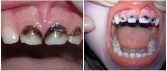
- The most common oral diseases are tooth decay, gum disease, infectious diseases and oral cancer.
- Caries and gum disease are highly treatable in the early stages.
- The causes of infectious diseases of the oral cavity vary - from viruses to poor diet and uncontrolled use of medications.
- For prevention, it is important to review “life hygiene” and regularly visit the dentist.
Anti-Aging Medicine Seminars Gain knowledge based on evidence-based medicine first-hand from the world's leading experts. As part of the Anti-Age Expert Modular School, in-person two-day seminars are held every month, where the intricacies of anti-age medicine are revealed to doctors of more than 25 specialties
Find out more
Prevention
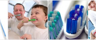
To prevent painful symptoms, experts recommend following the universal rules of oral hygiene:
- use properly selected toothbrushes, use them systematically, and also avoid bad habits, especially smoking.
- It is recommended to control your diet: in some cases, oral irritation may be caused by excessive consumption of oranges, lemons, etc.
- the habit of cleaning seeds with teeth rather than with hands can become unfavorable for the oral cavity.
Results and discussion
A study of the prevalence of TS showed that most often TS occurs in the first 1.5 months from the start of orthodontic treatment ( p
≤0.01), and subsequently the frequency decreases (Fig. 3).
Rice. 3. The incidence of traumatic stomatitis during orthodontic treatment in children and adults at various times during the active treatment period. Thus, according to the data we obtained, the incidence of TS was significantly less common than according to the literature [3–5], and amounted to 6.62–15.38% in children during different periods of the active period of orthodontic treatment with fixed appliances, and 4.69% in adults. -11.63% (see Fig. 2)
.
Before the start of treatment, TS of moderate and severe severity was detected in both children and young adults (Fig. 4, 5).
Rice. 4. The number of patients in the 1st and 2nd study groups depending on the severity of TS at different follow-up periods.
Rice. 5. The number of patients in the ٣-th and ٤-th study groups depending on the severity of traumatic stomatitis at different periods of follow-up. As a rule, the pain syndrome was mild, and the mucous membrane of the cheeks was most often affected, and less often the lips. Typically, in patients, regardless of age, with severe swelling and hyperemia, erosive and ulcerative lesions of varying severity were observed in the area of projection of orthodontic equipment (braces and arches).
The use of topical agents to treat TS has been effective in both children and adults. More pronounced positive dynamics of the reparative process of oral mucosa was noted when using gum gel with propolis in all age groups ( p
≤0.05) and observation period (
p
≤0.05).
At the same time, it should be noted that if in children on the 3rd day from the start of treatment TS of severe severity remained (see Fig. 4), then in young people (see Fig. 5) there was no such degree of TS ( p
≤0.05).
By this time, in adults, regardless of the drug used for the treatment of TS, there were no pathological changes in the mucous membrane in the area of the mucous membrane previously injured by the brace system ( p
≥0.05).
In children of groups 1 and 2, the effectiveness of treatment on the 3rd day was 34.66 and 48.96%, respectively ( p
≤0.01);
on the 5th day - 78.66 and 83.33%, respectively ( p
≤0.05). Similar data on the effectiveness of treatment of TS caused by injury to the mucous membrane of the cheeks and lips by braces and arches were obtained in young adults (Fig. 6).
Rice. 6. The effectiveness of treatment of traumatic stomatitis in patients of different groups at different periods of dynamic observation. Thus, in individuals included in the 3rd and 4th study groups, the effectiveness of treatment on the 3rd day was 60.29 and 68.03%, respectively ( p
≤0.05);
on the 5th day 91.18 and 95.08%, respectively ( p
≤0.05), which also indicated the greater therapeutic effectiveness of the gum gel with propolis for the treatment of TS caused by wearing fixed orthodontic appliances.
On the 10th day, no TS was detected in patients of all four study groups.
Literature
- Banchenko G.V. Combined diseases of the oral mucosa and internal organs. - M.: Medicine, 1979. - 190 p.
- Banchenko G.V., Maksimovsky Yu.M., Grinin V.M. Language is the “mirror” of the body: Clinical guide for doctors. - M., 2000. - 408 p.
- Borovsky E. V., Danilevsky N. F. Atlas of diseases of the oral mucosa. — 2nd ed., revised. and additional - M.: Medicine, 1991. - 320 p.
- Danilevsky N. F., Leontyev V. K., Nesin A. F., Rakhniy Zh. I. Diseases of the oral mucosa. - M., 2001. - 271 p.
- Diagnosis, treatment and prevention of dental diseases / V. I. Yakovleva, E. K. Trofimova, T. P. Davidovich, G. P. Prosveryak. — 2nd ed., revised and supplemented. - Mn.: Higher. school, 1994. - 494 p.
- Dmitrieva L. A., Glybina N. A., Glybina T. A. et al. Experimental substantiation of the use of a new antioxidant drug in the treatment of erosive and ulcerative lesions // Periodontology. — 2012, No. 3 (64). - pp. 52-58.
- Diseases of the mucous membrane of the mouth and lips / Ed. prof. E. V. Borovsky, prof. A. L. Mashkilleyson. - M.: Medicine, 1984. - 400 p.
- Diseases of the oral mucosa: Textbook / Ed. L. M. Lukinykh. - N. Novgorod: Publishing House of NGMI, 1983. - 212 p.
- Karakov K. G., Vlasova T. N., Oganyan A. V., Pisarev G. Yu. Optimization of complex therapy of lichen planus of the oral mucosa // Dentist-practitioner. - 2012, No. 1. - P. 35-37.
- Karakov K. G., Vlasova T. N., Oganyan A. V., Polyakova O. V. The role of complex therapy in the treatment of herpetic infection of the dental system // Practitioner dentist. - 2012, No. 2. - P. 48-49.
- Practical guide to anti-infective chemotherapy / Ed. L. S. Strachunsky, Yu. B. Belousov, S. N. Kozlov. - M.: Borges, 2002. - 384 p.
- Rybakov A. I., Banchenko G. V. Diseases of the oral mucosa. - M.: Medicine, 1978. - 232 p.
- Therapeutic dentistry: textbook: 3 hours / ed. G. M. Barera. - M.: GEOTAR-Media, 2055. - Part 3. - 288 p.
- Tretyakovich A. G., Borisenko L. G., Pishchinsky I. A. Differential diagnosis and principles of treatment of diseases of the oral mucosa: educational method. allowance. — 2nd ed., revised. and additional - Mn.: BSMU, 2005. - 66 p.
- Udzhukhu V. Yu., Minatulaeva M. A., Kubylinsky A. A. Pathogenetic rationale and clinical effectiveness of the use of Lavomax in patients with exudative erythema multiforme // Farmateka. - 2008, No. 9. - P. 60-62.
- Tsepov L. M., Nikolaev A. I. Medical tactics for erosive and ulcerative lesions of the mucous membrane of the mouth, tongue and lips (Training manual). - Smolensk: SGMA, 2005. - 16 p.
Material and methods
We studied the incidence of traumatic lesions of the oral mucosa (traumatic stomatitis - TS) in children 12-17 years old and young adults (18-32 years old) when using brace systems. Frequency T.S. assessed in 52 children and 43 adults for up to 1.5 months immediately after installation of the brace system, as well as in 136 children and 256 adults during orthodontic treatment, from 1.5 months to 1.5 years from the start of using fixed orthodontic equipment .
To evaluate the effectiveness of treatment, 4 groups of patients were formed, taking into account age and the drug used for local treatment of traumatic injuries of the oral mucosa caused by the use of fixed orthodontic appliances (Fig. 1).
Rice. 1. Distribution of patients into study groups.
The 1st group included children who used vinylin for internal use for the treatment of traumatic injuries of the oral mucosa (JSC ", Moscow region, Staraya Kupavna, Russia), in the 2nd group, children used a gum gel with propolis (JSC "VERTEX" ", Saint-Petersburg, Russia). The following groups included adult patients whose TS was treated with topical vinylin (group 3) or gum gel with propolis (group 4). All observed patients (children and adults) underwent orthodontic treatment using vestibular brace systems with correction in order to prevent re-injury of the oral mucosa during treatment.
Control examinations of patients of all groups suffering from TS were carried out on the 3rd, 5th and 10th day from the start of treatment using these drugs. Their local application in children was carried out by parents, and in young people - by the patients themselves, 3 times a day after meals. Along with this, all patients during dynamic observation for up to 10 days before local use of the drug for the treatment of TS, in order to increase the effectiveness of treatment, used the ASEPTA parodontal active mouth rinse (JSC VERTEX, St. Petersburg, Russia).
To ensure the comparability of the results obtained and their reliability, a developed semi-quantitative method was used, which consisted of a visual assessment of various clinical symptoms of TS, each of which was assigned one of three symbols indicating the absence or severity of a specific clinical symptom. Thus, based on an analysis of the symptoms of TS, an index method was proposed for assessing the severity of this pathology, taking into account the following clinical symptoms and their assessment in points:
1. Pathological sensations (pain): absent - 0; moderately expressed - 1; pronounced - 5.
2. The area of the focus of traumatic lesions of the oral mucosa (determined taking into account the damaged organs and
fabrics): TC is not determined - 0; lesion on the mucous membrane in the area of the brace system on one of the jaws - 1; lesion on the mucous membrane in the area of braces on both jaws - 5.
3. Localization of traumatic lesions
: the lesion is not determined - 0; visualized in the lips or cheeks - 1; visualized in the area of lips and cheeks - 5.
4. Presence of hyperemia and edema of the mucous membranes
: no hyperemia, mucous membranes are pale pink, no swelling - 0; moderate (mild) hyperemia and swelling of the mucous membranes - 1; pronounced hyperemia (bright red) and swelling of the mucous membranes - 5.
5. Clinical and morphological characteristics of the focus of traumatic lesions of the oral mucosa
: the lesion is not determined - 0; erosion or erosions are visualized - 1; erosive and ulcerative lesions are diagnosed - 5.
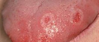
To establish the severity of the clinical picture of TS, the listed clinical symptoms are first diagnosed. After registering the symptoms of the pathology, the sum of points is calculated and the severity of TS is assessed based on the obtained sum of points as follows: 0 - no pathology; 1-4 points - mild severity of the vehicle; 5-9 points - average degree; 10-25 points - severe degree. The data obtained in each group of patients according to symptoms were arithmetically added, and the resulting average values were used for further statistical processing (Fig. 2).
Rice. 2. An erosive-ulcerative traumatic lesion in the area of the mucous membrane of the lower lip on the right of moderate severity, caused by wearing a brace system, in a 14-year-old patient (group 1, subgroup 1). Clinical manifestations: pathological sensations (pain): moderate - 1; area of the focus of traumatic lesion of the oral mucosa (determined taking into account damaged organs and tissues: lesion on the mucous membrane in the area of the brace system on one of the jaws) - 1; localization of traumatic lesions: visualized in the area of the lower lip - 1; presence of hyperemia and swelling of the mucous membranes: moderate (mild) hyperemia and swelling of the mucous membranes - 1; clinical and morphological characteristics of the focus of traumatic lesion of the oral mucosa: erosive and ulcerative lesion was diagnosed - 5 (total 9 points).
To objectify the result of treatment for TS, a method was used that involved determining the effectiveness of the therapy for the specified pathology of the oral mucosa, which was carried out according to the formula:
Efficiency (%)=100·(A–B):A,
where A is the sum of points in the clinical assessment of the severity of TS before the start of therapeutic measures; B - the sum of points for clinical assessment of the severity of the pathology in question on the 3rd, 5th and 10th day from the start of treatment.
The reliability of differences in the average values of independent samples was assessed using the parametric Student's test for normal distribution and the non-parametric Mann-Whitney test for differences from the normal distribution of indicators. Normality of distribution was checked using the Shapiro-Wilk test. For statistical comparison of proportions with an assessment of the significance of differences, the Pearson χ2 test was used, taking into account the Mantel-Haenszel likelihood correction. In all statistical analysis procedures, the achieved level of significance was considered ( p
), the critical significance level was equal to 0.05.