What is this disease?
Herpetic stomatitis is a pathological process that develops in the mucous membrane lining the oral cavity. The causative agent of this disease is herpes simplex virus type 1. In children under the age of five, this virus is detected in 60% of all cases. By adolescence, it is detected in the vast majority of people. Herpes stomatitis in children develops during the baby’s first contact with the virus. This occurs most often before the age of three.
The high incidence is explained by:
- low level of production of own antibodies;
- immaturity of cellular immunity;
- the fact that the baby does not receive antibodies from mother's milk;
- high reactivity of the child's body.
If the baby is bottle-fed, he may get sick in the first months of life. Viral infection often goes into a latent state. It persists in the nerve ganglia.
Treatment of ENT diseases in infants
The most important task in the treatment of otolaryngological diseases is to prevent the disease from becoming chronic. Therapeutic (drug, physiotherapeutic) methods are used in the treatment of ENT pathology. In recent years, minimally invasive laser and endoscopic methods have been actively used to treat otolaryngological pathologies.
Prevention of diseases of the nasopharynx, larynx and hearing organs in children must be applied from a very early age. A qualified pediatric ENT specialist will help you develop a plan of preventive measures that will help your baby avoid chronic colds and infectious diseases, as well as the risk of various complications.
Remember that, regardless of age and general condition of the body, a child requires constant attention. A pediatric ENT doctor will always help to diagnose the disease in a timely manner, establish its causes, and also prescribe appropriate treatment and prevent possible complications.
Causes
The cause of the development of the pathology is the herpes simplex virus type 1. It belongs to DNA viruses belonging to the Herpesviridae viral family. The pathogen multiplies rapidly in the epithelial cells of the oral mucosa. Then it enters nearby lymph nodes (submandibular) and continues to reproduce in them. Then it enters the blood and migrates to the parenchymal organs (spleen, liver, kidneys). There it multiplies and enters the bloodstream again. As a result, it again appears in large quantities in the epithelial cells of the skin and mucous membranes. They are massively defeated. It is localized in the oropharynx, oral cavity, nose, lips, and nearby skin. Stomatitis and herpes of the skin and mucous membranes develop when the infection generalizes simultaneously.
Children become infected with it as follows:
- through contact and everyday life (using shared utensils, toys, through kissing);
- airborne (coughing, sneezing);
- from a sick mother to the fetus through the placenta or during childbirth.
A baby can become infected from sick adults, children, and carriers.
The following factors contribute to the development of the disease:
- previously suffered inflammatory processes;
- previous antibiotic therapy;
- deficiency of microelements and vitamins in the body;
- mechanical damage to the skin and mucous membranes;
- insufficient fluid intake;
- poor oral hygiene.
In children, the virus is especially easily transmitted through contact. The infectious process quickly spreads to healthy areas.
Snot in a baby
Very often situations arise when a child has snot, but there are no signs of a cold. This type of runny nose is physiological in nature and can continue until the newborn is 2 months old. Factors that cause snot in newborns:
- Infection. Most often, colds occur when a viral infection enters the body, transmitted by airborne droplets. ARVI in infants progresses rapidly and manifests itself with pronounced symptoms.
- Allergy. Snot in children can also be allergic in nature. They occur when nasal inhalation of allergens such as dust, pollen of flowering plants, fluff, and wool. In such situations, the breathing process becomes more difficult, the baby begins to sneeze, and watery snot comes out of the nose. Vascular response to external stimuli. Very often, snot in newborns occurs when the nasopharyngeal vessels are highly sensitive to environmental factors. This process usually manifests itself with bouts of sneezing, alternating sinus congestion, and copious nasal discharge.
- Enlarged adenoids. A peculiarity of the physiology of the children's respiratory system is that at birth, adenoids begin to grow rapidly in children. They sometimes provoke the formation of snot, which has a greenish color. In such cases, it is necessary for the baby to drip a 1% solution of collargol into the nose.
Treatment of rhinitis in newborns is difficult, due to the narrow nasal passages. The course of rhinitis in newborns has its own characteristics, which is explained by the physiological and anatomical characteristics of the infant. The complexity of the course of the disease lies in the fact that infants cannot independently free their noses from accumulated mucus, and also do not know how to breathe through their mouths, which is especially dangerous during sleep and breastfeeding.
Many parents don’t know what to do when their newborn’s snot bothers their baby day and night. You can’t start drug treatment for your baby’s rhinitis on your own; therapy can only be prescribed by a specialist.
How to treat a runny nose in a newborn depends on the factors that caused this condition of the child’s nasal mucosa. Even before visiting a specialist’s office, parents can take steps to alleviate their baby’s condition. First of all, if a baby has a severe runny nose that makes nasal breathing difficult, it is necessary to clear the nasal passages of pathological secretions. Solutions based on sea water or regular saline are well suited for this procedure.
Humidifying the air should be another action for parents who don't know what to do when their baby has a runny nose. A well-ventilated room with moist air promotes faster recovery of the nasal mucosa. You can increase the humidity in a dry room by using a humidifier. The optimal air humidity in the room where the sick child is is 50% at a temperature of 20-21ºС.
Treatment of clear snot in a child should be carried out only as prescribed by a doctor, since such a symptom may indicate several diseases. Regardless of the cause of rhinitis, parents should regularly clean the baby’s nose, thereby improving nasal breathing. To do this, you can use a special device for suctioning mucus - a nasal aspirator. If the clear mucus in the nose is so thick that it is difficult to remove it from the nasal cavity, the mucus must first be thinned. Solutions based on sea water, as well as decoctions of herbs such as chamomile, are well suited for this. You need to drop a few drops into each nasal passage of the child, and then use an aspirator. It is important to adhere not to symptomatic treatment, but to take actions aimed at eliminating the cause of the disease. Parents should promptly contact specialists who will tell you how to treat clear snot in a child, having previously established an accurate diagnosis.
Pharynx
In children, near the median septum of the retropharyngeal cellular space, there are lymph nodes into which lymphatic vessels flow from the palatine tonsils, posterior parts of the nasal and oral cavities. With age, these nodes atrophy; in children they can fester, forming a retropharyngeal abscess.
Adenoids are common in children.
Larynx
In newborns and young people, the larynx is located slightly higher than in adults (in adults, the upper edge of the larynx is at the border of the IV and V cervical vertebrae).
In children, the Adam's apple is soft and not palpable.
In children, the subglottic cavity has a well-developed loose submucosal layer. Its inflammatory swelling is called false croup.
Outer ear
In a newborn and infant in the first 6 months of life, the entrance to the external auditory canal looks like a gap due to the fact that the upper wall is almost closely adjacent to the lower one.
In newborns, the temporal bone is not yet fully developed, so they do not have the bony part of the ear canal, there is only a bone ring to which the eardrum is attached. The bony part of the auditory canal is formed by the age of 4, and up to 12-15 years the diameter of the lumen, shape and size of the external auditory canal change.
Eardrum
In children, the eardrum has an almost round shape and is much thicker than in adults (0.1 mm), due to the inner and outer layers. Therefore, with acute otitis media in children, perforation of the tympanic membrane may not be observed.
Middle ear
The auditory (Eustachian) tube in children is wider and shorter than in adults.
Mastoid
In a newborn, the mastoid part of the middle ear looks like a small elevation behind the superoposterior edge of the tympanic ring, containing only one cavity - the antrum. The formation of the mastoid process ends by the beginning of the 7th year of the child’s life.
Classification
According to the flow, acute, chronic, and wave-like variants of the pathology are distinguished. The following degrees of severity of the pathological process are distinguished:
- Mild - it is typically characterized by a slight increase in body temperature, moderate inflammation of the mucous membrane in the mouth, and enlargement of regional lymph nodes. Rashes form on the mucous membrane and skin.
- Medium – high temperature rises. Severe weakness and sudden deterioration in health. The baby begins to vomit and the pain in the mouth increases. Significant rashes appear in the mouth and the skin around it.
- Severe - a severe headache is added to the pathological process. High temperature rises, severe muscle pain. Not only regional but also distant lymph nodes enlarge. The rashes are located not only in the oral cavity, but on the skin next to it. They appear on the mucous membrane of the eye, on the eyelids, and on other parts of the face.
The severity of the pathology depends on the viral load (the amount of virus in the body) and the general reactivity of the body.
Diagnosis of ENT diseases in infants
Examination and treatment of children differs markedly from working with adult patients. A young patient cannot always clearly explain what is bothering him; he does not know how to properly dissolve pills or gargle. The ability and skills of a good pediatric ENT doctor to find an approach to a sick child and establish psychological contact with him are no less valuable than the professional skills of an otolaryngologist. The physiological and anatomical characteristics of a small child’s body determine the specificity of treatment procedures, examination of ENT organs, and anesthesia (if necessary).
Modern methods for diagnosing ENT pathology include: clarification of parental complaints, questions of the diagnostic and treatment complex, etc., objective examination, laboratory tests, endoscopic and computer studies of the nose, throat, and ear, ultrasound.
Clinical manifestations
Stomatitis in children is characterized by a gradual onset of the disease. Clinical signs of the disease do not appear immediately after infection. The incubation period is typical for this pathology; it lasts from two days to three weeks.
Symptoms of herpetic stomatitis in children appear starting from the prodromal period.
Each period has its own clinic:
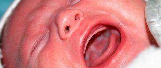
- Latent – lasts up to two weeks. The child's sleep is disturbed and he refuses to eat. The baby becomes restless and whiny. He has increased salivation, possible nausea and vomiting. The lymph nodes are enlarged and painful on palpation.
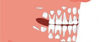
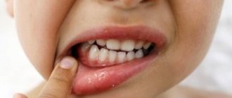
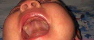
- The height of the disease - at this time rashes appear on the skin and mucous membranes. They are located on the soft, hard palate, gums, cheeks, lips and tongue. They are group or single, up to three millimeters in size. They are thin-walled bubbles filled with a clear liquid. Their formation lasts up to four days. The vesicles quickly open, then erosions and painful aphthae form. They are shallow ulcers covered with a white coating. The mucous membrane in the mouth is swollen and bleeding. The child develops a high temperature of up to 40 degrees. A runny nose, cough, and conjunctivitis occur.
- Fading - aphthae, erosions gradually heal and epithelialize. These formations heal without scarring. Often there is a wavy course of the disease. The periods of appearance of rashes alternate with rises in temperature.
The illness usually lasts up to two weeks. In children under one year of age, generalization of the process is possible. The development of sepsis, damage to all internal organs and meninges is likely.
Diagnostics
A patient suspected of having this disease is examined by a dentist. He asks the child’s parents about how the disease progressed. The diagnosis is established on the basis of the characteristic clinical picture of stomatitis, revealed during examination, anamnesis and characteristic complaints. Upon examination, typical mucosal lesions are revealed.
To confirm the diagnosis, laboratory testing of scrapings of the oral mucosa, the contents of ulcers, the patient’s saliva and blood is used.
The following are used for research:
- cytopolymerase chain reaction;
- immunofluorescence method;
- serological blood tests (RSC, ELISA, immunoglobulin M test);
- HSV test for the detection of immunodot G-specific glycoprotein.
These methods are used only for severe infections, as they are quite expensive.
Treatment
Treatment of herpetic stomatitis in children with mild and moderate forms is carried out on an outpatient basis. In severe cases and the development of complications, the baby is hospitalized. Treatment is carried out under the supervision of a pediatric dentist or periodontist.
Children are prescribed bed rest and a diet with pureed, non-irritating food. He is given separate hygiene items and dishes. Plenty of warm fluids are recommended.
The following drugs are prescribed:
- Non-steroidal anti-inflammatory drugs (Nise, Paracetamol, Nurofen) are used to relieve temperature and inflammatory reactions.
- Antihistamines (Clemastine, Loratadine) are used to relieve swelling of the mucous membranes.
- Antiviral drugs (Famciclovir, Acyclovir, Zovirax) are used at the beginning of treatment or in severe cases.
- Immunomodulators (Lysozyme, Gamma globulin, Thymogen) are used to enhance immunity.
During treatment, vitamin-mineral complexes and fish oil are used in monthly courses.
Local drugs that act directly on the mucous membrane are widely used.
For local treatment of herpetic stomatitis the following are used:
- antiseptics (Hexoral, Miramistin) - they are used to rinse the mouth every four hours for two weeks;
- ointments and gels with anesthetics (Kamistad, Lidochlor gel) are used for pain relief, they are lubricated with mucous membranes three times a day for up to two weeks;
- antiviral agents (Ganciclovir, Acicovir) - destroy the viral cell, gums are treated with these ointments five times a day, two weeks;
- proteolytic enzymes (Trepsin, Chymotrypsin) are used to cleanse the necrotic surfaces of ulcers; they are washed with these solutions twice a day.
- rinsing with decoctions of medicinal herbs (calendula, chamomile, sage) is carried out after each meal for up to two weeks;
- epithelializing ointments (Solcoseryl, Methyluracil) are used to enhance the healing of erosions and ulcers, used in the recovery stage, used up to four times a day for ten days.
Physiotherapy is applied locally. Irradiation of affected mucous membranes with ultraviolet and infrared rays is used.
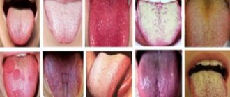
White plaque
The tongue and palate of an infant may be covered with a thin whitish coating. Main reasons:
- leftover food;
- mouth breathing;
- thrush.
Leftover food
Immediately after feeding, you can see the remains of breast milk or formula on the baby's tongue and palate. After some time, the plaque disappears on its own. Many pediatricians advise removing traces of food from the baby’s mouth, as they serve as a breeding ground for opportunistic microorganisms.
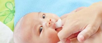
Cleaning methods:
- give the baby 1-2 teaspoons of boiled water immediately after feeding;
- wipe the epithelial membrane 2 times a day with soda solution or water (the procedure is described below).
If the plaque is easily removed or disappears on its own (it is not present before feeding), do not worry. But in cases where white marks on the tongue, palate and inner surface of the cheeks are constantly present, it is necessary to consult a doctor.
Mouth breathing
If a child breathes through his mouth, his mucous membrane dries out and a whitish coating forms on it. In children under one year of age, this phenomenon is most often caused by acute respiratory viral infections and dry air in the room, which leads to the formation of crusts in the nasal passages.
The only way to prevent the formation of white plaque is to restore nasal breathing in the baby. Basic methods:
- when the mucus dries out, instill saline and oil drops into the nose, as well as humidify the air;
- for a runny nose and congestion due to ARVI - similar measures, in addition, the doctor may recommend the use of vasoconstrictor drops (no longer than 5-7 days).
Prevention
Preventive measures are aimed at preventing infection. This presents certain difficulties, since the vast majority of the population is infected with it. It is better if the child gets sick from it at an older age.
For this it is recommended:
- avoid contact with infected people;
- will provide personal utensils and personal hygiene products for the child;
- Kissing people with herpetic rashes is prohibited;
- strengthening the baby's immune system.
It is recommended that the child be provided with adequate nutrition and regularly given vitamin and mineral complexes.