General information
Pyogenic granuloma is conditionally classified as a group of pyoderma , because previously it was believed that the provoking factor was a staphylococcal infection , but now it is defined as benign inflammatory hyperplasia of granulation tissue.
The pathology is usually localized on the skin of the feet, hands, red lip border, on the abdomen, oral mucosa, periungual ridges, in places of cuts, injections, burns, splinters, ingrown nails , in extremely severe cases in the gastrointestinal tract and is most often single.
Pathogenesis
Typically, pyogenic granuloma, also called telangiectatic granuloma, occurs at the site of a minor skin injury, such as a scratch or abrasion. The formation process is quite fast - it begins 1-2 weeks after a minor scratch and is benign; it can be tumor-like, granulomatous, rising above the skin and forming a structure the size of a lentil grain, cherry bone or nut.
The formation of pyogenic granuloma occurs in several stages:
- phagocytes accumulate in the area of minor injury ;
- then the transformation of cellular structures into macrophages and degeneration into granuloma occurs;
- Subsequently, an epithelioid pyogenic tumor is formed due to the transformation of macrophages and phagocytes into epithelioids, which additionally gives it the properties of a granuloma.
The structure is usually represented by granulation tissue in the form of an atypical proliferation of numerous small capillaries ( angioblastoma ), inflammatory infiltrate and endothelium; in later stages it can become lobulated. Then a regression stage is possible due to extensive fibrotic processes.
In most cases, botryomycoma occurs at the site of minor injuries, however, it can also occur without previous trauma; in this case, elevated tumor-like formations of a bright red or bluish color develop, which contain a large number of superficially located vessels, which gives it the features of a lobulated capillary hemangioma. Ulceration and damage to the tumor entail an increased risk of secondary infections and recurrent inflammation. It is important to remember that any benign neoplasm can degenerate into malignant.
Root cause
The neoplasm is called botryomycomoma, telangiectatic granuloma, benign pedunculated granuloma, pregnancy granuloma, lobular capillary hemangioma, and many others. A large number of synonyms and definitions of this disease suggests that there is no clear understanding of the root cause of the formation of the disease. This is why doctors sometimes have difficulty identifying, diagnosing, treating and predicting this disease.
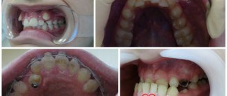
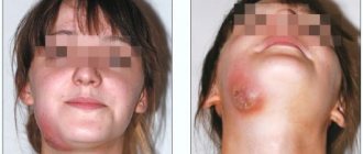
Observation of PG in a pregnant woman.
Observation of PG in a pregnant woman. Gestation period 9 months
Education appeared and grew in 1 month. The girl is 32 years old.
The term "botriomycosis" was proposed by the German veterinarian Otto Bollinger in 1887. 10 years later, the outstanding French surgeons Poncet and Dor for the first time at a congress of surgeons showed patients with characteristic, in their opinion, manifestations of equine botryomycosis.
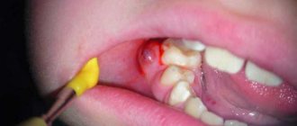
Granuloma appeared and grew within 2 months
Subsequently, it was proven that the surgeons’ opinion was erroneous, although the term “botryomycoma” is still used to this day.
Causes
The etiogenesis is usually based on minor injuries and a combined pyococcal polymicrobial infection (most often staphylococcal) with a virus. The initial idea of the fungal nature of the formations turned out to be erroneous.
Pathology can also develop against the background of such vascular formations as flaming nevus and juvenile hemangioma . At risk are young people, people who have taken oral contraceptives, those with dermatological or hormonal disorders, as well as pregnant women (in approximately 5% of cases).
Factors
Factors that can cause the manifestation of the disease can be injuries, pregnancy, infections, dermatosis. There have also been cases of granulomas appearing in places after a burn, after the use of oral contraceptives, protease inhibitors, and in the treatment of acne with the use of isotretinoin. After childbirth, PG disappears. In one of the previous studies in PG, a pregnant woman found an increase in the concentration of vascular endothelial growth; after pregnancy, their content was almost not detected.
Observation of PG in a pregnant woman. Gestation period 9 months
Symptoms
Usually botryomycoma does not cause significant discomfort, except for complexes regarding appearance. However, its injury and corresponding bleeding can be quite painful.
Pyogenic granuloma: distinctive appearance features
The appearance of pyogenic granuloma resembles millet grain, cherry bone or hazelnut, which is covered with crusts of a dirty brown color, in other cases with a papillomatous or smooth surface; a soft nodule is usually located under the neoplasm. It has a dense elastic consistency and an external smooth or, in most cases, uneven surface, which may have lobulated or papillary tissue patterns. The base can be thin or wide, bleed easily when injured, cause ulceration and necrotization processes with the formation of a hemorrhagic crust. Along the periphery at the base, a “collar” can be identified due to the exfoliating epidermis, as in the photo below.
Botriomycoma on the finger
Typically, neoplasms are single, having a soft-elastic structure with a characteristic painless or slightly painful organization. Externally, the formation is tumor-like, its size in rare cases is more than 1 centimeter in diameter, but can reach 3-4 cm.
Clinical case
Based on Lam S. et al, Eosinophilic granuloma/Langerhans cell histiocytosis: Pediatric neurosurgery update. 2015
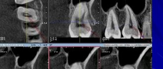
A 17-year-old young man was hospitalized due to an increasing scalp lesion over the past 6 weeks. The formation is painful on palpation and periodically bleeds due to ulceration, but no neurological deficit has been identified. CT and MRI revealed a large lesion in the frontal bone on the right, compressing the superior sagittal sinus. A total resection of the formation was performed, and the diagnosis of Langerhans cell histiocytosis of the skull bone was confirmed. At the outpatient stage, cytostatic therapy was carried out.
Figure 6. (a) CT examination without contrast - frontal scan (upper and middle part) and 3D reconstruction of the skull (lower part). (b) MRI scan. T1-weighted image in the coronal plane (top) and T2-weighted image in the sagittal plane
Sources
- Coppes-Zantinga A., Egeler RM The Langerhans cell histiocytosis X files revealed. Br J Haematol, 2002. - V. 116 - N. 1 - P. 3–9.
- Lam et al., Management of adult patients with Langerhans cell histiocytosis: recommendations from an expert panel on behalf of Euro-Histio-Net. Orphanet J Rare Dis, 2013 - V. 8. - N 72.
- Lam S., Reddy GD, Mayer R., Lin Y., Jea A. Eosinophilic granuloma/Langerhans cell histiocytosis: Pediatric neurosurgery update. Surg Neurol Int, 2015. - N. 6 (Suppl 17): S435 - S439.
- Langerhans' Cell Histiocytosis (Histiocytosis X). What is it? Harvard Medical School. Harvard Health Publishing. October, 2014. www.health.harvard.edu
- Sharma R., Singh R. et al. Langerhans cell histiocytosis (skeletal manifestations). Radiopaedia. https://radiopaedia.org/articles/langerhans-cell-histiocytosis-skeletal-manifestations-1
- Shea C. R, James W. D. et al. Langerhans Cell Histiocytosis. Medscape, 2022. https://emedicine.medscape.com/article/1100579‑overview
- Churilov L.P. Death on takeoff, or Who are you, Doctor Taratynov? Health is the basis of human potential: problems and ways to solve them, 2014. - T. 9 - No. 2 - P. 919–929.
- Yusupova L. A., Yunusova E. I., Garayeva Z. Sh., Mavlyutova G. I. Histiocytosis X. Practical Medicine, 2014 - T. 08 - No. 14.
Pyogenic granuloma in children
The prevalence of pyogenic granuloma among children and adolescents is quite high. It is very important that parents conduct regular visual examinations of the skin and mucous membranes of children and contact a specialist if they identify any papule-like or nodular formations of a conical, raised shape, as well as changes in pigmentation and growth of moles.
For children, a separate danger from botryomics is increased bleeding of the neoplasm, erosiveness and the addition of a bacterial infection, up to blood poisoning. Therefore, it is very important that the child knows the basic rules of asepsis and immediately contacts adults in the event of the slightest traumatic incident.
Botryomycomas are detected quite easily in children, because they are usually located on the hands, soles, face and lips, have a characteristic appearance and rapid growth.
Laboratory and instrumental diagnostics
A general blood test may show an increase in ESR, eosinophilia, leukocytosis, and a decrease in hemoglobin levels.
The use of various imaging methods (radiography, CT, MRI) allows us to identify foci of destruction up to 5 cm in size with clear boundaries without sclerotic changes, sometimes pathological fractures, flattening of the affected vertebrae (vertebra plana).
Pathomorphological examination is of decisive importance in the diagnosis of histiocytosis in adults and children. Microscopy reveals infiltrates of Langerhans cells (large oval cells with irregularly shaped nuclei), eosinophils, lymphocytes, and macrophages. Immunohistochemical analysis reveals the expression of CD1a, langerin, and S-100 proteins, characteristic of Langerhans cells. Electron microscopy allows one to see the Birbeck granules characteristic of Langerhans cells.
Differential diagnosis for EG is carried out with osteomyelitis, primary tumors or metastatic bone lesions, lymphoma, multiple myeloma, Papillon-Lefevre syndrome and bone cysts.
Figure 6. Birbeck granules, characteristic of Langerhans cells (electron microscopy data)
Diet for pyogenic granuloma (bothryomycoma)
Diet for skin diseases
- Efficacy: therapeutic effect after a month
- Time frame: three months or more
- Cost of products: 1400-1500 rubles per week
In order to avoid skin problems and maintain the body’s defenses against external negative factors, it is very important to adhere to the rules of a healthy lifestyle and follow a rational, balanced diet. In addition to the balance of BJU and additional intake of complex vitamin preparations, it is advisable to introduce into the diet:
- ginger is a product known for its antibacterial and tonic properties;
- turmeric is a healthy spice that has anti-inflammatory and antiseptic effects;
- various vegetables, including different types of cabbage and legumes;
- fresh juices and smoothies from various vegetables and fruits;
- nuts and sea fish, rich in omega-3 unsaturated acids, can prevent the development of inflammatory processes and strengthen the immune system ;
- garlic is another product with a powerful antimicrobial immunostimulating effect;
- Hot milk with honey is a great evening drink to help restore strength and improve sleep.