Tooth extrusion – what is this procedure? The essence of the method and indications for its use
Tooth extrusion is a separate dental procedure. Nowadays it is performed quite rarely, but in some clinical cases it is precisely this that makes it possible to preserve the root in case of severe destruction of the visible part of the tooth and subsequently, based on it, restore the crown using modern methods of restoration and prosthetics. Extrusion was first introduced to the world in 1973. The author of the technique was Dr. GS Heithersay, who proposed this procedure as an alternative to removing roots with fractures1. Read more about the essence of this method and how such treatment is carried out further in this article.
Why might extrusion develop?
Disc extrusion can begin if the protrusion has not received the necessary treatment for a long time. The main causes of this disease are usually osteochondrosis and related diseases. But this list is not limited to them. Here are other possible causes of extrusion:
- completeness;
- regular strong physical activity;
- age;
- metabolic problems;
- spinal injuries;
- lack of movement.
Strong physical exertion can lead to the development of extrusion
What is the essence of the procedure
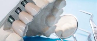
Tooth extrusion is the procedure of pulling a tooth or remaining root out of the gum in a vertical direction. According to this definition, extrusion allows a slight elevation of the root and creates more suitable conditions for a reliable and durable crown restoration. As part of this procedure, the tooth is usually pulled out by 3-4 mm. But to carry it out, it is important that at least the dentinal rim is preserved. Its thickness must be at least 1 mm, and its height must be at least 2 mm. If there is no such rim, the specialist may try to pull it out from under the gum.
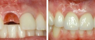
Tooth extrusion is a procedure for pulling out a tooth or remaining root from the gum.
This procedure in some cases allows you to avoid root removal and subsequently restore the crown on it. However, if the root is damaged, its complete extraction followed by replacement with an implant will be the best solution. By the way, extrusion promotes the formation of bone tissue, so the procedure can also be performed as an alternative to artificial expansion of the jawbone for subsequent installation of implants.
Tips for preventing illness
In the ranking of the importance of medical actions in relation to extrusion, one of the first places is occupied by the prevention of this disease.
To effectively prevent extrusion or its complications, certain measures can be taken:
- do physical exercise regularly. Overworking is harmful, but in order for the spine to be healthy, sufficiently trained back muscles are necessary;
- stop smoking and drinking alcohol;
- when working in a job that is harmful to the spine, it is better to change profession;
- take care of your posture, avoid wearing shoes with high (more than 40 mm) heels;
- organize your workplace wisely;
- take timely measures against infections and inflammations in the body;
- If you experience the slightest discomfort, consult a doctor immediately.
To prevent the disease from occurring, it is important to carry out prevention
Trying to treat yourself on your own is dangerous: it can lead to severe complications, as a result of which the person will lose his ability to work and may even remain disabled. At the first signs of discomfort, you should immediately contact a specialist.
Indications for extrusion
As mentioned above, this method is indicated for severe destruction and fracture of the crown of the tooth, as well as for a root fracture, but only if the fracture is in the gum. For reference: according to ICD-10, the root fracture is assigned the number S02.5. Among other indications for the use of this method, dental experts highlight retention. This diagnosis is made when the coronal part does not completely erupt or remains in the gum. This situation is especially typical for wisdom teeth.
“They performed extrusion on me. There was a tooth that was constantly being destroyed. The doctors increased it several times, but it became so dark and chipped all the time. When I once again came to the appointment, they again removed all the composite, they cleaned something there for a long time, and then they said that’s all, I can’t build it up anymore. Instead of removing it, they suggested pulling out the root a little. I agreed. The procedure went well and there was no pain. In the end, they installed a crown, and it’s holding up so far.”
Ira, review on the website www.32top.ru
This procedure can be life-saving if the carious cavity is localized below the gingival level. By removing the affected surface, it is possible to prevent the influence of harmful microorganisms on the mucous membrane and its inflammation.
Traction is also prescribed for retention
Another indication, which was already mentioned above, is insufficient thickness and height of the jaw bone to securely fasten the implant. Extrusion of the tooth root allows you to free up some space between it and the alveolus, which will promote the growth of bone tissue cells and connective epithelium.
Contraindications to the use of the method
Like any other dental procedure, extrusion has its contraindications. For example, it can only be carried out if the adjacent soft tissues and teeth are in satisfactory condition, since they will act as supports for fixing the traction structure.
The procedure will not be possible if the roots are too short or severely curved, and the condition of the canals and crown does not allow the installation of at least one of the elements of the corrective system: ligatures, a button or a pin with a hook.
Therefore, before prescribing extrusion, the doctor must assess the condition and length of the roots, their location. If the patient is diagnosed with an occlusion pathology in which there is no free space for traction, the procedure will not be possible. And one more thing: the method is indicated exclusively for pulpless teeth, so the nerves will first have to be removed if this has not been done previously.
How to recognize the disease
The symptoms of this disease are determined by which part of the spine begins to suffer from it. Extrusion involves incomplete prolapse of the central core of the disc, but this does not affect the nerve roots. So the disease usually does not cause noticeable symptoms such as pain. And only a pinched nerve will “inform” about the presence of problems and the need to consult a specialist.
In the case of cervical extrusion, if a nerve is pinched, the person will begin to have a headache, especially the back of the head. When turning the head, convulsions may occur. However, most often extrusion develops in the lower back. In this case, the symptoms of the disease are as follows:
- numbness of the lower extremities;
- tingling in the fingers of the lower extremities;
- buttocks or thighs go numb;
- pain is localized in a place “subordinate” to the affected nerve.
With this disease, sharp painful attacks occur quite rarely. The doctor makes a diagnosis based on completely different manifestations and symptoms. For example, testing the patient's knee reflex.
Back pain accompanied by the same sensation in the legs is a clear symptom of extrusion
Prices for orthopedic corsets and posture correctors
In most cases, a disc in the lumbar region falls out in fairly elderly people. At this age, the tissue becomes thinner, so extrusion is not uncommon in people over 60 years of age. At the same time, the thigh along the sciatic nerve may also hurt. The latter is characterized by an extremely high sensitivity to pain, so that the pain in the knees and legs can be very severe.
Young people often experience neck extrusion caused by poor posture when sitting at a desk. In this case, the following symptoms are often encountered:
Methods of carrying out the procedure
There are two main methods of extrusion. Let's look at each type in a little more detail:
- orthodontic is a more gentle method, which is often combined with correction with a brace system. This method is indicated for impacted teeth, but it requires more time and patience to achieve results. Pulling is carried out by installing an appropriate structure,
- surgical is a rather radical method, which is fraught with serious complications if errors are made on the part of the doctor. But if everything is done correctly, the result can be obtained much faster. The method does not require long-term wearing of the traction system. We will talk further about how extrusion is carried out in this case.
In the meantime, it should be noted that after surgical traction, the patient is given a temporary restoration for the period of tissue and ligament restoration. It allows you to quickly restore aesthetics.
Types of extrusion
Doctors perform two types of extrusion – orthodontic and surgical.
The orthodontic method is more gentle and can be combined with wearing braces; it is often used if there are impacted elements, but it requires more time and patience from the patient, as well as more visits to the clinic.
The surgical method is more radical, and if performed incorrectly, complications can occur. But with a technically purely performed operation, the result can be achieved faster. At the same time, you do not have to wear orthodontic devices, which during the treatment period violate the aesthetics of the smile and cause some discomfort.
During the period of tissue healing and ligament restoration after surgical extrusion, the patient is given a temporary restoration that makes the smile attractive - this is impossible with the orthodontic method.
Preparatory stage
Before the procedure, regardless of the chosen method, it is important to undergo a full diagnostic examination to ensure that the patient has no contraindications. You will need to take an X-ray or undergo a computed tomography scan, and a procedure performed by a prof. hygiene to remove plaque and deposits, as well as remove the nerve from the causative tooth if the crown is preserved and depulpation has not been performed previously.
Before the procedure, the patient should undergo a computed tomography scan
How is extrusion performed using the orthodontic method?
In this case, traction is carried out using a special orthodontic system. Its design includes the following components:
- arch or bracket - one of these parts is fixed on elements adjacent to the causative tooth using a composite or special orthodontic rings. If the patient already has braces, the bracket is not installed;
- a screw or a special pin with hooks is attached to the causative tooth, that is, to the remaining crown. If there is not enough hard tissue for this, the pin is fixed in the root canal using dental cement,
- traction – they combine the above-described structural elements and create a force effect.
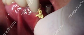
The photo shows the surgical method.
The system is fixed under local anesthesia, since to install it the doctor has to cut the fibers that connect the gum to the root. This manipulation is called fibrotomy. Due to this, the specialist gives the root mobility in order to speed up the pulling process and prevent it from returning to its original position.
Let's sum it up
Spinal disc extrusion can occur due to problems with metabolism or lack of exercise, so necessary to maintain health. If a person consults a doctor in time, when the disease has not yet “advanced,” then there is a high chance of getting rid of it with the help of conservative therapy. Therefore, it is very important not to hesitate to see a doctor - it determines how quickly relief will come and what the quality of life will be in the future.
How long does treatment last?
After the procedure, the patient will have to visit the orthodontist once a week so that he can monitor the process and adjust the tension of the rods. Usually, in one week, in this way it is possible to extend the root by about 1 mm. Therefore, to stretch it by 3-4 mm, you need to visit the doctor about 3-4 times, and the process itself can take up to a month.
But the treatment does not end there. It will take some time for the root and ligaments to recover. This process usually takes a couple of months. In the meantime, the patient is undergoing a temporary restoration with the removal of the causative element from the bite and its splinting - this is necessary to relieve the chewing load from it.
Expert recommendations during treatment
During the period of traction, orthodontic experts recommend increasing your oral care. You should brush your teeth not only in the morning and evening, but also every time after eating. It is also recommended to chew on the opposite side and try to eliminate any mechanical factors that could interfere with the healing process. If the structural elements rub the mucous membrane, you should resort to orthodontic wax.
Orthodontic wax will help protect the mucous membrane from damage
Corpus horizontal movement of the tooth. Elimination of three, diastema, protruding teeth.
Part 4.
aligner overview page here This article describes the solution to certain clinical situations in the oral cavity, in particular, the elimination of teeth and diastemas (gaps between teeth). The page is an excerpt from the work of Ryakhovsky A.N. and Arevadze T.Yu., for complete information, please see the source.
Most problems can be solved in different ways, the main thing is to choose the optimal one.
A rail attachment should also be used to close the diastema and trema (Photo 13).
Photo 13
Features of the surgical procedure
This method is most often used in cases where the crown part of the tooth is so damaged that it can no longer be restored in the classical way, or if it is necessary to re-prosthetize / replace the old restoration material. This procedure can also be performed if the patient wants to get results faster and without the need for temporary “aesthetic loss”.
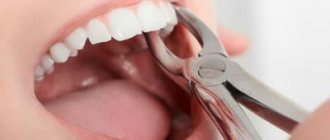
This operation is not classified as complex, but it requires appropriate experience and skill from a specialist. For pulling, the doctor uses a special scalpel, elevator and forceps. You could say he creates a dislocation and then does not completely remove the root, after which he fixes it with sutures.
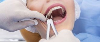
The photo shows the surgical method of the procedure
Hernia formation
A spinal hernia develops in four main phases. Each of them has its own characteristics. Their treatment is also different, and the later the phase of the disease, the more difficult it is to cure it.
Phase number one. Prolapsed intervertebral disc
Degenerative phase. Now the intervertebral disc has a deficiency of fluid - this leads to a drop in pressure in the disc, loss of elasticity, softening, and greater pliability. Although there is little fluid, the volume of disc tissue, of course, does not disappear anywhere, as does the axial load. Moreover, the load may later increase altogether, for example, if the body tends to be overweight or if a person does not eat properly. Because of this, a gradual protrusion of the disc wall begins, and it protrudes beyond the limits of the spine allocated by nature. This is just the first stage of the development of the disease.
Prolapse of the intervertebral disc is the first stage of hernia formation
There are still no significant symptoms. Only slightly higher back fatigue and accelerated fatigue from physical activity are noticeable. There is no pain yet, so patients usually do not pay any attention to this phase. They feel that there is no need to seek the help of a specialist.
Phase number two. Intervertebral disc protrusion
The nucleus pulposus has almost no previous shock-absorbing properties, the fibrous ring is also weakened, and all for the same reasons - due to insufficient fluid and nutrients. However, both the core density and the load on the spine remained the same. So now the nucleus begins to shift from its nature-prescribed position, and it puts pressure on the annulus fibrosus. Then everything follows the principle of a jackhammer: under any, even the smallest load, the nucleus “beats” the fibrous ring. Of course, this is far from a full-fledged blow, this is just an increase in pressure, but gradually it brings its results. One day, the annulus fibrosus begins to recede. This leads to an increase in protrusion of the intervertebral disc.
Protrusion is the second stage of development of intervertebral hernia
Before pain appears, the only symptom that can be noticed is fatigue. Often people, wanting to help a patient, can recommend that he play sports to restore his back. However, in most gyms, trainers understand “back rehabilitation” as something completely different. As a result, the patient greatly accelerates the development of the disease with incorrectly selected exercises.
Eventually the core finds a weakness in the already severely damaged annulus fibrosus and then “wedges” into it. Because of this, the second stage of the disease begins - protrusion of the intervertebral disc. We especially note: the fibrous ring is still intact, and the nucleus pulposus is still within its boundaries. So far there is only a strong protrusion of the disc.
How can protrusion be recognized?
In cases where the bulging disc does not come into contact with the nerve roots, this phase passes without any symptoms. If there is contact, the nature of the pain, as well as where it will be felt, will be determined by the location where the bulge occurs. The back, neck, and limbs may hurt. Sometimes the spaces between the ribs or the back of the head may ache. The muscles in the areas “subordinate” to the pinched root may become slightly weaker, and this is accompanied by problems with the sensitivity of these areas.
The main symptom of protrusion is severe pain
Protrusion has other symptoms:
Recommendations after surgery
As mentioned above, after the operation the tooth is covered with a temporary restoration. About a week after surgery, the sutures are removed. Over the next 2-3 weeks, the patient must adhere to a special diet - it is recommended to eat only soft, warm food, not too cold or hot. You will also need to strengthen your oral hygiene and treat soft tissues using an antiseptic solution, for example, Chlorhexidine, as prescribed by your doctor.
To treat the mucous membrane, you can use Chlorhexidine
1.5-3 months after the operation, the root acquires sufficient stability, which allows endodontic treatment and prosthetics to begin. Some experts insist that it is better to install a permanent prosthesis only 1-2 years after traction in order to prevent the development of complications. It will take quite a long time for the periodontal ligaments to fully recover.
How impacted elements are pulled out
Extrusion in this case has its own characteristics. It can be carried out in two different ways, but each of them necessarily includes both surgical and orthodontic stages. These are the two methods:
- delayed - first the doctor dissects the gum and exposes the crown. At the next appointment, 2-3 days later, he attaches a special orthodontic button and rods to the impacted element, after which he installs brackets on the adjacent supporting elements (if there are braces, there is no need for brackets),
- one-step – in one visit, the doctor cuts the gum, fixes the orthodontic system and applies sutures.
With the one-step method, the structure is installed immediately.
During rehabilitation, the specialist prescribes antibiotics and antiseptics to the patient. Again, you will have to adhere to a special diet and exclude any traumatic factors. Pulling in this way can last up to 1 year or even longer.
Advantages and disadvantages of the method
The main advantage of the method is the ability to preserve the root and subsequently carry out prosthetics based on it. Here is a list of other obvious advantages of this procedure:
- the ability to avoid expensive treatment, for example, prosthetic bridges or implantation,
- the ability to extend the life of a living tooth for up to 5-10 years and even more,
- the ability to avoid traumatic surgery to build bone tissue before installing implants.
The main thing is that an experienced, highly qualified doctor undertakes the treatment, since much here depends on the quality and accuracy of the manipulations performed. If a specialist makes mistakes, they can result in resorption and a decrease in root stability, and the development of ankylosis.
Among the disadvantages, patients highlight the duration of treatment and the inconvenience associated with it. Partly, it is the fact that pulling takes quite a long time, during which you have to adhere to strict dietary restrictions and be careful not to accidentally damage the system. But the result of such a correction can be considered the preservation of a living root, the opportunity to avoid more expensive and, by the way, no less lengthy implantation.
1Persii L.S. Orthodontics. Diagnosis and treatment of dental anomalies: a guide for doctors, 2004.