Fissure sealing is an extremely effective method of caries prevention and the main etiotropic method for the prevention of fissure caries. It involves the prevention of caries of the chewing surface of molars by filling fissures and other anatomical recesses of healthy teeth with adhesive materials - sealants in order to create a barrier to external cariogenic factors (microorganisms and carbohydrates)
Sealing tasks:
— eliminating the local risk of caries;
— creating conditions for the death of microorganisms remaining in the deep areas of the fissures; — elimination of potential reservoirs for the accumulation of cariogenic microorganisms; — acceleration of enamel mineralization in the fissure area when using glass ionomer cements.
Currently, glass ionomer cements, compomers, and composites are used for sealing.
Based on their chemical composition, sealants can be divided into groups:
1. Composite sealants: - classic, - sealants-ormokers, - flowable composite materials. 2. Compomers. 3. Glass ionomer cements Composite sealants can be: self-hardening and photo-hardening, for example: Estiseal LC, Fissurit, Helioseal.
Composite sealants and compomers are used to seal the fissures of teeth that have completely erupted.
Glass ionomer cements can be used for prophylactic coating of fissures in teeth at the eruption stage (Fuji VII, Fuji Triage).
Techniques for using sealants: - cleaning teeth with a regular brush, - cleaning with a circular brush with antiseptics, - applying material to fissures, - correction of teeth closure, - insulating the glass ionomer surface with varnish, - monitoring the safety of the sealant every 3-6 months.
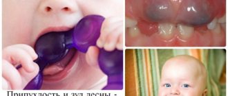
Toothpastes for children
Requirements for children's toothpastes: Fluoride content. Daily use of fluoride toothpaste in combination with proper brushing techniques is the basis for caries prevention! The use of low fluoride toothpastes containing less than 500 ppm F in the formula as a preventive measure is considered unsatisfactory. One of the problems associated with the use of fluoride toothpaste in children is the ingestion of the toothpaste by children with the subsequent risk of dental fluorosis.
Parents are advised to monitor the amount of toothpaste and the process of brushing teeth in children under seven years of age. Low abrasiveness. For temporary teeth (baby teeth) and newly erupted permanent teeth, the use of gel pastes is optimal. The RDA value for baby toothpastes should not exceed 50.
The absence of flavoring additives that can make a child want to eat pasta. It is preferable to use neutral, mint or fruit flavors that do not cause rejection in the child.
Examples of toothpastes for children aged 2 to 3 years: - Blend-a-med (0.005% NaF-250 ppmF) - My first Colgate (NaF) - Elmex enfant (aminofluoride - 250) - Lacalut Baby (aminofluoride – 250 ppm F, vitamins A, E) – Lacalut Kids 4-8 (aminofluoride – 500 ppm F, vitamins A, E) – Oral-B Stages fruity ( NaF – 500 ppm F) – Pokemon ( Na2PO3F- 500 ppm F)
Toothpastes for children from 2 to 6 years old: - Blend-a-med Junior Gel - Colgate junior (0.15% NaF- 680 ppm F-) - Colgate junior super star (0.76% Na2PO3F - 1000 ppm million F-) - Lacalut Kids 4-8 (aminofluoride - 500 ppm F-) - Lacalut Teens 8+ (aminofluoride and NaF -1400 ppm F-) - Dental dream for children (0.5% Na2PO3F -660 ppm F-) – Four Fruit – Mildfresh junior (0.76% Na2PO3F – 1000 ppm F) – Sanino Junior – New Pearl Junior 7-12 years (0.76% Na2PO3F – 1000 ppm F, oil tea tree) – Children's pearl complex – Prodent for teenagers
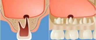
Cavity formation in caries of natural pits
Cavity formation in caries of natural pits
The natural pits on the chewing teeth include the depressions present on the buccal surface of the molars, as well as blind pits formed by the dental tubercles on the palatal surface of the front teeth (see Fig. 6, b, c).
Among the anterior teeth, blind fossae are more common on the small incisors, less common on the central incisors of the upper jaw, and almost never occur on the canines and lower incisors.
If the carious process that arose in the natural fossa on the buccal surface of the molar has not affected the entire fissure up to the chewing surface, then it is possible to create a box-shaped cavity without bringing it to the chewing surface. It is necessary that the cavity, as in all cases, be located not only in the enamel layer, but must penetrate into the dentin. During work, you should remember the proximity of the pulp chamber.
The carious process on the buccal surface of the molars creates a symmetrical round or diamond-shaped destruction of the tooth tissue. When forming a cavity for the tab, it is necessary to make it asymmetrical in order to facilitate the correct insertion of the tab. To do this, it is best to give it the shape of a trapezoid with rounded corners (Fig. 18, a). In some cases, it is enough to additionally include a nearby fissure in the cavity, which will serve as a guide when introducing the insert into the cavity (Fig. 18, b).
Formation ends with the creation of a fold along the entire edge of the cavity, which is best done with a small carborundum head.
The direction of wax removal should be perpendicular to the buccal surface of the tooth. Consequently, all work must be done with a fissure bur in a contra-angle handpiece and the direction of this bur must be perpendicular to the buccal surface of the tooth.
In cases where caries does not spread towards the pulp chamber, you can use a bur to follow the path of development of the carious process, excising
softened dentin. In this case, the drill can be given a direction slightly deviating from perpendicular, then all the walls of the cavity should be created at the same angle. The wax model should be drawn out in the same direction, and then the finished inlay should be inserted.
When the carious process spreads along the buccal fissure of a molar, a cavity is formed depending on the strength of the enamel roof hanging over it. If the roof remaining on the chewing surface above the cavity is thin, then in order to avoid breakages under the force of chewing pressure, it may be recommended to bring the cavity to the chewing surface.
Often the fissures of the chewing surface of the molars and the fossa on the buccal surface are simultaneously affected by the carious process. In this case, it is necessary to connect all the affected areas of the tooth into one common cavity. The direction of wax removal should be the same as for fissure caries, i.e., parallel to the long axis of the tooth (Fig. 18, c).
In this case, the contra-angle handpiece with the fissure bur should be held strictly parallel to the long axis of the tooth. Having created a cavity on the chewing surface, as described above, they move to the buccal surface of the tooth. The working part of the bur in this case will be the side surface.
While sparing the tooth tissue, you need to deviate from the strictly vertical direction of the bur, giving it a slightly inclined position, parallel to the groove on the buccal surface of the tooth (Fig. 18, d).
A fold is created along all edges of the cavity on both the chewing and buccal surfaces of the tooth.
When forming a cavity on the palatal surface of the upper anterior teeth, care must be taken, since in this area the pulp is located close to the surface of the tooth. Nevertheless, the rule of cavity formation not only in the enamel layer, but also in dentin, remains in force.
By excising tooth tissues affected by caries, they simultaneously create an asymmetrical cavity shape, for which they use natural depressions that are often present on the palatal surface of the incisors. These depressions can serve as retention areas where secondary caries may occur.
In its finished form, such a carious cavity on the palatal surface of the incisors most often represents a rounded cavity with one or two spurs (Fig. 18e).
The direction of wax removal should always be perpendicular to the palatal surface of the incisors. It is best to form a cavity using a fissure bur No. 1-3 in an angled tip directed perpendicular to the palatal surface of the tooth.
When, simultaneously with caries of the blind fossa on the palatal surface, there are carious cavities on the proximal surface of the incisor, it is advisable to combine both cavities into one common one or connect them together with a bridge.
The fold is made only on those edges of the cavity or bridge that extend onto the palatal surface of the tooth.
Anatomy: Internal base of the skull (basis cranii interna)
The inner base of the skull (basis cranii interna) has a concave, uneven surface corresponding to the shape of the base of the brain. There are three cranial fossae: anterior, middle and posterior. The posterior edges of the lesser wings (ala minor) and the tubercle of the sella turcica of the sphenoid bone (tuberculum sellae turcicae ossis sphenoidalis) separate the anterior cranial fossa (fossa cranii anterior) from the middle one (fossa cranii media).
The border between the middle and posterior cranial fossa (fossa cranii posterior) is the upper edges of the pyramids of the temporal bones (margines superiores partis petrosae) and the back of the sella turcica of the sphenoid bone.
When examining the internal base of the skull, numerous openings for the passage of arteries, veins, and nerves are visible.
Cranial fossae. The inner base of the skull is deepened; there are three cranial fossae: anterior, middle and posterior. These depressions deepen from the forehead to the back of the head, forming terraced structures. • The anterior cranial fossa is formed by the orbital parts of the frontal bones, the cribriform plate of the same bone and the large wings of the sphenoid bone (and is limited from the middle fossa by the small wings of the sphenoid bone and the tubercle of the sella). • The middle cranial fossa is formed by the body and greater wings of the sphenoid bone, the anterior surfaces of the pyramids and the squamosal parts of the temporal bone. • The posterior cranial fossa is formed by the occipital bone, the posterior surface of the pyramids and the internal surfaces of the mastoid processes of the temporal bones, the posterior part of the body of the sphenoid bone (dorsum sella).
1. The anterior cranial fossa (fossa cranii anterior) is formed by the orbital parts of the frontal bone (pars orbitalis ossis frontalis), on which the cerebral eminences and finger-like impressions are well defined, and by the ethmoidal plate of the ethmoid bone (lamina cribrosa ossis ethmoidalis), through the openings of which numerous bundles pass olfactory nerve fibers. In the center of the cribriform plate rises the cock's crest (crista galli), in front of which there is a blind foramen (Moran's foramen, foramen caecum), surrounded by the pterygoid processes of the cock's crest of the ethmoid bone and the legs of the frontal crest (Morand Sauveur Francois, 1697-1773 - French surgeon and anatomist), and frontal crest.
Near the crest of the ethmoid bone there is Palfyn's sinus - a space connecting to the frontal and ethmoidal cells (Palfyn Jean, 1650-1730 - French physician and anatomist).
2. The middle cranial fossa (fossa cranii media) is much deeper than the anterior fossa. The walls of the middle fossa are formed by the body and large wings of the sphenoid bone (corpus et alae majores ossis sphenoidalis), the anterior surface of the pyramids and the scaly part of the temporal bones (facies anterior partis petrosae et pars squamosa ossis temporalis). In the middle cranial fossa, the central part and lateral sections can be distinguished. The central part is occupied by the sella turcica with its pituitary fossa. At the bottom of the pituitary fossa of the body of the sphenoid bone there may be a non-permanent formation (found in 0.3% of adults) - Landucert's canal (syn.: craniopharyngeal canal, canalis craniofaryngealis). It penetrates the body of the sphenoid bone and opens on its lower surface (near the junction of the wings of the vomer) with a “pharyngeal” opening.
The canal contains a continuation of the dura mater of the brain in the form of a fibrous coupling containing connective tissue and blood vessels (veins) (Landuzert Fedor Pavlovich (1833-1889) - professor of the St. Petersburg Medical-Surgical Academy).
Anterior to the pituitary fossa, the chiasm groove (sulcus hiasmatis) is visible, leading to the right and left optic canals (canalis opticus), through which the optic nerves pass. On the lateral surface of the body of the sphenoid bone there is a well-defined carotid groove (sulcus caroticus), and near the apex of the pyramid an irregularly shaped foramen (foramen lacerum) is visible. Here, between the lesser and greater wings and the body of the sphenoid bone, there is the superior orbital fissure (fissura orbitalis superior), through which the oculomotor, trochlear and ophthalmic nerves pass into the orbit. Posterior to the superior orbital fissure there is a round foramen for the passage of the maxillary nerve, then an oval foramen for the mandibular nerve.
At the posterior edge of the greater wing of the sphenoid bone, the foramen spinosum is visible, through which the middle meningeal artery passes into the skull. On the anterior surface of the pyramid of the temporal bone there is a trigeminal depression (impressio trigemini) - Meckel's fossa (Meckel Johan Friederich (senior), 1724-1774 - German anatomist), lateral to it is the cleft of the canal of the greater petrosal nerve ( hiatus canalis nervi petrosi majoris) - Taren's foramen - an opening on the anterior surface of the pyramid of the temporal bone, through which the greater petrosal nerve exits, and the groove of the petrosal nerve (Tarin Pierre (1725-1761) - French physician and anatomist). Even more lateral and anteriorly there is a cleft (hole) of the canal of the lesser petrosal nerve and a groove of the lesser petrosal nerve.
Lateral and posterior to these formations, the roof of the tympanic cavity (tegmen tympani) and the arcuate eminence (eminentia arcuata) are visible. Between the carotid canal and the trigeminal ganglion - the Gasserian ganglion (syn. ganglion of the trigeminal nerve, ganglion trigeminale) on the pyramid of the temporal bone there is Gruber's notch (syn.: jugular notch, inciscura jugularis), covered with a thin bone plate (Gasser Johann Laurentius, 1723 -1769) - Austrian doctor and anatomist; Gruber Wenceslau Leopoldovich (Gruber WL, 1814-1890) - Austrian anatomist who worked in Russia). In the pyramid of the temporal bone, under the dura mater of the brain, there is a Dorello canal formed by it and the groove of the inferior petrosal sinus - a canal through which passes the inferior petrosal sinus, vessels and abducens nerve leading to the cavernous sinus (Dorello Paolo, born in 1872 .) - Italian anatomist). In the area of the apex of the pyramid of the temporal bone there is Princeteau's tubercle - an elevation to which the superior petrosal sinus adjoins (Princeteau Laurent (1858-1932) - French doctor and anatomist).
The topographic-anatomical landmark during surgical interventions on the labyrinth, less often on the cerebellum, is Trautmann's triangle - the region of the skull limited behind by the sigmoid sinus of the dura mater of the brain, in front - by the posterior semicircular canal of the inner ear, above - by the upper edge of the petrous part of the temporal bone (Trautmann Moritz ( Trautmann Moritz F., 1832-1902) - German surgeon).
3. The posterior cranial fossa (fossa cranii posterior) is the deepest. It is formed by the occipital bone, the posterior surface of the pyramids and the inner surface of the mastoid processes of the right and left temporal bones, as well as the posterior part of the body of the sphenoid bone and the posteroinferior angles of the parietal bones. In the center of the fossa there is a large (occipital) foramen, in front of it is the Blumenbach clivus (syn. slope of the skull, clivus), formed by the bodies of the sphenoid and occipital bones fused in an adult, on which the bridge (brain) and medulla oblongata lie (Blumenbach Johann ( Blumenbach Johann Friedrich, 1752-1840) - German physician and anatomist, zoologist and anthropologist).
Between the bodies of the occipital and sphenoid bones there may be an additional bone - Albrecht's bone (Albrecht Karl Martin Paul, 1851-1894 - German anatomist). In the posterior edge of the large foramen of the occipital bone, during development, the Kerckring bone stands out - the ossification point of the occipital bone (Kerckring Theodor, 1640-1693 - Dutch physician and anatomist).
Posterior to the foramen magnum (occipital) along the midline are the internal occipital crest (crista occipitalis interna) and the cruciate eminence (eminentia cruciformis). On the back surface of the pyramid, on each side, the internal auditory opening (porus acusticus in tern us) is visible, leading to the internal auditory canal (meatus acusticus internus). In its depths, the facial canal begins, in which the facial nerve passes. The vestibulocochlear nerve emerges from the internal auditory opening. At the bottom of the posterior cranial fossa behind the pyramids there is a paired jugular foramen (foramen jugulare), through which the glossopharyngeal, vagus and accessory nerves pass, and medial from it is the hypoglossal canal for the nerve of the same name. Through the jugular foramen, the internal jugular vein also exits from the cranial cavity, into which the sigmoid sinus continues, lying in the groove of the same name.
On the surface of the cranial vault, 3 cm posterior and above the upper edge of the external auditory canal, there is a Keen point, which is a topographic and anatomical landmark for puncture of the lower horn of the lateral ventricle of the brain (Keen William Williams, 1837-1932 - American surgeon).
On the inner base of the skull, in the region of the posterior cranial fossa, there is a Mouret zone - an area of the skull bounded above by the inferior petrosal sinus of the dura mater, behind - by the transverse sinus, in front and inside - by the internal auditory canal on the pyramid of the temporal bone; this area is a common site for cerebellar abscesses.
The boundary between the vault and the inner base of the skull in the region of the posterior cranial fossa is the groove of the transverse sinus (sulcus sinus transversi), which passes on each side into the groove of the sigmoid sinus (sulcus sinus sigmoidei).